Spina Bifida: Causes, Symptoms, Diagnosis, and Treatment

Spina bifida is a complex neural tube defect that happens during the initial stages of foetal development, where the spinal cord and the bones surrounding it do not develop properly. Depending on its severity, this condition can lead to a spectrum of challenges, from mild physical differences to significant neurological impairments. With its varying types and presentations, spina bifida remains a key topic of study in paediatrics and neurodevelopmental medicine.
For NEET PG aspirants, it is important to have a clear understanding of this condition. This guide offers a well-organised and easy-to-follow overview to help build both strong academic knowledge and practical skills.
What is Spina Bifida?
Spina bifida is a congenital condition that affects the spine. It occurs at an early stage of foetal development, whereby the neural tube that develops to comprise the brain and spinal cord fails to close properly.
As a result, the spinal cord is not encased by bones as it is supposed to be. In some cases, this may cause brain functioning and learning challenges.
What are the Different Types of Spina Bifida?
The types of spina bifida are mentioned below:
- Meningocele
This rare form is characterised by the development of a sac filled with fluid in the spine’s opening gap.
- Myelomeningocele
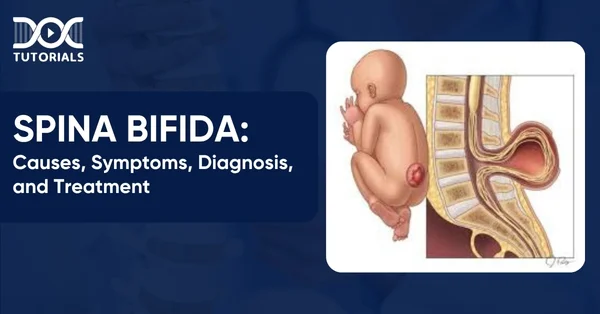
It is considered the most serious of spina bifida types. Myelomeningocele features a sac that contains parts of the spinal cord and nerves, protruding through the spinal gap.
- Spina Bifida Occulta
This is the most widespread form, which manifests itself in a narrow spinal gap without the development of a fluid-filled cavity.
What are the Causes of Spina Bifida?
Spina bifida causes can be genetic and environmental. The following is a detailed overview:
- Genetic Predisposition
The likelihood of having another child with spina bifida is around 4% if a parent or sibling is affected by the condition.
- Folate Deficiency
Vitamin B9, or folate, is crucial for healthy foetal development. In its synthetic form, folic acid is found in fortified foods and supplements. A lack of folate has been linked to neural tube defects, including spina bifida.
- Family History of Neural Tube Defects
Couples who already have one child with a neural tube defect can have another child with the same condition. This risk increases further if both previous children were affected. Additionally, women born with neural tube defects have a greater likelihood of having children with spina bifida.
- Medication Use
Certain drugs, especially anti-seizure medications such as valproic acid, have been associated with an increased risk of neural tube defects. These medicines may interfere with the ability of an individual’s body to utilise folate and folic acid properly.
- Diabetes
Women with poorly managed diabetes during pregnancy are at greater risk of giving birth to a baby with spina bifida.
- Obesity
A higher pre-pregnancy body weight is associated with an elevated risk of neural tube defects, including spina bifida.
- Alcohol Misuse
Excessive alcohol intake can lead to macrocytosis, a condition that damages folate. Recovery of bone marrow function after macrocytosis can take several months once alcohol consumption has ceased.
- Raised Body Temperature
Studies suggest that hyperthermia in early pregnancy may slightly increase the risk of spina bifida. This elevated temperature may result from fever or the use of saunas and hot tubs.
What are the Risk Factors of Spina Bifida?
The following are the risk factors of spina bifida:
- Inadequate folic acid intake during pregnancy
- Use of certain medications, such as specific anti-seizure drugs
- Existing health conditions, including diabetes or obesity
- High body temperature or fever in early pregnancy
- Personal or family history of neural tube defects, including having the condition oneself or a previously affected child
What are the Symptoms of Spina Bifida?
With spina bifida occulta, the only external indication can be a patch of hair or a spina bifida occulta birthmark over the area of the spine affected. For meningocele and myelomeningocele, a noticeable sac may protrude from the infant’s back. In meningocele, this sac is often covered by a thin layer of skin.
Myelomeningocele, on the contrary, usually has no skin cover, and the tissue of the spinal cord is exposed. This form is usually accompanied by additional spina bifida symptoms, such as:
- Weakness in the leg muscles, which in some instances may result in an inability to move the legs
- Abnormalities in the lower limbs or spine, including misshapen feet, uneven hips, or spinal curvature (scoliosis)
- Hydrocephalus, where fluid builds up in the brain’s cavities, potentially leading to an enlarged head, a bulging fontanelle, and impaired brain function
- Seizure activity
- Difficulties with bladder or bowel control
How to Diagnose Spina Bifida?
Spina bifida may be detected before birth through a range of prenatal screening methods, although these tests are not entirely conclusive. A positive result does not always indicate the presence of the condition, and a negative result does not completely rule it out. Discussions with a medical professional are essential to understand the benefits, limitations, and implications of each test.
They may include the following:
- Blood Tests
The following are the blood tests that need to be carried out:
- Maternal Serum Alpha-Fetoprotein (MSAFP): This test involves analysing a sample of maternal blood for levels of alpha-fetoprotein (AFP), a protein produced by the foetus. Elevated AFP levels may indicate a neural tube defect such as spina bifida, although they can also result from other factors.
- Confirmatory Blood Testing: If AFP levels are unusually high, further blood tests may be carried out to verify the results. Incorrect gestational age or multiple pregnancies can also influence AFP levels.
- Combined Screening: MSAFP is usually conducted with two or three other blood tests to detect conditions such as Down syndrome (trisomy 21).
- Ultrasound
Ultrasound is believed to be the best mode of identifying spina bifida even before birth. It can be done either in the first trimester (11 and 14 weeks) or, more accurately, in the second trimester (18 and 22 weeks) when anatomical structures are more defined. An advanced ultrasound may identify signs such as an open spine or brain abnormalities and can help determine the severity of the condition.
- Amniocentesis
In case spina bifida is proven by means of ultrasound results, an amniocentesis can be suggested. In this process, a needle is inserted into the amniotic sac, sampling a fluid which will then be analysed for genetic conditions. Although helpful, amniocentesis carries a small risk of pregnancy loss and should be carefully considered with medical guidance.
What are the Treatment Options for Spina Bifida?
Mild cases of spina bifida may not require any medical intervention. Nevertheless, more severe forms usually involve surgical procedures, along with ongoing therapies and support to manage associated complications and enhance mobility and daily functioning.
They may include the following:
- Foetal Surgery
The surgery can be done at an early stage when the baby is in the womb, which can be before the 26th week of pregnancy in some instances. In this process, the opening above the growing spinal cord is closed. While this early intervention may reduce the severity of disability, it carries significant risks for both the mother and baby, such as an increased chance of premature birth.
- Postnatal Surgery
Surgery may be carried out within the first one to two days following birth. In infants who have meningocele, the spinal hole is sealed up and the membrane that covers the spinal cord is repositioned. In cases of myelomeningocele, the exposed spinal cord and tissues are placed back inside the body and covered with skin.
- Management of Hydrocephalus
When there is a buildup of cerebrospinal fluid (CSF) in the brain ventricles, it can lead to a condition called hydrocephalus. Doctors may need to insert a ventricular shunt, which is a thin tube placed in the brain to divert excess fluid to another part of the body, where it can be reabsorbed.
- Subsequent Surgeries
Additional procedures may be required to address orthopaedic issues that include the feet, hips, or spine, or to replace a brain shunt if necessary. Progressive tethering may also require surgery, and the result is an abnormal attachment of the spinal cord inside the spinal canal. If untreated, it may result in muscle weakness or difficulties that make the child unable to control bowel or bladder functioning.
- Mobility Support
Early physiotherapy can be done to assist infants in developing more leg strength to use braces or crutches. Other children can use walkers or wheelchairs for mobility at a later age.
FAQs About Spina Bifida
- What is spina bifida?
Spina bifida is a congenital condition where the spinal cord and spine do not develop properly due to incomplete closure of the neural tube during early foetal development.
- What are the symptoms of spina bifida?
The symptoms of spina bifida vary by type and include:
- A visible sac on the back (in meningocele or myelomeningocele)
- Misshapen feet or spine (scoliosis)
- Leg weakness or paralysis
- Seizures
- Bowel or bladder dysfunction
- Hydrocephalus
- Is it possible to reduce the risk of spina bifida before and during pregnancy?
Yes, women can significantly lower spina bifida before and during pregnancy with proper preventive measures. They should take folic acid supplements prior to conception and throughout early pregnancy for effective results. In addition, avoiding known risk factors such as specific anti-seizure medications, managing diabetes, and maintaining a healthy body weight also contribute to reducing the risk.
Conclusion
Spina bifida may be complicated, but it is not fatal when detected early and well handled. It is imperative that NEET PG aspirants have a good knowledge of its types, causes, diagnostic approach, and treatment. Recognising risk factors and clinical features such as neural tube defects or hydrocephalus is crucial for accurate diagnosis and patient care.
To help you excel in both exams and clinical practice, DocTutorials offers high-yield video lectures, concise study notes, Quick Revision Programmes (QRPs), and mock tests.Join our NEET PG courses today and strengthen your medical foundation!
Latest Blogs
-

INI CET Exam 2025: Your Roadmap to Success – Key Topics, Strategies, and Lessons from Last Year’s Papers
The INI CET exam is more than just a test; it’s a significant milestone for many medical students aiming to…
-

INI CET Exam Success: Previous Year Question Papers & Ultimate Guide – INI CET PYQ
One can feel overwhelmed while preparing for the INI CET (Institute of National Importance Combined Entrance Test). A vast syllabus,…
-

INI CET Exam Pattern 2024: A Complete Guide with Subject-Wise Weightage
The Institute of National Importance Combined Entrance Test (INI CET) is your key to entering some of the most prestigious…


