Baker’s Cyst | Causes, Symptoms, Diagnosis, and Treatment
Baker’s cyst, also referred to as a popliteal cyst, is a widely studied knee pathology, the prevalence of which has been reported to be between 5% to 41% in adults who are subjects of knee MRI or ultrasound. The prevalence rates are lower in community or asymptomatic middle-aged cohorts, at 11.7%, in the middle- and older-aged populations, as determined through ultrasound screening.
As a NEET PG candidate, you must know that initial care is aimed at treating the underlying pathology of the knee (e.g., arthritic condition or meniscus injury), whereas symptomatic treatment can be reached through aspiration, steroid injection, or, rarely, excision. The majority of the ruptured cysts are treated conservatively with rest and elevation in case of no complications.
Keep reading to learn the causes, symptoms, diagnosis, and treatment options for a Baker’s cyst!
What is a Baker’s Cyst?
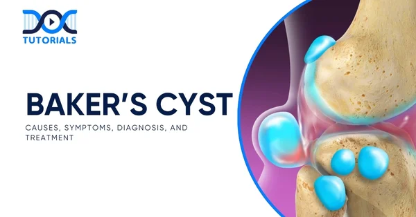
A Baker’s cyst (popliteal cyst) is a synovial fluid–filled sac in the posterior knee, usually arising from intra‑articular pathology like osteoarthritis or meniscal tear. It may cause swelling or tightness behind the knee, sometimes calf pain if ruptured, and is typically confirmed via ultrasound or MRI.
This condition is a distension of the gastrocnemio-semimembranosus bursa with synovial fluid, which is usually in continuity with the knee joint. It closely relates to intra-articular pathologies, especially osteoarthritis, meniscal injury, rheumatoid arthritis, and joint effusions.
Symptoms that patients report typically include stiffness of the back of the knee or discomfort, accompanied by a demonstrable mass or enlargement of the popliteal fossa, which manifests as swelling.
What are the Causes of Baker’s Cyst?
Check out a detailed overview of the Baker’s Cyst causes in the section below:
- Osteoarthritis
Degenerative joint diseases, such as osteoarthritis, serve as a major contributor to Baker’s cyst. Disintegration of cartilage due to osteoarthritis is accompanied by inflammation, which irritates the joint lining and triggers the production of more synovial fluid as a protective mechanism. This added fluid may drain into the popliteal bursa at the back of the knee, causing the formation of a cyst.
- Rheumatoid Arthritis
Being an autoimmune disease, rheumatoid arthritis leads to a continuing inflammation of the synovial membrane. This inflammation encourages the knee joint to produce excessive synovial fluid, after which the joint is not able to cope with the excess fluid levels.
As a result, the fluid may accumulate in the popliteal area, causing a cyst.
- Meniscal Tears
The cartilage (meniscus) cushion between the tibia and femur may undergo tears, which may interfere with joint stability and mechanics. This tends to bring about irritation and swelling within the knee joint, which promotes synovial effusion, which can develop into a Baker’s cyst.
- ACL Injuries
An ACL tear, either a partial or complete one, is a prevalent sports injury causing knee destabilisation. It usually causes lining bleeding and inflammation, which promotes swelling, resulting in the accumulation of synovial fluid in the bursa.
Baker’s cysts in young patients and children are chiefly idiopathic (they develop in the absence of a discernible underlying disease of the joint/injury), as opposed to adult patients. These cysts can be spontaneous, and often they are asymptomatic.
What are the Symptoms of a Baker’s Cyst?
A Baker’s cyst is often asymptomatic and may go unnoticed. In many cases, it’s detected incidentally during imaging tests, such as an MRI performed for unrelated knee issues.
However, if Baker’s cyst symptoms do occur, they may include:
- Pain localised to the back of the knee
- Stiffness or a feeling of tightness in the joint
- A sense of fullness or swelling behind the knee, especially noticeable during knee extension
- A soft lump that disappears on flexion of the knee
These symptoms typically worsen with prolonged standing or physical activity. When the cyst occurs alongside an underlying intra-articular pathology, like osteoarthritis, patients may also report generalised knee pain or crepitus.
In some cases, complications such as cyst enlargement or rupture may lead to additional signs, including:
- Warmth
- Redness
- Tingling
- Numbness
How to Diagnose a Baker’s Cyst?
When evaluating knees suspected of having popliteal cysts, various imaging techniques can be employed. These include:
- Plain Radiographs (X-rays)
Early in the diagnostic process, obtaining plain radiographs is helpful. Recommended views include:
- Posteroanterior Rosenberg view
- Lateral view
- Patellofemoral axial view
These X-rays help detect other knee conditions often associated with popliteal cysts, such as:
- Osteoarthritis
- Inflammatory arthritis
- Loose bodies within the joint
Occasionally, loose bodies may also be visible inside the Baker’s cyst on radiographs.
- Direct Arthrography
Baker’s cysts were identified with the help of direct arthrography in the past. It can be done by injecting the knee joint with gas or iodinated contrast. The joint is then manoeuvred to force the contrast into the cyst, after which it will be checked on radiography or by use of a fluoroscope to detect the presence of the contrast in the cyst.
Drawbacks of direct arthrography are:
- It is invasive due to the need for contrast injection.
- It exposes the patient to ionising radiation.
Because of these disadvantages, arthrography has largely been replaced by other imaging modalities.
- Ultrasound
Ultrasound is now often preferred over arthrography due to several advantages:
- It is noninvasive and does not involve radiation exposure.
- It is cost-effective and widely available.
- Ultrasound can detect Baker’s cysts with nearly 100% sensitivity.
However, it has the following limitations:
- It is highly dependent on the operator’s skill and experience.
- It cannot reliably differentiate Baker’s cysts from other cystic lesions like meniscal cysts or myxoid tumours.
- It does not provide detailed images of other intra-articular knee structures that are often involved in these cases.
- Magnetic Resonance Imaging (MRI) – The Gold Standard
MRI is considered the most accurate imaging modality for diagnosing Baker’s cysts. It excels in:
- Differentiating Baker’s cysts from other similar cystic lesions, such as meniscal cysts.
- Visualising soft tissue abnormalities comprehensively.
- Evaluating associated intra-articular disorders like meniscal tears, synovitis, or arthritis.
Despite its high diagnostic accuracy, MRI is expensive and not always necessary as a first-line investigation. In cases where detailed evaluation of the knee joint’s internal structures is not required, ultrasound remains a practical and effective screening tool.
What are the Treatment Options for Baker’s Cyst?
Not every case of Baker’s cyst has to be treated actively. The cyst is usually self-resolving without treatment, and it is seen in most cases, particularly in children.
However, in instances where Baker cyst forms due to a certain underlying knee condition, e.g., arthritis or meniscal injury, symptoms tend to clear up when the correct management of such a condition has been done.
Other Baker’s cyst treatment options include:
- Pain Relief: Over-the-counter painkillers can help manage discomfort.
- Steroid Injection: A hydrocortisone injection directly into the knee joint may reduce inflammation and relieve pain.
- Physiotherapy: Exercises aimed at strengthening the muscles around the knee can improve joint stability and reduce symptoms.
- Aspiration: Removing excess fluid from the cyst using a needle can provide temporary relief.
- Surgery: In rare cases, surgery may be needed to fix damage within the knee joint. However, surgical removal of the cyst itself is uncommon.
It’s essential to note that Baker’s cysts may take several months or even years to resolve fully.
FAQs About Baker’s Cyst
- Is a Baker’s cyst contagious?
A Baker’s cyst is a sterile, fluid‑filled swelling behind the knee due to joint fluid leakage, not infection, so it’s not contagious. It arises from intra‑articular issues like arthritis or meniscal tears, not transmissible agents.
- Can I live a normal life with a Baker’s cyst?
Yes. Most cysts are asymptomatic or produce mild discomfort. Treating the underlying knee issue (e.g., arthritis, meniscal tear) and conservative measures (rest, physiotherapy, NSAIDs) usually allow full daily activities. It rarely requires surgery.
- Can children get a Baker’s cyst?
Yes, though rare in children aged 4–8 years. Paediatric cysts are typically primary, asymptomatic, and unrelated to joint pathology. They usually resolve spontaneously over months to years without intervention.
- Is a Baker’s cyst normal?
A Baker’s cyst is benign and not dangerous. In adults, it often signals underlying pathology. In children, it’s generally harmless. It is a common clinical finding and does not imply malignancy.
- How do I know that I have a Baker’s cyst?
Diagnosis begins with examination, and visible or palpable swelling behind the knee is more obvious when standing. Confirmation is done by ultrasound or MRI. Transillumination may also help.
Conclusion
Recognising symptoms such as sudden swelling of the knee, tightness, and pain behind the knee could be an indication of a Baker’s cyst, particularly in people with related joint diseases like arthritis or meniscal injuries. It is necessary to diagnose the problem early and provide appropriate management to avoid complications, suppress the symptoms, and retain joint functionality.
For NEET PG aspirants, having a clear understanding of Baker cyst is not only essential for covering the syllabus but also for performing accurate diagnoses in clinical settings. To assist students in this regard, DocTutorials offers high-quality video lectures, concise high-yield notes, mock tests, and more, helping you take your exam prep to the next level.
Check out our NEET PG courses today!


Latest Blogs
-

INI CET Exam 2026: Your Roadmap to Success – Key Topics, Strategies, and Lessons from Last Year’s Papers
The INI CET exam is more than just a test; it’s a significant milestone for many medical students aiming to…
-

INI CET Exam Success: Previous Year Question Papers & Ultimate Guide – INI CET PYQ
One can feel overwhelmed while preparing for the INI CET (Institute of National Importance Combined Entrance Test). A vast syllabus,…
-

NEET PG 2026 Preparation with Live Power Pack: Your Path to Success in 2026
Preparing for the NEET PG 2026 can be an overwhelming journey because of its vast syllabus and the pressure to…


