Muscles of the Gluteal Region: Anatomy, Functions, and Disorders

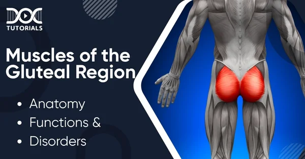
The gluteal region comprises the region located posterior to the pelvic girdle of the body and overlaps the side of the pelvis as well. This part consists of multiple muscles that support the human body’s erect posture. The gluteal muscles have a key role in supporting the trunk against gravity and are a must-know topic for every NEET-PG aspirant.
DocTutorials makes sure that you never miss out on any high-yield topic like gluteal muscles. Thus, we have developed this guide, which contains a detailed overview of the gluteal muscles, their anatomy, functions, disorders, and other crucial aspects.
Keep reading!
Which Muscles Make the Gluteal Region?
The gluteal area consists of multiple muscles that stabilise the hip and help to move the torso. These muscles can be divided into two major groups:
- Superficial Muscles
This group comprises the abductors and extensors that abduct and extend the femur bone. The muscles include:
- Gluteus Maximus
- Gluteus Medius
- Gluteus Minimus
- Tensor Fascia Lata
- Deep Muscles
These muscles include the lateral rotators that majorly work on the internal rotation of the femur. They include:
- Piriformis
- Gemellus Superior
- Gemellus Inferior
- Obturator Externus
- Obturator Internus
- Quadratus Femoris
The superior and inferior gluteal arteries, branches of the internal iliac artery, facilitate the blood supply to these muscles. Venous drainage follows along the arterial supply.
What are the Anatomy & Functions of the Gluteal Muscles?
- Gluteus Maximus
This is the heaviest muscle in the human body and makes up the major chunk of the gluteal region.
- Attachment: The muscle originates from the gluteal surface of the ilium, sacrum, and coccyx. It inserts into the femur’s iliotibial tract and gluteal tuberosity.
- Actions: Its main function is the extension and lateral rotation of the thigh. This muscle is also used in strenuous exercises like running or climbing.
- Innervation: The muscles are innervated by the inferior gluteal nerve.
- Gluteus Medius
A fan-shaped muscle, lying between the gluteus maximus and minimus.
- Attachment: This muscle originates from the gluteal surface of the ilium and inserts into the lateral surface of the greater trochanter of the femur.
- Origin: Abducts and medially rotates the lower limb. It also facilitates the stabilisation of the pelvis during movements.
- Innervation: The muscle is innervated by the superior gluteal nerve.
- Gluteus Minimus
It is the smallest of the superficial muscles and functions similarly to the gluteus medius.
- Attachment: The muscle originates from the ilium and converges to form a tendon. Its insertion is at the anterior side of the greater trochanter.
- Actions: It has a similar function to the gluteus medius.
- Innervation: Innervation is by the superior gluteal nerve.
- Tensor Fascia Lata
It is a small muscle that tightens the fascia lata.
- Attachment: The muscle originates from the crest anterior superior iliac spine and inserts into the iliotibial tract, which in turn attaches to the lateral condyle of the tibia.
- Actions: Its major function is to assist the gluteus medius and minimus in facilitating medial rotation and abduction of the leg. It plays a key role in maintaining the gait cycle.
- Innervation: By the superior gluteal nerve.
- Piriformis
The piriformis is a key landmark muscle for identifying the deep muscles. It is the most superficial of all the deep muscles.
- Attachment: This muscle originates from the anterior surface of the sacrum and finds its insertion at the greater trochanter of the femur.
- Actions: Facilitates lateral rotation and abduction of the leg.
- Innervation: The anterior rami of S1 and S2 spinal nerves form the nerve supply of this muscle.
- Gemelli Muscles (Gemellus Superior and Inferior)
The gemelli muscles are two triangular muscles that are separated by the tendon of the obturator internus.
- Attachment: The superior muscle originates from the ischial spine, while the inferior originates from the ischial tuberosity. Both these muscles are inserted into the greater trochanter of the femur.
- Actions: They act the same as the other deep muscles of the gluteal region.
- Innervation: The superior muscle is innervated by the nerve to the obturator internus, while the inferior muscle is innervated by the nerve to the quadratus femoris.
- Obturator Externus
A triangular-shaped muscle that covers the outer surface of the anterior wall of the pelvis.
- Attachment: The muscle originates from the outer surface of the obturator membrane and the bony margins of the obturator foramen. It inserts into the trochanteric fossa by forming a tendon.
- Actions: Same as the other deep muscles.
- Innervation: By the posterior division of the obturator nerve.
- Obturator Internus
This muscle is the major forming block of the lateral wall of the pelvic cavity.
- Attachment: The origin is from the pubis, and the insertion is at the greater trochanter.
- Actions: Same as the other deep muscles.
- Innervation: By the nerve to obturator internus.
- Quadratus Femoris
The quadratus femoris is a flat, square-shaped muscle. It is the deepest muscle of the gluteal region.
- Attachment: It originates from the upper part of the outer border of the ischial tuberosity and gets inserted into the quadrate tubercle and the area below it.
- Actions: Same as other deep muscles of the gluteal region.
- Innervation: By the nerve to the quadratus femoris.
What are the Disorders of the Gluteal Muscles?
Disorders of the gluteal muscles need immediate attention as they affect the entire locomotion process. Some of the common disorders of the gluteal muscles include:
- Trendelenburg Gait
An injury to the superior gluteal nerve paralyses the gluteus medius, gluteus minimus, and the tensor fascia lata. It leads to the loss of certain movements like abduction and rotation of the hip. It also prevents the stabilisation of the pelvis while walking, and the pelvis drops to the unsupported side. This gait disorder is known as Trendelenburg gait.
- Glute Muscle Weakness
The gluteus maximus only activates when doing a heavy exercise like running or climbing. Long-term inactivity can lead to the weakening of the muscles and a longer time to activate. Some conditions like the dead butt syndrome, hamstring pain, and lower back pain can also lead to weakening of the glutes.
FAQs about Gluteal Muscles
- Which 6 muscles make up the deep gluteal muscles?
The deep gluteal muscles comprise the piriformis, obturator internus, obturator externus, superior and inferior gemelli, and quadratus femoris.
- What is meant by the gluteal muscles?
The gluteal muscles are a group of muscles that form the buttocks area.
- Which part of the body is defined as the buttocks?
The posterior part of the hip, located between the lower back and the perineum, is known as the buttocks.
- Which is the biggest muscle in the body?
The gluteus maximus is the largest and heaviest muscle in the body.
Conclusion
The gluteal muscles make up an important part of the muscles involved in locomotion. They are extremely crucial for daily activities and make up an important topic for the upcoming NEET PG exam. Knowing about the important aspects of gluteal muscles, like their anatomy, function, and disorders, is quintessential to covering the exam syllabus and achieving a high score.
To assist students in their preparation, DocTutorials offers NEET PG courses comprising informative video lectures, faculty-reviewed question banks, NEET PG test series, and more. Join our courses today and ace your medical exams!
Latest Blogs
-

NEET PG Exam 2025- Date, Pattern, Marking Scheme, Subject Wise Weightage, and Exam Mode
NEET PG Exam 2025 is the ultimate gateway for medical graduates aspiring to pursue postgraduate courses in medicine, including MD,…
-

INI CET Exam 2025: Your Roadmap to Success – Key Topics, Strategies, and Lessons from Last Year’s Papers
The INI CET exam is more than just a test; it’s a significant milestone for many medical students aiming to…
-

INI CET Exam Success: Previous Year Question Papers & Ultimate Guide – INI CET PYQ
One can feel overwhelmed while preparing for the INI CET (Institute of National Importance Combined Entrance Test). A vast syllabus,…


