Important Topics for Anatomy in MBBS: Strengthen your Foundation
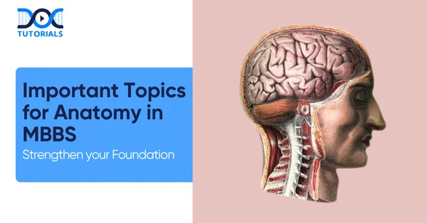
Anatomy forms the foundation of a future doctor’s career, helping them understand the human body better. Hence, students need to clarify their concepts and review the essential topics with significant clinical importance.
Having a good grip on anatomy not only helps them to manage patients smoothly but also prepares them for their medical career and clinical practice. Continue reading to learn about the important topics with exam-focused areas and essential questions of anatomy.
MBBS Anatomy Exam Pattern
The MBBs anatomy theory paper has been divided into two papers from the 2019 batch, each of which is worth 100 marks. The papers consist of:
- Short answer questions.
- Long answer questions.
- Application and case-based questions.
However, in NEET-PG, you can expect a weightage of around 10 to 15 questions, while in INI-CET, you can expect around 10 questions.
High-Yielding Topics and Important Questions for Anatomy in MBBS
Anatomy is divided into several essential branches, each containing high-yielding topics that need to be emphasised by students. The following section lists the issues along with important topics and essential questions that have been repeated over the years:
1. Neuroanatomy
- Nerve Supply
- Venous Drainage of any Region
- Cavernous Sinus
- Sciatic Nerve
- Saphenous Vein
- Arterial Supply
- Third Ventricle
- Weber’s Syndrome
- Parts of the Diencephalon
Important Questions
- Enumerate:
- Ascending tracts, along with the sensations carried by them.
- Arteries supplying the spinal cord.
- Branches of the vertebral artery.
- Branches of the cerebral part of the internal carotid artery.
- Recesses of the third ventricle.
- Draw Labelled Diagrams of:
- Circle of Willis.
- Transverse section of medulla at the level of pyramidal decussation.
- Floor of the fourth ventricle.
- Superolateral surface of cerebrum showing important sulci, gyri, and functional areas.
- Transverse section of midbrain.
- Write Short Notes on:
- Arterial supply of cerebrum.
- Blood-brain barrier.
- White matter cerebrum.
- Lateral ventricle
- Hydrocephalus.
- Lateral spinothalamic tract.
- Long-Answer Questions:
- Write the difference between upper and lower motor neuron paralysis.
- Describe the internal capsule under:
- Parts
- Arterial Supply
- Constituent Fibres
- Applied
- Write the anatomical basis for:
- Syringomyelia
- Pontine Haemorrhage
- Argyll Robertson Pupil
- Parkinson’s Disease
- Arnold-Chiari Malformation
2. Histology
- Adrenal Gland
- Thymus
- Uterus Histology
- Thin and Thick Skin
- Goblet Cells
- Karyotyping
- Types of Epithelium
- Serous Salivary Gland
- Spinal Cord
- Elastic Artery
- Liver Histology
- Thyroid and Parathyroid Gland
- Lymph Node
- Sweat Gland
Important Questions
- Define Meissner’s Plexus.
- Define Microvilli.
- Microscopic structure of retina.
- What are the three layers of the Mucosa?
- Which type of epithelium lines the thyroid follicles?
- Microscopic anatomy of the spiral ganglion.
- Microscopic anatomy of the prostate gland.
- How do you identify Barrett’s oesophagus?
- Microscopic structure of the ileum.
- Where is the dense, irregular connective tissue found?
3. Thorax
- Subclavian Artery
- Lung Hilum
- Diaphragm with Embryology
- Blood Supply
- Broncho-Vascular Segments of the Lung
Important Questions
- Enumerate:
- Intercostal muscles and their actions.
- Differences between the right and left lungs.
- Medial branches from the thoracic part of the sympathetic chain/trunk.
- Contents of the costal groove from above downwards.
- Tributaries of the coronary sinus and thoracic duct.
- Sites of constrictions of the oesophagus.
- Branches of the arch of the aorta.
- Branches of the right and left coronary artery.
- Draw a labelled diagram of:
- Structures passing through the hilum of the right and left lungs.
- Bronchopulmonary segments of the right and left lung.
- Contents of a typical intercostal space.
- Branches of a typical intercostal nerve.
- Arterial supply and Venous Drainage of the heart.
- Write a short note on:
- Pleural recesses.
- Transverse and oblique sinus of the heart.
- Thoraco-abdominal diaphragm.
- Azygos system of veins.
- Thoracic duct.
- Superior and Posterior Mediastinum.
- Long-Answer Questions:
- Describe the thoracic duct under:
- Origin.
- Course.
- Tributaries and areas drained.
- Applied aspect.
- Describe the arterial supply of the heart under:
- Origin and course of the coronary arteries.
- Branches and distribution.
- Cardiac dominance.
- Applied.
- Write the anatomical basis of:
- Oesophageal Varices.
- Achalasia Cardia.
- Thoracic Inlet Syndrome.
- The prognosis of Coronary artery disease is better in older people.
4. Abdomen
- Spermatic Cord Contents
- Ureter
- Urethra
- Perineum
- Pudendal Canal
- Rectum-Anal Canal Anatomy
Important Questions
- Enumerate:
- Muscles of the anterior abdominal wall.
- Structures lying at the transpyloric plane.
- Structures crossed by the mesentery.
- Retroperitoneal organs.
- Arteries supplying the stomach.
- Posterior relations of the Caecum.
- Draw a labelled diagram of:
- Formation of the rectus sheath.
- Vertical disposition of the peritoneum.
- Arterial supply of the stomach.
- Various positions of the apex of the appendix.
- Lymphatic drainage of the stomach.
- Cross-section of the spermatic cord.
- Write a short note on:
- Eploic Foramen.
- Second part of the Duodenum.
- Appendix.
- Caecum.
- Extrahepatic biliary apparatus.
- Lymphatic drainage of the stomach.
- Subphrenic and sub-hepatic spaces.
- Long answer questions:
- Describe the rectus sheath under:
- Formation.
- Contents
- Applied.
- Describe the stomach under:
- Blood Supply.
- Stomach Bed.
- Applied Anatomy.
- Location and parts.
- Lymphatic drainage.
- Describe the pancreas under:
- Location and parts.
- Relations.
- Arterial supply.
- Applied anatomy.
- Write the anatomical basis of:
- Varicocele is more commonly seen on the left side.
- Caput Medusae.
- The lesser curvature has a higher incidence of gastric ulcers.
- Cholecystitis pain is seen on the right side of the tip of the right shoulder.
5. Upper and Lower Limbs
- Muscles of the sole
- Axillary Artery
- Dermatomes of the upper and lower limbs
- Arches of the Foot
- Brachial Plexus
Important Questions
- Enumerate:
- Contents of the Axilla.
- Structures attached to the coracoid process of the scapula.
- Muscles supplied by the median nerve in the hand.
- The muscles responsible for the eversion of the foot.
- Branches of the popliteal artery.
- Structures passing deep to the flexor retinaculum from the medial to the lateral side.
- Draw a labelled diagram of:
- Adductor canal.
- Femoral sheath.
- Boundaries and contents of the popliteal fossa.
- Boundaries and contents of the femoral triangle.
- Brachial plexus.
- Anastomosis around the elbow.
- Cutaneous innervation of the hand.
- Anastomosis around the scapula.
- Write a short note on:
- Arches of the foot.
- Popliteal fossa.
- Adductor Canal.
- Lymphatic drainage of the mammary gland.
- Elbow joint.
- Lumbricals.
- Long answer questions:
- Describe the brachial plexus under:
- Formation and parts.
- Branches.
- Applied aspect.
- Describe the shoulder joint under:
- Types and articular surfaces.
- Applied aspect.
- Capsule and ligaments.
- Movements and muscles producing them.
- Describe the sciatic nerve under:
- Origin and course.
- Structures innervated.
- Applied.
- Describe the hip joint under:
- Types of joint and articular surfaces.
- Capsule and ligaments.
- Movements and muscles responsible.
- Applied anatomy.
- Write the anatomical basis of:
- Foot drop.
- Sleeping foot.
- Meralgia Paresthetica.
- Erb’s and Klumpke’s paralysis.
- Student’s elbow.
- Tennis elbow.
- Pulsating scapula.
6. Embryology
- Cleft Lip
- Varicose Veins
- Annular Pancreas
- Placenta Previa
- Tetralogy of Fallot
- Polycystic Kidney
- Rptation of Midgut
- Ectopic Pregnancy
- Neural Crest Derivatives
- Hemorrhoids
- Development of RA
- Congenital Anomalies of the Kidney
- Imperforate Anus
- Tracheoesophageal Fistula
- Germ Layers
7. Joints
- Temporomandibular Joint
- Ankle Joint
- Hip Joint: Relations and Ligaments
- Knee Joints: Capsule, Menisci, and Locking Unlocking
8. Osteology
- Orbital Bone
- Lateral Wall of the Nose
- Skull Bones
- Types of Switchers
- Middle Ear Anatomy
- Sphenoid Bone
- Types of Joints
9. Reproduction
- Spermatogenesis
- Fertilisation and Implantation
- Results of Fertilisation
- Abnormal Sites of Implantation
10. Pelvis and Perineum
- Haemorrhoids.
- Perineal Body.
- Urogenital Triangle.
- Support of the Uterus.
- Pelvic Diagram.
- Pudendal Canal
- Descent of the Testis
- White Line of Hitton
- External anal Sphincter.
- Internal Iliac Artery Brancjes
- Sphincters of the Anal Canal.
- Lateral and Anterior Relation of the Ovary.
Important Questions
- Enumerate:
- Contents of the broad ligament.
- Supports of the rectum.
- Muscles forming the pelvic diagram.
- Posterior relations of the rectum.
- Structures opening into the prostatic urethra.
- Contents of the ischiorectal fossa.
- Draw a labelled diagram of:
- Normal position of the uterus.
- Interior of the anal canal.
- Venous drainage of the rectum and anal canal.
- Lymphatic drainage of the uterus.
- Muscles attached to the perineal body.
- Write a short note on:
- Pudendal canal.
- Perineal body.
- Supports of the uterus.
- Broad ligament.
- Nerve supply of the urinary bladder.
- Pelvic diagram.
- Long answer questions:
- Describe the urinary bladder under:
- Parts and relations.
- Internal features.
- Nerve supply.
- Applied.
- Describe the uterus under:
- Parts and relations.
- Supports.
- Lymphatic drainage.
- Applied.
- Describe the rectum under:
- Extent and curvatures.
- Relations.
- Blood supply.
- Applied.
- Write the anatomical basis of:
- Internal haemorrhoids.
- Prolapse of the uterus.
- Atonic bladder.
- Ischioanal hernia.
- Pudendal nerve block.
- Autonomous bladder.
- Anorectal fistula.
11. Important Derivatives
Derivatives of:
- Ectoderm
- Paramesonephric duct
- Mesonephric duct
- Midgut
- Secondary Yolk Sac
12. Important Diagrams
- Floor of the fourth ventricle
- Sensory nerve supply of the face
- Cubital fossa
- Brachial Plexus
- Lateral Wall of Nose
- TS of:
- Midbrain at the level of the superior and inferior colliculus
- Medulla at the level of the sensory decussation
- Brainstem at the lower level of the pons
- Medulla at the level of the pyramidal decussation
- Digastric Triangle: Boundaries, contents
- Horizontal Sec of the brain through the anterior and posterior horns of the lateral ventricle
13. Important Lobes to Retain
- Heart: Blood Supply, Coronary Sulcus, and RA
- Arches of the Foot
- Lungs: Pleural Recesses, Pleura and Bronchopulmonary Segments
- Duodenum: 2nd and 3rd Part
- Urinary Bladder: Location, relation, Nerve Supply, Anatomy, and Gross
- Portal Vein and Portocaval Anastomosis
- Diaphragm: Origin, Insertion, Opening, and Developmental Basis of Innervation
14. Other Important Topics
- Sciatic Opening
- Psoas Major
- Azygous Vein
- Profunda Femoris
- Anal Canal
- Gluteus Maximum
- Diaphragmatic Opening
- Spermatic Cord Contents
- Fontanelles
- Circle of Willis
- Ape Thumb Deformity
- Bells Palsy
- Fontanelles
- Cortico Spinal Tract
- Flexor Retinaculum
FAQs about Important Topics for Anatomy in MBBS
- What are the most critical topics in anatomy?
Some of the important topics in anatomy are:
- Various systems such as cardiovascular, skeletal, nervous, etc.
- Neuroanatomy.
- Embryology.
- Terminologies.
- Is anatomy high weightage in NEET PG?
Being a pre-clinical subject, anatomy has lesser weightage, and hence, there are few questions from anatomy in NEET-PG.
- What are the seven ways to study anatomy?
The seven effective ways to learn anatomy are:
- AV aids.
- Retention.
- Group Study.
- Anatomical terminologies and clearing the basics.
- Take tests.
- What are the common mistakes in anatomy learning?
The most common mistake students make when learning anatomy is that they focus solely on memorising the points rather than understanding the core concepts, which leads to difficulties later.
Conclusion
Having a clarity of the concepts in anatomy lays the foundation for a stronger approach towards clinical practice. Students have a vivid understanding of the various essential systems in the human body, which helps them to diagnose and treat patients effectively in the future.
With a proper study plan and reliable resources, students can prepare with DocTutorials. We offer CBME-aligned syllabus, animated video lectures, previous year questions (PYQs), and structured mentorship to help student unlock their true medical potential.
Join DocTutorials today and explore our MBBS curriculum to excel in your medical career.
Latest Blogs
-

INI CET Exam Pattern 2025: A Complete Guide with Subject-Wise Weightage
The Institute of National Importance Combined Entrance Test (INI CET) is your key to entering some of the most prestigious…
-

NEET PG Registration 2025: An Essential Guide For Exam Prep
The NEET PG registration, which is conducted online, is a crucial step in the exam process. Filling out the NEET…
-

NEET PG Syllabus 2026: A Must-Have Complete Guide for Exam Success
The NEET PG Syllabus acts as one of the foundation stones for aspiring postgraduate medical students like you who are…
