Teres Minor Muscle: Anatomy, Function, and Clinical Importance
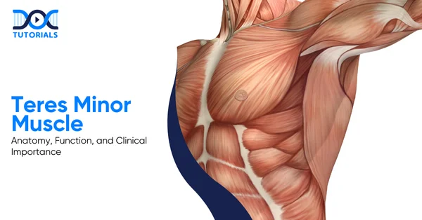
The teres minor is the smallest posterior muscle out of the 4 muscles in the rotator cuff (a cluster of four muscles that permits wide mobility of the human shoulder) that connects the scapula to the humerus and primarily assists in external rotation of the arm.
This muscle, although less discussed than other muscles of the rotator cuff, plays a crucial role in stabilising the shoulder, particularly during overhead and rotational movements.
Read below to know more about the anatomy, functions, clinical relevance, and common pathologies of the teres minor muscle in detail.
What is the Teres Minor Muscle and its Anatomy?
The teres minor is a small but essential posterior shoulder muscle that forms part of the rotator cuff and connects the scapula to the humerus. It plays a key role in stabilising the shoulder joint and enabling external rotation and adduction of the arm.
Teres minor is an intrinsic muscle of the shoulder region. It is a crucial part of the rotator cuff muscle group. It is a posterior muscle of the shoulder, which connects the scapula to the humerus.
It, along with other muscles like the supraspinatus, infraspinatus, and subscapularis, helps in controlling shoulder movements. Although it is a small muscle, it cannot be overlooked, since it also plays a prominent role in providing shoulder joint stability.
Rotator muscles of the cuff work cooperatively to hold the head of the humerus fixed inside the glenoid cavity while moving in various directions. Every rotator cuff also has its own set of functions, and among them, the teres minor has its own role in external rotation and adduction of the arm.
What is the Origin of the Teres Minor Muscle?
The teres minor has its origins from the back of the scapula, more precisely from the upper two-thirds part of its lateral border. From there, the muscle fibres turn superolaterally to attach to the greater tubercle on the humerus. The teres minor can cause rotation on the humerus, thus facilitating the control of the movement of the arm.
- Relations
The teres minor muscle lies superficial to the long head of the triceps brachii and between the large shoulder muscles. The medial aspect of the muscle is partly concealed by the trapezius muscle, and the deltoid muscle covers the superolateral part of the muscle. It relates to the infraspinatus muscle superiorly and to the teres major muscle inferiorly.
As it passes across the shoulder joint, the tendon of teres minor intermingles with the glenohumeral joint capsule on its inferoposterior aspect. This feature is valuable as it stabilises the joint. The muscle also defines the limits of two important axillary spaces.
It forms the superior boundary of the quadrangular space, which carries the axillary nerve and a posterior circumflex humoral artery, and also the superior boundary of the triangular region, through which passes the circumflex scapular artery.
Thus, any injury/compression injury related to this region should not be underestimated because of the critical nerves and blood vessels present.
- Innervation
The teres minor muscle receives innervation from the axillary nerve, which also innervates the deltoid muscle and derives from nerve roots C5 and C6.
As the teres minor and deltoid muscles share a common nerve, injury to the former may compromise the stability or rotation of the shoulder joint, making weakness of the external rotation an important point not to miss during clinical examination.
- Blood Supply
The supply of the teres minor muscle is from the circumflex scapular and posterior circumflex humoral arteries.
These arteries provide sufficient supply to this muscle so that it can carry out its roles in stabilising and rotating the shoulder joint during regular movements.
What is the Function of the Teres Minor Muscle?
The teres minor muscle has a major role in shoulder stability as well as shoulder movements. This muscle, together with all the rotator cuff muscles, is associated with shoulder joint stability, as well as maintaining the head of the humerus within a stable position in the glenoid cavity.
The functions of the teres minor muscle are explained in detail below:
1. Shoulder Stability
The muscle forms a functional musculotendinous unit that surrounds the shoulder joint like a sleeve. They work independently to produce specific movements. When they work together, they form a mechanism called concavity compression.
This compresses the humeral head into the glenoid fossa, thereby preventing dislocation. The teres minor supports the posterior aspect of the joint. Thus, the teres minor prevents the posterior displacement of the humeral head.
Without these supports, the shoulder cannot stabilise during the rapid movements that involve elevating the limbs.
2. External Rotation of the Arm
In the anatomical position, the teres minor is a lateral rotator of the shoulder joint. It assists in pulling the greater tubercle posteriorly, along with infraspinatus, in bringing about the rotation of the arm externally.
This is significant because it assists with shoulder functions and exerts force during the pre-load phase of throwing. The muscle contracts eccentrically at the end of this process so that the limb will not suddenly drop.
3. Arm Adduction
When the arm is lifted, the action of arm adduction is aided by the teres minor. Along with the latissimus dorsi and pectoralis major, the teres major assists in lowering the arm from an abducted position.
Clearly, this movement has particular significance in overhead activities and movements such as pulling or throwing, where controlled adduction of the arm helps in efficiently completing these acts.
What are the Pathologies Related to the Teres Minor Muscle?
Pathologies involving the teres minor muscle are relatively uncommon when compared with other muscles of the rotator cuff. However, in the context of an injury, shoulder stability and external rotation are greatly compromised.
The teres minor muscle hasn’t garnered much attention in the context of related pathologies. This is primarily because tears involving the teres minor are far less frequent than those affecting the supraspinatus, infraspinatus, or subscapularis muscles.
Additionally, the teres minor can be involved in specific cases of shoulder denervation injuries, a condition we’re still learning about.
- Tears of the Teres Minor
Tears of the teres minor generally occur as part of large or massive rotator cuff tears involving multiple tendons. With failure of other rotator cuff tendons, the remaining muscles are exposed to increased mechanical stress, and thus, the teres minor might tear either insidiously or after acute trauma.
These tears can involve the muscle belly, tendon, or the broad muscular attachment to the proximal humerus, and they often present with teres minor pain during shoulder movement. Traumatic tears may also result from shoulder dislocation or subluxation, especially posterior shoulder instability.
In these situations, injury to the teres minor is often linked with posterior capsular disruption and posterior labral tears. Isolated teres minor tears do occur, but are less frequent; most commonly to posterior shoulder trauma.
Diagnosis of this problem is sometimes difficult, and cannot be based on the appearance of the tendon alone, as the tendon may be intact while the muscle belly is torn.
- Injuries with Shoulder Dislocation
Although anterior shoulder dislocation is more common, the association between injury to the teres minor and posterior shoulder dislocation or subluxation has been well established. Posterior instability events have been documented in studies to involve both the teres minor and the posterior capsule frequently.
In many patients, injury to the teres minor can occur in the absence of injury to other rotator cuff tendons and thus necessitates clinically generated suspicion. These injuries typically have posterior shoulder pain and weakness in external rotation.
Given that posterior instability is not always clinically evident, imaging is highly important for delineating these injury patterns.
- Denervation Injuries of the Teres Minor
Denervation injury to the teres minor is more common than structural tears. Denervation, often related to axillary nerve injury, is frequently seen on MRI (magnetic resonance imaging) and, depending on the stage of injury, may manifest as musoedema or fatty atrophy.
This has been attributed to quadrilateral space syndrome, in which the axillary nerve is compressed in the quadrilateral space. However, isolated denervation of the teres minor has now been increasingly recognised as a separate entity.
This occurs because the nerve branch to the teres minor follows a vulnerable course along the joint capsule and inferior glenoid rim. This nerve branch can be injured by trauma, repetitive microtrauma from overhead activity, or glenohumeral instability and still spare the deltoid muscle.
The absence of involvement of the deltoid does not exclude a nerve-related pathology.
What are the Clinical Tests for the Teres Minor Muscle?
The tear in the teres minor muscle needs to be evaluated carefully, as its function is important for shoulder joint function. Although imaging studies can help to some extent, they may not always give correct results; thus, clinical examinations remain important and cannot be ignored altogether.
The teres minor should be assessed in patients with massive rotator cuff tears because its integrity strongly influences shoulder function and surgical outcomes. Imaging is helpful, but not always reliable.
Clinical examination still matters in many regards, especially when considering management options. Various tests are used in practice; however, their accuracy varies.
The following table provides a quick overview of the most appropriate clinical tests for assessing the function of the teres minor:
| Clinical Test | How the Test is Performed | What a Positive Test Means | Clinical Relevance |
| External Rotation Lag Sign (>40°) | Patient sits with elbow flexed to 90° and shoulder slightly elevated in the scapular plane. • The examiner places the arm in near-maximal external rotation and asks the patient to hold it. | Inability to maintain position with a lag >40° | • Most accurate test for teres minor insufficiency. • High specificity and sensitivity, hence strongly predictive of dysfunction. |
| Drop Sign | • Arm abducted to 90° in the scapular plane, elbow flexed to 90°, shoulder externally rotated to 90°. • The patient is asked to hold the position against gravity. | Arm drops into internal rotation. | • Useful for assessing posterosuperior cuff function. • Also helps identify the teres minor weakness when the infraspinatus is compromised. |
| Patte Test | • The arm was abducted to 90° in the scapular plane with the elbow flexed. • Patient performs external rotation against resistance. | External rotation strength < MRC Grade 4 | Only test that specifically helps isolate the teres minor strength, especially when the infraspinatus is deficient. |
| Hornblower’s Sign | • Arm elevated to 90° in the scapular plane, elbow flexed. • Patient attempts resisted external rotation (forming a “field-goal” position). | Inability to externally rotate | Suggests teres minor tear or dysfunction. Often used as a quick screening tool. |
FAQs about the Teres Minor Muscle
1. What conditions frequently affect the teres minor muscle?
Rotator cuff tears, tendinitis, and shoulder impingement syndrome may also affect the teres minor. It might also be involved in quadrangular space syndrome due to compression of the axillary nerve or posterior circumflex humoral artery.
These conditions more often than not present with shoulder pain and weakness, especially with overhead activities.
2. What is the primary function of the teres minor muscle?
It externally rotates the arm at the shoulder joint. The teres minor also assists in stabilising the humeral head within the glenoid cavity, especially during movements that involve medial rotation.
3. What nerve provides innervation to the teres minor muscle?
The teres minor is innervated by the axillary nerve. This arises from the posterior cord of the brachial plexus. It passes through the quadrangular space before supplying the muscle.
4. What blood vessels supply the teres minor muscle?
Teres minor is supplied by the circumflex scapular artery and by the posterior circumflex humeral artery. Both arteries arise from branches of the axillary artery. These vessels pass through defined anatomical spaces near the muscle.
5. How is an injury to the teres minor clinically evaluated?
Hornblower’s sign is utilised to examine the function of the teres minor. The test is considered positive when the patient is unable to externally rotate the shoulder against resistance, thus indicating possible pathology of the teres minor or its involvement in the rotator cuff.
Conclusion
The teres minor muscle is a small but functionally important part of the rotator cuff, contributing to shoulder stability, external rotation, and controlled arm movements. Having a detailed knowledge of the teres minor is essential for MBBS students, as it forms an important basis for evaluating rotator cuff pathology and shoulder dysfunction.
To understand such clinically important anatomy concepts with ease, DocTutorials can be your learning partner. We provide 3D video explanations and structured expert mentorship, and CBME-based video lectures to help MBBS students understand the concepts.
Join DocTutorials today, and explore our MBBS curriculum to excel in your medical career!


| > Bed Sores | > Pruritus |
| > Polycythemia Vera | > Corticosteroids |
| > Biomedical Waste Management | > Hot Flashes |
| > Tinea Versicolor | > Xerostomia |
| > Kwashiorkor | > Narcolepsy |
Latest Blogs
-

INI CET Exam 2026: Your Roadmap to Success – Key Topics, Strategies, and Lessons from Last Year’s Papers
The INI CET exam is more than just a test; it’s a significant milestone for many medical students aiming to…
-

INI CET Exam Success: Previous Year Question Papers & Ultimate Guide – INI CET PYQ
One can feel overwhelmed while preparing for the INI CET (Institute of National Importance Combined Entrance Test). A vast syllabus,…
-

NEET PG 2026 Preparation with Live Power Pack: Your Path to Success in 2026
Preparing for the NEET PG 2026 can be an overwhelming journey because of its vast syllabus and the pressure to…


