Hangman’s Fracture: A Comprehensive Guide for PG Residents
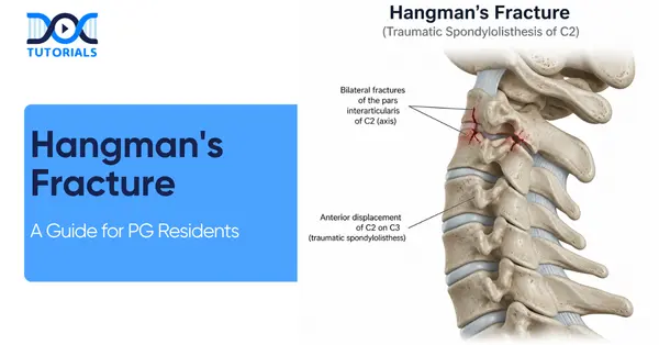
Welcome to this in-depth, high-yield rapid revision guide on Hangman’s Fracture. Tailored explicitly for Orthopaedic PG residents, this blog distills essential concepts regarding cervical spine trauma into actionable knowledge. From deciphering nuanced radiological landmarks to navigating complex surgical decision-making matrices, this guide provides the foundational and advanced insights necessary for both clinical excellence and board exam preparation.
Get High Quality, High Yield Orthopedics PG Residency Notes for Free!
Definition and Pathoanatomy
Hangman’s Fracture is defined strictly as a traumatic spondylolisthesis of the C2 vertebra over the C3 vertebra. The term itself is derived from the Greek words “spondylos” meaning vertebra, and “listhesis” meaning slip.
In orthopaedic pathology, this injury is synonymous with the traumatic disruption of the C2 vertebra originating specifically from the pars interarticularis. It is crucial to remember that the primary anatomical failure point defining this specific injury pattern is the bilateral C2 pars interarticularis.
While the nomenclature carries a historical weight—coined and originally described by Horton—the term is clinically a misnomer in modern traumatology. Today, traumatic spondylolisthesis is most commonly caused by road traffic accidents (RTAs), rather than judicial hanging.
Radiological Landmarks for Diagnosis
Initial assessment of a suspected Hangman’s Fracture heavily relies on precise imaging. The lateral cervical spine X-ray serves as the primary and most vital diagnostic modality for the initial evaluation of this injury.
Understanding normal cervical anatomy is a prerequisite for identifying pathology. In a normal lateral radiograph, the anterior margin of most vertebral bodies aligns perfectly in a straight vertical line.
When analyzing Hangman’s pathology, the physician must look for the disruption of this anterior vertebral line precisely at the C2-C3 junction. The affected C2 vertebra demonstrates noticeable anterior translation, which visually confirms the spondylolisthesis. Loss of this straight anterior vertebral body line at C2 is considered the hallmark radiological sign of spondylolisthesis.
The Levin & Edwards Classification System
To properly manage these injuries, PG residents must rely on established classification systems. Just as you might study the Seinsheimer classification of subtrochanteric fractures to master complex lower limb trauma, thoroughly understanding the Levin & Edwards classification is non-negotiable for navigating Hangman’s Fracture scenarios.
The primary factor differentiating stable from unstable injury patterns in this classification is a critical threshold of 3mm of displacement.
Stable and Minimally Displaced Injuries
Type I injuries involve angulation or translation primarily driven by extension forces. In these cases, there is a substantial injury to the C2-C3 interspace, but the structural alignment remains relatively stable, presenting with less than 3mm of displacement.
Unstable and Highly Displaced Injuries
Type II injuries are categorized by more than 3mm of displacement. These fractures present with significant anteroposterior (AP) and angular deviation caused by flexion. Type II is considered an unstable injury and frequently requires surgical intervention.
The Star & Eismont Modification
The Star & Eismont modification introduces the Type IA category, which falls within Type I injuries due to having less than 3mm of displacement. However, a portion of the posterior C2 body remains in physical continuity with a pars fracture fragment.
This specific anatomical arrangement is highly dangerous. The posterior fragment can directly compress the spinal canal. Despite minimal translation, Type IA injuries carry a highly elevated incidence of neurological deficit.
Why is the Type IA Hangman’s Fracture often referred to as a “wolf in sheep’s clothing”?
Type IA is considered a “wolf in sheep’s clothing” because, on standard imaging, it presents with minimal displacement (less than 3mm), mimicking a benign Type I injury. However, because a portion of the posterior C2 body remains in continuity with the fractured pars fragment, it directly compresses the spinal canal, carrying a deceptively high risk of spinal cord compression and severe neurological deficit.
Classification Breakdown Matrix
| Classification Type | Displacement / Features | Stability & Mechanism | Key Clinical Note |
| Type I | < 3mm displacement | Extension injury; relatively stable | Substantial injury to C2-C3 interspace |
| Type IA | < 3mm displacement | High risk of canal compression | Portion of posterior C2 body remains continuous with pars fragment |
| Type II | > 3mm displacement | Flexion injury; considered unstable | Significant AP and angular deviation |
| Type IIA | No anterior translation | Horizontal/oblique fracture line | Significant angular deviation; highly unstable |
| Type III | C2 pars fracture + facet dislocation | Extreme instability | Requires operative stabilization |
Non-Operative Management Principles
Conservative, non-operative management is the preferred route exclusively when a thorough assessment confirms the absence of neurological compromise or deficit. Stable Hangman’s Fracture patterns rarely present with neurological deficits, making conservative treatment the standard protocol for the majority of non-displaced cases.
Before committing a patient to a 6-8 week cervical collar regimen, clinicians must always rule out concomitant cervical spine injuries and hidden neurological deficits.
For Type I and Type IA injuries, application of a rigid cervical collar for 6 to 8 weeks is standard, provided no other injuries exist. For Type II fractures, the management protocol dictates initial reduction via traction, which is subsequently followed by halo brace immobilization.
The Critical “No Traction” Rule
Traction must be applied with extreme caution and is strictly contraindicated in Type IIA fractures. Applying traction to a Type IIA fracture actively worsens the injury, increasing the deformity and potentially causing catastrophic iatrogenic spinal cord injury and further neurological compromise.
Because Type IIA fractures are highly unstable due to their oblique or horizontal fracture lines, reduction must be achieved exclusively through extension and compression maneuvers using a halo apparatus.
Operative Management and Indications
When conservative measures fail or extreme instability is present at diagnosis, surgery becomes necessary. A failure of Halo immobilization—such as a loss of reduction or the development of non-union—in Types II and IIA immediately upgrades the management directly to surgical fusion.
Type III injuries serve as an absolute indication for surgery. Type III cannot be managed conservatively because they always present with facet dislocation. Surgery is the singular indication for Type III fractures due to these dislocated facets and the severe lack of continuity between the C2 articular processes and the main C2 vertebral body.
The standard operative intervention for unstable Hangman’s Fracture cases is a posterior C1-C3 fusion utilizing pedicular screws. Anterior reduction maneuvers are notably difficult in Type III scenarios because the articular process is completely disconnected from the C2 body.
Surgical Approaches Comparison Matrix
| Approach | Stabilization Levels | Advantages | Disadvantages | Indications |
| Anterior Fusion | C2-C3 | Preserves C1-C2 motion (maintains lateral rotation) | Less rigid/strong than posterior lateral mass fixation | Indicated for Type II or IIA if non-operative treatment fails |
| Posterior Fusion | C1-C3 | Strongest construct for total stabilization | Sacrifices atlantoaxial mobility (permanently locks lateral neck rotation) | Ideal for Type III; preferred when anterior reduction is impossible |
Frequently Asked Questions (FAQs)
1. What is the most common cause of a Hangman’s Fracture today?
Despite its name, road traffic accidents (RTAs) vastly outnumber judicial hanging as the primary cause of this fracture in modern practice.
2. What anatomical structure is primarily fractured in this injury?
The hallmark failure point is the bilateral C2 pars interarticularis.
3. What is the primary imaging modality for diagnosing this fracture?
The lateral cervical spine X-ray is the primary modality used for initial assessment.
4. What radiological sign confirms the diagnosis?
The loss of the straight anterior vertebral body line at C2, demonstrating anterior translation, is the hallmark sign.
5. What is the displacement threshold between Type I and Type II fractures?
The critical threshold differentiating these fracture types is 3mm of displacement.
6. Why is traction strictly contraindicated in Type IIA fractures? Traction increases deformity in the oblique/horizontal fracture lines of Type IIA, risking catastrophic iatrogenic spinal cord injury.
7. How should a Type IIA Hangman’s Fracture be reduced?
Reduction for Type IIA must be achieved by applying extension and compression via a halo apparatus.
8. What defines a Type III Hangman’s Fracture?
Type III is exclusively defined by any C2 pars fracture combined with a dislocation of the C2-C3 facet joint.
9. Can a Type III fracture be managed conservatively?
No, Type III cannot be managed conservatively; the presence of facet dislocation means surgery is the only indication.
10. What is the primary disadvantage of a Posterior C1-C3 fusion?
While providing maximum stability, it permanently eliminates lateral neck rotation by locking the atlantoaxial joint.

Latest Blogs
-

NEET SS Exam 2024: Analysis, Key Dates, Counselling
The NEET SS 2024 exam kicked off on March 29, 2025. Over two days and two slots, candidates across 13…
-

INI SS Topper: 1 Dream, 1 Study Buddy, 1 Rank – The Journey of Dr. Prithivi Raaj, CML 1 & (AIIMS) AML 1 in DM Cardiology
Achieving the top rank in a competitive exam like the INI SS DM Cardiology requires tireless dedication, a structured approach,…
-

INI SS Results: Merit List, Exam Insights, and What Comes Next
The INI SS results for October 2024 are just around the corner. This is a life-changing moment for thousands of…