Cartilage: Meaning, Function, Types, and More
Cartilages are essential connective tissues that support movement, protect joints, and maintain the structure of several body parts. These tissues may get damaged or become degenerative due to several factors, which can have a significant impact on mobility and quality of life. As a result, cartilages have become a topic of significant medical research and clinical practice.
Among medical aspirants, particularly those preparing for NEET PG, it is essential to clearly understand cartilages. Knowledge of its meaning, functions, types, associated disorders, methods of diagnosis, and treatment can aid proper clinical evaluation and sound decision-making.
Read on to gain an in-depth understanding of cartilage and its importance in the human body!
What are Cartilages?
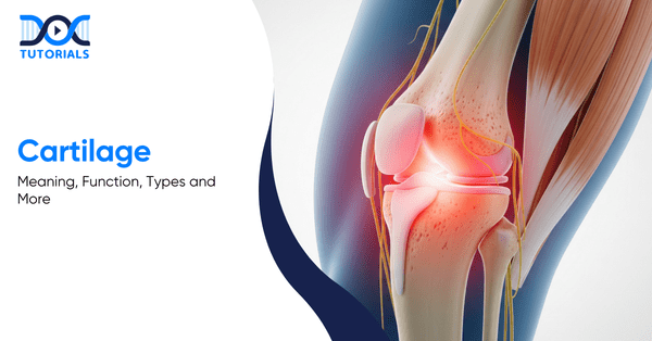
Cartilage is a smooth, resilient connective tissue that supports joints, absorbs shock, and allows friction-free movement. Found in structures like joints, ears, and the spine, it plays a key role in flexibility and skeletal stability.
Cartilage is a kind of flexible connective tissue existing in different parts of the body, including joints, ears, nose, and spacing between different bones of the spine. It gives joints structure, support, and protection, allowing them to move easily.
The cartilages consist of specialised cells known as chondrocytes, which synthesise collagen, proteoglycans, and other proteins that give the tissue its strength and resilience.
What are the Functions of Cartilages?
Cartilage cushions joints by absorbing shock during movement, reducing friction between bones for smooth motion, and maintaining joint shape and stability. It also provides structural support and attachment points for muscles, tendons, and ligaments, helping the body move efficiently without joint damage.
Cartilage is very critical in cushioning bones and joints. The function of cartilage is pointed out below:
- Shock Absorption
Cartilage is a natural shock absorber during movement. It cushions the bones when you walk, run, or jump, as sports shoes do for your feet.
- Reducing Friction
It enables the bones to move more easily against each other by reducing friction at the joints. This enables smooth movement with no grinding and reduces wear and tear.
- Structural Support
Cartilage helps joints maintain their shape while in motion. It also provides support and connection to other tissues, offering muscle, tendon, and ligament attachments in the entire body.
What are the Different Types of Cartilages?
The human body has three types of cartilage: hyaline, elastic, and fibrocartilage. Hyaline cartilage provides smooth joint movement, elastic cartilage offers flexibility and shape retention, and fibrocartilage delivers strong cushioning in weight-bearing areas such as the spine and knees.
Three primary types of cartilage are present in the human body, and they are listed as follows:
- Elastic Cartilage
Elastic cartilage is found in the outer ear, the throat epiglottis, parts of the nose, and the windpipe. Its key role is to provide flexibility along with strength, allowing these structures to maintain their shape while bending easily.
- Fibrocartilage
Fibrocartilage is found in the menisci, which are special cushioning pads in the knee and between the spinal column in the spine. It aids in the alleviation of friction and pressure in weight-bearing joints. Fibrocartilage is the harder of the two types because it has thick sheets of stiff collagen fibres.
- Hyaline Cartilage
Hyaline cartilage can be found in the larynx, nose, trachea, and ribs. It can also be found in the thin, smooth covering at the ends of bones in joints, called articular cartilage, which cushions movement. Hyaline cartilage is the weakest of the three types despite the fact that it consists of fine collagen fibres that give it strength.
What are the Common Conditions that Affect Cartilages?
Cartilage can be affected by injuries, degenerative diseases, and spinal conditions. Some of the common issues are tearing due to sports activities, age-related wear resulting in osteoarthritis, herniated discs in the spine, and osteochondritis dissecans.
Below are some common conditions that affect cartilages:
- Injuries
Accidents, impacts during sports activities, or sudden trauma can tear or damage cartilage. Common examples include meniscus tears in the knee or shoulder injuries that affect joint cartilage. Damage to a joint may also lead to conditions such as osteochondritis dissecans, which affects the ends of bones in joints.
- Osteoarthritis
This is the most widespread form of arthritis. Cartilage in the joints gradually breaks down as people grow older. As a result, joints may become painful, stiff, and inflamed with less cushioning and lubrication.
- Herniated Discs
The condition is also known as the slipped, bulging or ruptured disc that occurs when the cartilage disc that separates the spinal vertebrae ruptures or becomes damaged. This can have a strain on the other nerves near and bring about discomfort or pain.
What are the Symptoms of Cartilage Damage?
Cartilage damage may cause joint pain, stiffness, swelling, and tenderness over time. Affected individuals may also experience limited motion, inability to perform daily tasks, and joint locking or instability, particularly in weight-bearing joints like the knees and hips.
Damage to cartilages might not be accompanied by immediate or distinct symptoms. Nevertheless, certain common symptoms can develop over time, such as:
- Pain and Stiffness
Affected weight-bearing joints, such as the knees or hips, may experience discomfort and stiffness during movement.
- Swelling or Tenderness
Inflammation around the affected area may occur, making the joint feel swollen or sensitive to touch.
- Restricted Movement
Cartilage damage can limit how freely a joint moves, leading to difficulty with everyday activities.
- Joint Locking or Instability
In some situations, affected joints can become unstable, or they may even lock up, which can significantly impact movement.
How to Diagnose Cartilage Damage?
Physical examination, imaging tests such as X-rays and MRI scans, and arthroscopy are the methods used by doctors in the diagnosis of cartilage damage.
In cases of cartilage damage, physicians apply an integrated approach in order to determine the problem. The tests they perform include the following:
- Physical Examination
The physician will examine the joints to assess swelling, tenderness, and pain due to movement.
- Imaging Tests
Scans like X-rays and MRI images would assist in visualising the joint structure, such as cartilage, as well as surrounding tissues, to identify any abnormalities.
- Arthroscopy
It includes inserting a small camera into the joint, enabling the doctor to view the cartilage in question and assess the amount of damage.
What are the Treatment Options for Cartilages?
Treatment for cartilage-based conditions is based on the extent of the damage and can involve physiotherapy, medications, and lifestyle modifications in mild cases. Severe damage may require surgical procedures to restore joint function.
Cartilage issues can be treated based on the cause and severity of damage. The treatment typically starts with a conservative approach and could be followed by surgery in case it is necessary.
Non-Surgical Treatments
- Physiotherapy involves activities that strengthen the muscles surrounding the affected joint in order to minimise pressure on the cartilage.
- Anti-inflammatory drugs are commonly prescribed to relieve pain and swelling.
- Lifestyle changes, such as maintaining a healthy body weight and doing frequent low-impact exercise, can be used to reduce the load on the joints and maintain their long-term health.
Surgical Treatments
The most common treatment methods include:
- Abrasion Arthroplasty
A surgical device operates at a very high speed to scrape away the damaged cartilage with some tiny openings in the underlying bone and induce the body to grow new cartilage.
- Autologous Chondrocyte Implantation
This is a two-stage procedure. Healthy cartilage cells are first collected and grown in a laboratory. The cartilage affected by the disease is removed later via surgery and replaced with the lab-grown cells.
- Microfracture
In this operation, the surgeon takes out the damaged cartilage and drills holes in the bone underneath. This improves blood flow, which consequently aids normal healing and cartilage growth.
- Drilling
Similar to microfracture, drilling creates small channels in the underlying bone to improve blood supply and support the growth of new cartilage.
- Osteochondral Autograft Transplantation
Healthy cartilage is removed from the patient’s non-weight-bearing joint and implanted into the injured region. This alternative is typically appropriate in small regions because of the lack of donor tissues.
- Osteochondral Allograft Transplantation
In this procedure, cartilage tissue is sourced from a donor. Allografts are commonly used when there are larger defects and the tissue of the patient is inadequate.
FAQs about Cartilages
- Can cartilage repair itself?
The specialised cells, known as chondrocytes, do not regenerate easily, and that is why the cartilage damage rarely heals without medical intervention.
- What can athletes do to mitigate the occurrence of cartilage injuries?
Some of the ways athletes can reduce cartilage injury risk are by wearing proper protective gear, using proper training methods, and including strength and conditioning activities in their exercise programmes.
- What helps keep cartilage healthy?
The cartilage can be kept healthy by consuming a well-balanced, nutritious diet, engaging in low-impact workouts, and maintaining a healthy body weight, which will help avoid imposing unnecessary loads on the joints.
Conclusion
Being an NEET PG candidate, it is important to have clear and detailed knowledge of the cartilage, as it is a major determinant of joint health, movement, and several clinical conditions. It will help build your clinical reasoning and equip you to tackle orthopaedic and rheumatological cases with a lot of confidence.
DocTutorials supports your preparation with high-yield video lectures, concise notes, and expert-led discussions tailored for NEET PG.
Start exploring our NEET PG Course today and take a decisive step towards securing your dream rank!


| > Bed Sores | > Pruritus |
| > Polycythemia Vera | > Corticosteroids |
| > Biomedical Waste Management | > Hot Flashes |
| > Tinea Versicolor | > Xerostomia |
| > Kwashiorkor | > Narcolepsy |
Latest Blogs
-

INI CET Exam 2026: Your Roadmap to Success – Key Topics, Strategies, and Lessons from Last Year’s Papers
The INI CET exam is more than just a test; it’s a significant milestone for many medical students aiming to…
-

INI CET Exam Success: Previous Year Question Papers & Ultimate Guide – INI CET PYQ
One can feel overwhelmed while preparing for the INI CET (Institute of National Importance Combined Entrance Test). A vast syllabus,…
-

NEET PG 2026 Preparation with Live Power Pack: Your Path to Success in 2026
Preparing for the NEET PG 2026 can be an overwhelming journey because of its vast syllabus and the pressure to…


