Gametogenesis: Stages, Types, and Importance
Gametogenesis is the complex biological process that produces gametes, or reproductive cells. It is an extremely important process of human reproductive embryology and biology since it results in the production of egg and sperm cells for use in fertilisation and the development of new life. Knowing about gametogenesis not only makes us aware of the intricacy of reproductive biology but also allows us to avoid diseases of reproduction and fertility.
For medical students preparing to take the NEET PG exam, an accurate understanding of the steps, categories, and clinical relevance of gametogenesis is essential. It also forms a foundation to study more general points such as hormonal control, the development of the embryo, and associated diseases.
Want to learn more? Keep reading for detailed insight!
What is Gametogenesis?
Gametogenesis definition refers to the biological process by which gametes or sex cells, in animals are developed. This crucial process takes place in reproductive organs called gonads: testes in males and ovaries in females. Sperm in males and ova in females are produced through gametogenesis in dioecious animals. During fertilisation, the gametes combine to produce genetically distinct offspring.
In addition to the decrease of chromosomes during meiosis, gametogenesis entails structural and functional changes during gamete development. This ensures that the gametes of both sexes carry half the quantity of genetic information, thereby eliminating chromosomal doubling within the offspring.
Spermatogenesis is the developmental process in males that turns diploid spermatogonia into mobile sperm cells. In females, oogenesis yields large, immotile egg cells that are rich in nutrients and crucial molecules for early embryonic development. Both are under hormonal control to coordinate the maturation and release of gametes with the reproductive cycle.
Knowledge of gametogenesis illuminates different reproductive processes and aids in the diagnosis and treatment of human fertility disorders.
What are the Various Stages of Gametogenesis?
There are various stages of gamete development, which emerge step by step, from the very formation of primordial germ cells to the formation of mature sperm and egg cells. The following is a detailed overview:
- Primordial Germ Cells and Their Migration
Primordial germ cells (PGCs) are the germ cells that mature into gametes. They emerge early in embryogenesis and migrate to the gonadal ridges, from which ovaries or testes develop later. This process of migration is essential during development, as it positions germ cells in the correct anatomical position.
- Mitotic Proliferation and Differentiation
Upon reaching the gonads, the PGCs are replicated by mitosis. In men, they are transformed into spermatogonia, while in women, they become oogonia. This is where the gamete creation process starts.
- Meiotic Division and Its Special Features
The process of meiosis causes cells to divide from diploid to haploid, reducing the number of chromosomes. Meiosis I and II are the two stages of the process. Features like crossing over and independent assortment give genetic variation.
Homologous chromosomes form pairs and swap genetic material during meiosis I. Meiosis II is similar to mitosis, with the only exception that sister chromatids are being divided, resulting in four haploid cells from one diploid cell.
What are the Various Types of Gametogenesis?
Gametogenesis can be divided into various categories depending upon the organism as well as the type of gametes formed. The two primary types in human beings are spermatogenesis and oogenesis in males and females, respectively.
Here is a detailed overview:
- Spermatogenesis
- In the Beginning
During puberty, which typically occurs between the ages of 10 and 16, males start to produce sperm. Approximately 200 million sperm are produced daily by biological males. This makes it more likely that sperm will get to the egg following ejaculation.
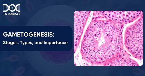
Sperm production occurs in the seminiferous tubules of the male testes. To keep the tubules separate from the systemic circulation, a blood-testis barrier develops in the testicles.
- Protecting the Sperm
Sertoli cells form the blood-testis barrier. This is critical in preventing chemicals found in blood from harming the growing sperm. These products could comprise hormones or waste materials.
It is also significant because it stops the male’s immune system from recognising the sperm as foreign. The sperm are genetically distinct from the male and will express different surface antigens.
- Developing Functional Sperm
The initial pool of diploid cells, spermatogonia, divides by mitosis to create two identical cells, with one maintaining the stem cell pool, specifically the A1 spermatogonium. The second one, type B spermatogonium, continues to mature into sperm. The type B cells proliferate and mature into primary spermatocytes, which remain diploid and are linked by cytoplasmic bridges.
Primary spermatocytes undergo meiosis in the following manner:
- After meiosis I, two haploid secondary spermatocytes are produced.
- During Meiosis II, four haploid spermatids are produced.
- Maturation
Spermiation is the process by which the cytoplasmic bridges disintegrate, and the spermatids are released into the seminiferous tubule lumen. As spermatids migrate from the seminiferous tubules to the epididymis, they undergo spermiogenesis, which is the process of differentiation and remodelling into adult spermatozoa.
The rete testis will get cells from the seminiferous tubule. This removes excess fluid, which helps to “concentrate” the sperm. The cells then proceed to the epididymis, where the sperm is stored and matures.
Spermatogenesis takes around 70 days; consequently, for sperm production to be continuous rather than intermittent, numerous spermatogenic processes occur concurrently within the same seminiferous tubule, with new groups of spermatogonia forming every 16 days (spermatogenic cycle). Each of these groups of spermatogenic cells will be at a distinct stage of sperm development.
- After Ejaculation
Once inside the female reproductive tract, sperm cell terminal maturation occurs by a process known as capacitation. It is a process in which glycoproteins and cholesterol are removed from the sperm head so that the sperm may adhere to the zona pellucida of the egg.
2. Oogenesis
In contrast to spermatogenesis, oogenesis takes place in the fetus before birth. Originating in the yolk sac of the embryo, primordial germ cells go to the primordial gonad’s cortex. Mitotic replication peaks at around 7 million by the middle of pregnancy, which is around 20 weeks.
After this peak, cell death occurs, leaving about 2 million cells. Meiosis I creates the primary oocytes and begins before birth. Therefore, the supply of ova is limited.
Clusters of primary oocytes develop in the gonads. The primary follicle, which consists of flattened epithelial cells, envelops them.
Atresia, or cell death, happens during childhood, leaving about 40,000 eggs at puberty. Only one of the several main oocytes (15–20) that mature each month when puberty starts fully matures to become an oocyte.
The main oocytes develop in three stages:
- Pre-antral
- Antral
- Pre-ovulatory
- Pre-Antral Stage
The primary oocyte is still in meiosis I, but it will rapidly expand during this stage. A stratified cuboidal epithelium is created by the development and multiplication of the follicular cells.
These cells, which we now refer to as granulosa cells, secrete glycoproteins. The zona pellucida, which envelops the primary oocyte, is created by the combination of these substances.
Theca folliculi, a specialised layer of surrounding cells that react to LH and secrete androgens under its influence, are also formed by the differentiation of surrounding connective tissue cells.
- Antral Stage
During the antral stage, fluid-filled spaces develop between granulosa cells, eventually forming a central fluid-filled region known as the antrum.
We now refer to the follicles as secondary follicles. During each monthly cycle, one of these secondary follicles becomes dominant and grows further due to the impact of FSH, LH, and oestrogen.
- Pre-Ovulatory Stage
The LH surge initiates this stage, and meiosis I is now complete. Two unequally sized haploid cells develop inside the follicle. One of the daughter cells receives significantly less cytoplasm than the other, resulting in the formation of the first polar body, which does not develop into an ovum.
Another haploid cell, known as the secondary oocyte, develops, and both daughter cells then go through meiosis II. After this, an initial polar body will replicate to produce two polar bodies. However, just 3 hours prior to ovulation, the secondary oocyte stops in the metaphase of meiosis II.
- Ovulation
The follicle, which is now mature and larger, is known as a Graafian follicle.
An LH surge develops, resulting in increased collagenase activity. This is an enzyme that degrades collagen. As a result, the follicular wall becomes weaker.
This, along with muscle contractions of the ovarian wall, causes the ovum to be expelled from the ovary, which is then transferred into the fallopian tube via the fimbriae.
- Fertilisation
The secondary oocyte only completes meiosis II when there is fertilisation. In this phase, it ejects a third polar body and develops into a fertilised ovum. In case fertilisation does not occur, the oocyte, which is in an arrested form of meiosis II, starts degenerating after 24 hours of ovulation.
After fertilisation, the fertilised egg is transported towards the uterus through the peristaltic movement of the fallopian tube. It eventually reaches the uterus and implants itself into the uterine posterior wall.
What is the Significance of Gametogenesis?
Gametogenesis is essential for reproduction, as it yields sperm and eggs for fertilisation. The quantity and quality of these gametes determine fertility and reproductive success. Any disruption in gametogenesis can lead to infertility or genetic disease. Additional knowledge of this process is also needed in embryology and assisted reproductive techniques.
FAQs About Gametogenesis
- Where does gametogenesis occur?
Gametogenesis occurs in the gonads. In males, this process, called spermatogenesis, takes place in the testes. In females, it is called oogenesis and occurs in the ovaries.
- What hormones are involved in gametogenesis?
The release of gonadotropin-releasing hormone (GnRH) by the hypothalamus signals the anterior pituitary to secrete follicle-stimulating hormone (FSH) and luteinising hormone (LH), both of which are essential for regulating the formation of gametes in the reproductive organs.
- What is the role of testosterone in gametogenesis?
Testosterone supports several key processes in spermatogenesis. It helps maintain the blood-testis barrier, supports meiosis, promotes the attachment of spermatids to Sertoli cells, and facilitates the final release of sperm.
Conclusion
The biological process by which reproductive cells—eggs in females and sperm in males—are created is known as gametogenesis. Mature gametes that are ready for fertilisation are produced by a sequence of meticulously controlled cell divisions. This process plays a vital role in human reproduction, and ongoing research continues to uncover the complex mechanisms behind gametogenesis, paving the way for significant advancements in reproductive medicine.
In addition to mastering reproductive biology, a broad understanding of high-yield topics, such as anatomy, pharmacology, obstetrics, etc., is crucial for NEET PG candidates. DocTutorials offers comprehensive NEET PG study materials, top-grade video lectures, and Quick Revision Programmes (QRP) to help students stay ahead of the curve. Want to secure a top rank? Enrol in our NEET PG course today!


Latest Blogs
-

INI CET Exam 2026: Your Roadmap to Success – Key Topics, Strategies, and Lessons from Last Year’s Papers
The INI CET exam is more than just a test; it’s a significant milestone for many medical students aiming to…
-

INI CET Exam Success: Previous Year Question Papers & Ultimate Guide – INI CET PYQ
One can feel overwhelmed while preparing for the INI CET (Institute of National Importance Combined Entrance Test). A vast syllabus,…
-

NEET PG 2026 Preparation with Live Power Pack: Your Path to Success in 2026
Preparing for the NEET PG 2026 can be an overwhelming journey because of its vast syllabus and the pressure to…


