Muscles of the Hand: Anatomy, Functions, and Disorders
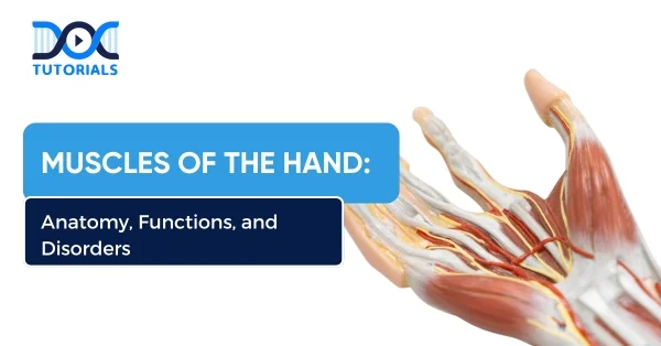
The human hand is designed for gripping, moving with accuracy, and sensing. There are 27 bones in the human hand, along with a lot of muscles, tendons, and ligaments that work together to make it quite flexible. The muscles in the hand are important for all the different things the hand can do, from soft to stronger grips.
When we engage in regular activities, the hand muscles play a crucial role. The muscles in the hand’s functional capability can be essential for maintaining strength and function in the hand. If you are a medical student preparing for the NEET PG exam, this topic is crucial to understand.
Read on to learn about the muscles in your hand, what they do, how they are built, and what problems they can cause.
What are the Different Muscles in the Hand?
The muscles in your hands are important for everyday tasks. They help with accuracy in tasks, keep grip strength, and control complicated finger movements. There are 2 types of hand muscles: extrinsic and intrinsic.
It is essential to know how the muscles in the hand work in order to correctly diagnose and treat a number of illnesses. Below is a table of the hand muscles, along with where they originate from, where they attach, and what nerves supply them.
| Muscle Group | Muscles | Origin of Muscles | Insertion of Muscles | Nerve Supply of Muscles |
| Thenar Muscles | Abductor pollicis brevis | Flexor retinaculum, scaphoid, trapezium | Lateral base of the proximal phalanx of the thumb | Median nerve |
| Flexor pollicis brevis | Superficial: flexor retinaculumDeep: trapezoid, capitate | Lateral base of the proximal phalanx of the thumb | Superficial: MedianDeep: Ulnar | |
| Opponens pollicis | Flexor retinaculum, trapezium | Lateral side of the 1st metacarpal | Median nerve | |
| Abductor pollicis | Oblique: 2nd/3rd metacarpals & capitateTransverse: 3rd metacarpal | Medial base of the proximal phalanx of the thumb | Ulnar nerve | |
| Hypothenar Muscles | Abductor digiti minimi | Pisiform | Medial base of the proximal phalanx of the 5th digit | Ulnar nerve |
| Flexor digiti minimi brevis | Hook of hamate, flexor retinaculum | Medial base of the proximal phalanx of the 5th digit | Ulnar nerve | |
| Opponens digiti minimi | Hook of hamate, flexor retinaculum | Medial border of the 5th metacarpal | Ulnar nerve | |
| Palmaris brevis | Palmar aponeurosis and flexor retinaculum | Skin of the medial hand | Ulnar nerve | |
| Lumbricals | 1st and 2nd lumbricals | The lateral two tendons of the FDP | Extensor expansions of digits 2 and 3 | Median nerve |
| 3rd and 4th lumbricals | The medial three tendons of the FDP | Extensor expansions of digits 4 and 5 | Ulnar nerve | |
| Interossei Muscles | Palmar interossei | Palmar surfaces of metacarpals 2, 4, 5 | Extensor expansions of the same digits | Ulnar nerve |
| Dorsal interossei | Adjacent sides of metacarpals 1–5 | Extensor expansions and proximal phalanges of digits 2–4 | Ulnar nerve |
What are the Functions of the Hand and its Anatomy?
You need your hand muscles to do a lot of different things. They help with fine motor abilities and give you strong, precise grips. These muscles are grouped by where they are, what they do, and how they are structured. They work with tendons and ligaments to make sure that each finger can move on its own. Here are their main functions:
- Extrinsic Muscles of the Hand
The muscles start in the forearm and connect to the hand. These muscles are in charge of movements like bending and straightening the fingers and wrist. The following muscles are part of this group:
- Flexor Digitorum Superficialis: They bend the proximal interphalangeal joints and metacarpophalangeal joints of fingers 2–5.
- Flexor Digitorum Profundus: They bend the distal interphalangeal joints of fingers two through five.
- Extensor Digitorum: They stretch the fingers at the MCP (metacarpophalangeal joints), PIP (proximal interphalangeal joints), and DIP (distal interphalangeal joints) joints.
- Intrinsic Muscles of the Hand
These muscles start in the hand and attach to other muscles in the hand area. These muscles are in charge of small, precise actions like delicate grips. These muscular groups make up:
- Thenar Muscles:
These muscles are near the base of the thumb and let you do complicated things like grasping and pinching.
- Abductor Pollicis Brevis: Moves the thumb away from the body
- Flexor Pollicis Brevis: Bends the thumb at the MCP joint.
- Opponens Pollicis: Allows the thumb to move against anything
- Adductor Pollicis: Pulls the thumb towards the palm
- Hypothenar Muscles:
These muscles are located at the bottom of the little finger. They look like the thenar group, but they don’t govern the fifth digit.
- Abductor Digiti Minimi: Pulls the little finger away from the rest of the hand
- Flexor Digiti Minimi Brevis: Bends the little finger’s MCP joint
- Opponens Digiti Minimi: Allows the fifth digit to oppose itself
- Palmaris Brevis: Tighten the skin on the hypothenar eminence
- Lumbricals in the Hand:
The tendons of the flexor digitorum profundus connect to four lumbrical muscles. The lumbrical muscles (1-4) in the hand help bend the MCP joints while also straightening the PIP and DIP joints. This is important for keeping a steady grasp.
- Interossei Muscles
The interosseous muscles are located between the metacarpal bones and can be broken down into the following groups:
Palmar Interossei: They pull the fingers together.
Dorsal Interossei: They control the movement of the finger away from the middle finger (they also help with flexing the MCP joint and extending the interphalangeal joint).
What are the Common Disorders of Muscles in the Hand?
The hand has a lot of supporting structures, which makes it quite easy for a range of muscle and tendon problems to happen. Here are some frequent problems that can happen with the muscles of the hand:
- Dupuytren’s Contracture
This happens when the ulnar side of the palmar aponeurosis gets inflamed. The aponeurosis is getting thicker and tighter, which makes it hard to straighten the fingers and causes the proximal and lateral middle phalanx to bend.
- Claw Hand
The metacarpophalangeal joints are hyperextended, and the interphalangeal joints are flexed, which affects the ring and little fingers. It also causes loss of sensation in the medial 1.5 fingers, including the nail bed, and is frequently caused by damage to the ulnar nerve.
- Carpal Tunnel Syndrome
Carpal tunnel syndrome happens when the median nerve gets pinched in the carpal tunnel of the wrist. This syndrome causes motor, sensory, vasomotor, and trophic symptoms in the hand. The examination shows that the thenar eminence is withering away, and the fingers on the side are feeling different.
- De Quervains Tenosynovitis
This ailment causes pain in the thumb tendons, especially the abductor pollicis longus and extensor pollicis brevis, and makes it hard to move the thumb. When the doctor examines the patient, they usually present with discomfort and swelling near the base of the thumb.
FAQs About Muscles of the Hand
- What muscle groups are present in the hand?
The thenar muscles, hypothenar muscles, lumbricals of the hand, and interossei (both palmar and dorsal interossei) are all muscles that are connected to the hand. Each one has a particular job when it comes to movement and grip.
- What is the function of the lumbricals of the hand?
The lumbricals help bend the metacarpophalangeal (MCP) joints and stretch the interphalangeal (IP) joints. Because of this, they are very important for tasks that need fine motor abilities, like writing and grabbing things.
- What innervates the thenar and hypothenar muscles?
The median nerve mostly sends signals to the thenar muscles, while the ulnar nerve sends signals to the hypothenar muscles.
- What are the commonly occurring disorders of the hand muscles?
Carpal tunnel syndrome, claw hand (ulnar nerve damage), Dupuytren’s contracture, and De Quervain’s tenosynovitis are all frequent disorders that affect the hand in different ways.
Conclusion
It’s important to grasp the topic of hand muscles to understand anatomical and clinical principles. DocTutorials is a comprehensive platform for NEET PG preparation with expert guidance and high-yield content.
It provides you with structured videos, MCQs, and trusted mentors to help you become exam-ready with confidence. Join DocTutorials today to explore our NEET PG course and take the next step towards your medical success.
Latest Blogs
-

NEET PG Exam 2025- Date, Pattern, Marking Scheme, Subject Wise Weightage, and Exam Mode
NEET PG Exam 2025 is the ultimate gateway for medical graduates aspiring to pursue postgraduate courses in medicine, including MD,…
-

INI CET Exam 2025: Your Roadmap to Success – Key Topics, Strategies, and Lessons from Last Year’s Papers
The INI CET exam is more than just a test; it’s a significant milestone for many medical students aiming to…
-
INI CET Exam Success: Previous Year Question Papers & Ultimate Guide – INI CET PYQ
One can feel overwhelmed while preparing for the INI CET (Institute of National Importance Combined Entrance Test). A vast syllabus,…
