Neurocysticercosis: Causes, Symptoms, Diagnosis, and Treatment
Neurocysticercosis is a serious brain infection caused by the pork tapeworm Taenia solium. This condition develops when parasitic larvae invade the central nervous system, causing seizures, headaches, and neurological symptoms. It is a leading cause of acquired epilepsy worldwide, especially in areas with less sanitation. Hence, early diagnosis and treatment are required to prevent damage.
This guide explains its causes, symptoms, diagnosis, and treatment with in-depth details, giving medical students and professionals a clear insight into managing one of the common parasitic brain infections worldwide.
Read on to get more insights!
What is Neurocysticercosis?
Neurocysticercosis is the most common parasitic infection of the CNS. It occurs when the larval forms of the pork tapeworm Taenia solium invade the brain and spinal cord to form cysts (cysticerci). It is a major cause of seizures and neurological impairments in various parts of the world.
The infection develops when a person ingests eggs or segments (proglottids) of Taenia solium. Inside the intestines, they mature into adult tapeworms ranging in length from 2 to 8 metres. Each worm generates thousands of proglottids, each carrying up to 50,000 eggs.
What are the Stages of Neurocysticercosis?
Neurocysticercosis progresses through five distinct stages, reflecting changes in the parasite’s life cycle and the body’s reaction to it. Below is a list of the stages:
- Noncystic Stage: It is not visualised on imaging at an early stage, like other stages. However, it may show areas of swelling. These areas progress to lesions in the coming months as the embryo starts to form cysts.
- Vesicular Stage: The cysts have thin, translucent walls filled with clear fluid containing the living parasite. Usually, the immune system does not react during this stage, and the person remains symptom-free.
- Colloidal Vesicular Stage: Cysts develop thicker walls while the fluid within becomes cloudy. This is when an intense inflammatory response starts, as the parasite begins to dissolve away, either naturally or due to treatment. Imaging usually reveals cystic lesions with surrounding swelling at this stage. Seizures are potentially common in this period.
- Granular Vesicular Stage: As the cysts degenerate, the swelling around them starts to disappear. However, seizures may still be provoked through inflammation in the brain tissues around the dying parasite.
- Calcific Stage: This is the final stage after the parasite has expired. The calcification of cysts occurs, leaving behind the residues of a parasitic membrane. However, they may still invoke an immune response, albeit rarely, causing seizures.
What are the Causes and Risk Factors of Neurocysticercosis?
Improperly cooked pork containing Taenia solium larvae, if ingested, causes neurocysticercosis. Once ingested, the larvae develop into adult tapeworms in the intestines and release eggs that might be dispersed to the brain and other tissues.
Areas with poor sanitation and where the pigs roam freely and come in contact with human faeces have a higher incidence. Especially, rural farming communities are at greater risk. Also, when there is contamination in the soil or water, such parasitic eggs can be carried further.
What are the Symptoms of Neurocysticercosis?
Seizures are the most common symptom, occurring in about 50-70% of patients with the condition.
Other common symptoms include:
- Chronic headaches
- Increased pressure in the brain (intracranial hypertension)
- Perilesional oedema (swelling around the cysts)
- Confusion or memory problems
- Difficulty with balance and coordination
- Sensory disturbances
In some rare and serious instances, growing cysts can exert pressure on brain tissues, resulting in multiple neurological problems or syndromes.
How is Neurocysticercosis Diagnosed?
Neurocysticercosis demands a clinical evaluation as well as some advanced investigations to confirm the presence of Taenia solium inside the central nervous system. They may include the following:
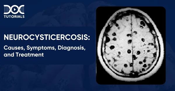
- Neuroimaging Techniques: MRI or CT scans are used to check the presence of cysts or lesions in the brain and spinal cord. They provide information regarding the location, size, and number of cysts and are essential for making the right diagnosis.
- Stool Examination: A stool sample may help diagnose an intestinal tapeworm by detecting proglottids. This test, however, cannot confirm whether the infection is causing neurological symptoms.
- Blood Tests: Enzyme-linked immunosorbent assays (ELISA) look for antibodies against the parasite, which can help determine if the infection is active. Such antibody-specific blood tests are commonly used to support neuroimaging findings.
- Cerebrospinal Fluid Analysis: Depending on the scenario, a lumbar puncture may be conducted to detect cysticercal antigens or antibodies in the cerebrospinal fluid.
What are the Neurocysticercosis Treatment Options?
Treating neurocysticercosis is about getting rid of the parasite and managing the symptoms like seizures, brain swelling, and intracranial hypertension. The choice of treatment depends on the location, size, number, and stage of the cysts and the overall condition of the patient.
Here is an overview of the neurocysticercosis treatment options:
- Antiparasitic Therapy: For patients with active noncalcified cysts, doctors may prescribe anthelmintic drugs to kill the parasite.
- Anti-inflammatory Treatment: Corticosteroids are used along with antiparasitic therapy to reduce swelling and prevent complications from the immune response as the cysts break down.
- Seizure Management: Anticonvulsant medications to control seizures, which are a common symptom of neurocysticercosis.
- Surgical Intervention: In some cases, surgery is needed to remove cysts that are pressing on the brain or blocking cerebrospinal fluid flow.
- Preventive Measures: In endemic areas, treating pigs with vaccines and antiparasitic drugs will break the transmission cycle.
FAQs About Neurocysticercosis
- How is neurocysticercosis diagnosed?
Diagnosis is based on symptoms, medical history, serological tests, and brain imaging (MRI or CT scan). Pathology findings also support the diagnosis in some of the cases.
- What is the best treatment for neurocysticercosis?
Treatment is the management of symptoms and targeting the parasite. Albendazole and praziquantel are the two main drugs used to kill or reduce cysticerci in the brain.
- How can neurocysticercosis be prevented?
It can be prevented by hand hygiene, cooking pork thoroughly, and treating people who carry intestinal tapeworms to break the cycle of transmission.
Conclusion
Neurocysticercosis is the most common parasitic disease of the central nervous system, which can cause serious neurological problems like seizures, chronic headaches, and cognitive changes. It is a major cause of epilepsy in developing regions, so early diagnosis, tailored treatment, and improved preventive measures are needed to reduce its impact on individuals and communities.
For NEET PG students, having a clear idea of neurocysticercosis is essential for covering the syllabus as well as for clinical diagnoses. To help them in this regard, DocTutorials provides a variety of topics, high-quality video lectures, question banks, test series, and expert guidance to strengthen your medical knowledge.


Latest Blogs
-

INI CET Exam 2026: Your Roadmap to Success – Key Topics, Strategies, and Lessons from Last Year’s Papers
The INI CET exam is more than just a test; it’s a significant milestone for many medical students aiming to…
-

INI CET Exam Success: Previous Year Question Papers & Ultimate Guide – INI CET PYQ
One can feel overwhelmed while preparing for the INI CET (Institute of National Importance Combined Entrance Test). A vast syllabus,…
-

NEET PG 2026 Preparation with Live Power Pack: Your Path to Success in 2026
Preparing for the NEET PG 2026 can be an overwhelming journey because of its vast syllabus and the pressure to…


