Popliteal Fossa: Anatomy, Boundaries & High-Yield Concepts for MBBS
The popliteal fossa is a diamond-shaped depression at the back of the knee that serves as a significant point of transition for vessels and nerves passing between the leg and the thigh. Although it may appear as a simple depression, it contains several structures that are critical for lower-limb function.
For MBBS students, a clear explanation of its layout, relationships, and clinical significance is necessary. Let’s take an in-depth look at the popliteal fossa and its key anatomical and clinical features. Keep reading to learn more.
What is the Anatomy of the Popliteal Fossa?
The popliteal fossa contains the popliteal vessels, sciatic nerve branches, and the short saphenous vein. Its muscular boundaries provide stability, support knee movement, and protect these vital structures.
The popliteal fossa allows the passage of large nerves, arteries, veins, and lymphatics as they travel between the thigh and leg. Some of the structures passing through this region include the terminal branches of the sciatic nerve, the popliteal artery and vein, and the short saphenous vein.
It has a number of muscles that form its borders, and when combined, they form a stable yet flexible area that aids knee movement and protects the fine neurovascular elements. Knowledge of such muscular and fascial boundaries is the basis for clinical examination and surgery.
What are the Boundaries of the Popliteal Fossa?
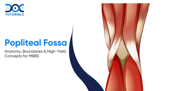
The popliteal fossa is bordered superiorly by the semimembranosus, semitendinosus, and biceps femoris, and inferiorly by the heads of the gastrocnemius. Its floor is formed by the distal femur, posterior capsule, oblique popliteal ligament, and popliteus. The popliteal fascia forms the roof and is pierced by the short saphenous vein and sural nerve.
Several muscles contribute to its borders, and together they create a stable yet flexible region that supports knee movement while safeguarding delicate neurovascular elements.
Understanding these muscular and fascial boundaries forms the foundation for clinical examination and surgical approaches:
- Superior Boundaries
The superior boundaries of the popliteal fossa consist of:
- Superomedial: Formed by the semimembranosus and semitendinosus muscles
- Superolateral: Formed by the long head of the biceps femoris
- Inferior Boundaries
The inferior boundaries of the popliteal fossa are:
- Inferomedial: Medial head of the gastrocnemius
- Inferolateral: Lateral head of the gastrocnemius (with plantaris occasionally contributing)
- Floor
The floor is composed of several structures arranged from superior to inferior:
- The posterior aspect of the distal femur
- The posterior capsule of the knee joint
- The oblique popliteal ligament
- The popliteus muscle
- Roof
The roof is formed by the strong popliteal fascia, which continues upward as the fascia lata and downward as the deep fascia of the leg. This tough fibrous layer acts as a protective cover while maintaining the structural integrity of the fossa.
The short saphenous vein and sural nerve pierce this fascia, making them easily identifiable during surgical procedures.
What are the Nerves in the Popliteal Fossa?
The popliteal fossa contains major superficial nerves that are prone to injury. The tibial nerve runs centrally, supplying posterior leg muscles and contributing to the sural nerve. The common fibular nerve follows the biceps femoris and curves around the fibular neck, where its exposed position makes it especially vulnerable before dividing into superficial and deep branches.
Although the fossa itself contains abundant fatty tissue, it is also home to several significant nerves. These are arranged superficially in the fossa, making them vulnerable to compression or trauma.
The primary nerves include:
- Tibial Nerve
The sciatic nerve divides into the tibial and common fibular nerves at the upper angle of the popliteal fossa. The tibial nerve, being the larger branch, travels vertically down the middle of the fossa before entering the posterior compartment of the leg.
It innervates several muscles, including the:
- Soleus
- Gastrocnemius
- Plantaris
- Popliteus
One of its branches, the medial sural cutaneous nerve, unites with the lateral sural cutaneous branch from the common fibular nerve to form the sural nerve, which supplies the posterolateral aspect of the leg and foot.
- Common Fibular Nerve
This nerve courses along the medial border of the biceps femoris before curving around the head and neck of the fibula. Due to its superficial position in this region, it is especially prone to injury. It eventually divides into 2 terminal branches: the superficial and deep fibular nerves.
What are the Blood Vessels of the Popliteal Fossa?
The popliteal artery runs deep in the fossa, giving off the genicular branches that form the knee’s vascular anastomosis. Superficial to it lies the popliteal vein, which becomes the femoral vein after receiving major tributaries.
The vascular structures lie deeper than the nerves, and they include the following:
- Popliteal Artery
The popliteal artery represents the continuation of the femoral artery after it exits the adductor hiatus. Within the popliteal fossa, it travels downward and laterally, giving off several branches collectively known as the genicular arteries.
These include:
- Superior medial genicular artery
- Superior lateral genicular artery
- Middle genicular artery
- Inferior medial genicular artery
- Inferior lateral genicular artery
These arteries participate in the genicular anastomosis, an elaborate network that ensures continuous blood flow around the knee joint, particularly during flexion. The artery also gives muscular branches to the gastrocnemius, soleus, plantaris, and hamstring muscles.
- Popliteal Vein
Lying superficial to the popliteal artery, the popliteal vein forms a part of the deep venous system. As it exits the fossa superiorly, it transitions into the femoral vein. It receives blood from the posterior tibial and peroneal veins as well as the short saphenous vein.
- Short Saphenous Vein
This superficial vein ascends along the posterior leg, enters the fossa through the popliteal fascia, and ultimately drains into the popliteal vein. Its course makes it clinically important in venous access and varicose vein assessment.
What is the Lymphatic Drainage for the Popliteal Fossa?
The popliteal fossa contains superficial and deep lymph nodes. The superficial nodes lie in the subcutaneous tissue and drain the lateral foot and leg via vessels accompanying the short saphenous vein. The deep nodes surround the popliteal vessels and receive lymph from deeper leg and foot structures before draining toward the deep inguinal nodes.
Two groups of lymph nodes are located within the popliteal fossa:
- Superficial Popliteal Nodes
Situated within the subcutaneous tissue, they drain lymph from the lateral foot and leg through vessels accompanying the short saphenous vein.
- Deep Popliteal Nodes
These nodes surround the popliteal vessels and receive lymph from the deeper structures of the leg and foot. From here, lymph drains toward the deep inguinal lymph nodes.
What is the Clinical Relevance of the Popliteal Fossa?
Swelling or pain in the popliteal fossa often results from a Baker’s cyst or a popliteal artery aneurysm. A popliteal aneurysm causes compression of nearby structures, especially the tibial nerve, leading to impaired plantar flexion and sensory changes, often accompanied by a pulsatile mass or bruit.
Swelling or pain in the popliteal fossa can represent a wide range of underlying conditions. Two of the most common causes include:
1. Baker’s Cyst
The popliteal cyst, also known as Baker’s cyst, develops as a result of the enlargement of the semimembranosus bursa, which is frequently caused by chronic inflammation or degenerative changes in the knee joint, such as osteoarthritis.
Even though most cysts spontaneously improve, ruptured Baker’s cysts may be imitations of deep vein thrombosis, and they manifest in the form of calf pain and swelling.
2. Popliteal Artery Aneurysm
A popliteal aneurysm represents a localised dilation of the artery. Due to the non-stretchable nature of the popliteal fascia, any enlargement compresses adjacent structures—particularly the tibial nerve.
Patients may experience:
- Reduced or absent plantar flexion
- Numbness or tingling along the foot and posterolateral leg
A pulsatile mass or an audible arterial bruit strongly suggests an aneurysm.
FAQs about the Popliteal Fossa
- Which popliteal fossa structure holds the greatest clinical significance?
The popliteal artery is of paramount importance, as trauma or aneurysmal dilation of this vessel can rapidly compromise perfusion to the lower limb. - Why is the common fibular nerve particularly susceptible to injury?
Its superficial course around the neck of the fibula renders it highly exposed, predisposing it to injury from fractures, external compression, or even habitual leg-crossing. - How is a popliteal aneurysm generally diagnosed?
Diagnosis is typically based on the presence of a palpable pulsatile mass, the detection of a bruit on auscultation, and confirmation via Doppler ultrasonography. - What conditions most commonly lead to the formation of Baker’s cysts?
Baker’s cysts generally arise in the context of chronic intra-articular inflammation, most often associated with osteoarthritis, rheumatoid arthritis, or meniscal pathology. - What is the optimal position for palpating the popliteal artery?
Palpation is most effective when the knee is gently flexed, a position that brings the artery closer to the surface and facilitates examination.
Conclusion
The popliteal fossa, though often overlooked, is one of the most clinically significant regions of the lower limb. It contains vital nerves, blood vessels, and lymphatics that contribute to the stability, movement, and vascular integrity of the leg.
For MBBS students, mastering the anatomy and the clinical relevance of this region is indispensable. For added guidance, DocTutorials can be your study partner. We offer 3D & 2D animated videos, CBME-based video lectures, and expert mentorship.
Join DocTutorials today and explore our MBBS curriculum to excel in your medical career!


| > Bed Sores | > Pruritus |
| > Polycythemia Vera | > Corticosteroids |
| > Biomedical Waste Management | > Hot Flashes |
| > Tinea Versicolor | > Xerostomia |
| > Kwashiorkor | > Narcolepsy |
Latest Blogs
-

INI CET Exam 2026: Your Roadmap to Success – Key Topics, Strategies, and Lessons from Last Year’s Papers
The INI CET exam is more than just a test; it’s a significant milestone for many medical students aiming to…
-

INI CET Exam Success: Previous Year Question Papers & Ultimate Guide – INI CET PYQ
One can feel overwhelmed while preparing for the INI CET (Institute of National Importance Combined Entrance Test). A vast syllabus,…
-

NEET PG 2026 Preparation with Live Power Pack: Your Path to Success in 2026
Preparing for the NEET PG 2026 can be an overwhelming journey because of its vast syllabus and the pressure to…


