Sarcoidosis: Causes, Symptoms, Diagnosis, and Treatment
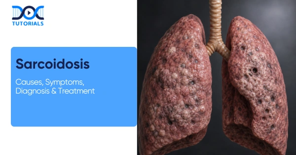
Sarcoidosis is a systemic inflammatory disease which is characterised by the formation of noncaseating granulomas in various organs. Typical presentations are bilateral hilar lymphadenopathy and changes in the interstitial lung, although sarcoidosis may occur in the skin, eyes, heart, and other organs.
This NEET-PG pathology review covers sarcoidosis aetiology, clinical features, diagnostic criteria, management, and complications, emphasising information relevant for medical students. Keep reading to know more.
What are the Causes of Sarcoidosis?
The exact cause of sarcoidosis is unknown. It is thought to have an exaggerated response of the immune system to unknown antigens. Risk is genetically predisposed, affected by race and environment.
There is no single known cause of sarcoidosis, and its formation is associated with a mixture of genetic and environmental factors, as well as immune-related factors:
- Unknown Aetiology: No specific trigger is found. Sarcoidosis is likely an autoimmune response (primarily Th1-mediated) to microbial or environmental antigens. Exposures (beryllium, insecticides, mould), infections (mycobacteria, Propionibacterium acnes) are investigated, but none of them can explain all the cases.
- Genetic Factors: Genetic predisposition is indicated by familial clustering and HLA (Human Leukocyte Antigen). There are also specific HLA-DR alleles and ACE (Angiotensin-Converting Enzyme) gene variants associated with susceptibility, which point to heritable immune hyperreactivity.
- Demographics: Most prevalent among adults between 20 and 40 years. It occurs more in African Americans than in whites, and the morbidity among African American females is the highest. Women tend to experience extrapulmonary disease, whereas men tend to exhibit cardiac involvement.
- Geographic/Seasonal Factors: Seen worldwide but more common in northern latitudes and urban areas. There is some seasonal variation, but there are no definite prevention measures.
- Immune Factors: Patients have an exaggerated cellular immune response at disease sites, characterised by CD4+ T-helper cell accumulation and inverted CD4/CD8 ratios in affected tissues.
High concentrations of cytokines (IL-2, IL-6, TNF-α, IFN-γ) favour the development of granuloma. These are immunologic pathways which play a central role in the pathogenesis of sarcoidosis.
Pathogenesis and Histopathology of Sarcoidosis
The pathology of sarcoidosis is characterised by an over-response of the immune system, which results in the production of compact noncaseating granuloma in tissues. Granulomas are composed of epithelioid histiocytes and giant cells.
The pathology of sarcoidosis is characterised by the dysregulated immune response, resulting in the appearance of distinct noncaseating granulomas, which are the key features of the disease and may be localised to various organs.
- Granuloma Formation: The characteristic lesion is the noncaseating epithelioid granuloma, with tight clusters of activated macrophages and Langhans-type giant cells. These do not have central necrosis, although there may be rare fibrinoid necrosis.
The presence of surrounding lymphocytes (primarily CD4+ T cells) and fibroblasts may ultimately result in fibrosis.
- Cellular Details: Epithelioid histiocytes release enzymes and cytokines, such as ACE, lysozyme, and calcitriol. The overproduction of calcitriol may lead to hypercalciuria and hypercalcemia.
Giant cells are the result of fusion and may include Schaumann or asteroid bodies- nonspecific but supportive findings.
- Organ Involvement: Granulomas can affect almost any organ, most commonly the lungs and intrathoracic lymph nodes (up to 90%). They may also affect skin, eyes, heart, liver, spleen, bone marrow, and nervous system, affecting normal tissue (e.g., fibrosis in lungs, arrhythmias in heart).
- Pathological Variants: Granulomas appear in skin lesions (e.g., lupus pernio), but acute sarcoidosis (Löfgrens syndrome) may have erythema nodosum without granulomas. Fibrosis is present in chronic disease, and granulomas in neural tissue in neurosarcoidosis.
- Non-Specific Findings: Granulomas can increase the ACE levels and lead to hypercalcemia. Pulmonary granulomas and lymphadenopathy tend to be seen with imaging.
Histopathology of sarcoidosis must be interpreted with caution. Since granulomas can be caused by a large number of diseases (e.g. tuberculosis, fungal infections, lymphoma), the most important diagnostic measure is to rule out other causes.
Special stains for acid-fast bacilli and fungi should be negative in sarcoidosis, confirming the granulomas are noncaseating and not due to infection.
Clinical Manifestations or Symptoms of Sarcoidosis
Sarcoidosis symptoms can vary widely depending on the organ affected, and most patients have no symptoms. Respiratory (dry cough, dyspnea), skin (erythema nodosum, lupus pernio), eye (uveitis), and constitutional (fatigue, fever) symptoms are common. Multisystem disease may cause weight loss and lymphadenopathy.
Patients with sarcoidosis may present with any combination of the following features:
- Respiratory: Mostly impacts the lungs with a result of dry cough, chest pain, and increasing dyspnea. The test can be normal, but bilateral hilar lymphadenopathy is usually seen on imaging. Chronic disease may lead to fibrosis and respiratory failure.
- Constitutional: Other non-specific symptoms are fatigue, low-grade fever, night sweats, and weight loss. Fever, hilar adenopathy, erythema nodosum, and polyarthritis (Löfgren syndrome) have a good prognosis and generally resolve.
- Skin: Prevalence of approximately 10 -35%. Involves erythema nodosum and lupus pernio, together with papules or nodules, which can have granuloma on biopsy.
- Eye: Observed in as many as 50% cases. Involves uveitis, conjunctivitis and copious tears, in which the eyes exhibit signs of redness, pain, and vision alterations. The untreated cases may result in cataracts or glaucoma.
- Lymphatic: Lymphadenopathy usually occurs, including hilar and peripheral nodes. In systemic disease, splenomegaly and hepatomegaly could be present.
- Musculoskeletal: It may involve arthralgia or arthritis (usually ankle) and bone lesions. Sarcoid arthritis is generally acute and migratory. Lytic lesions may be rarely caused by granulomas in bone.
- Cardiac: Cardiac sarcoidosis can either present as palpitations, syncope or heart failure because of granulomatous infiltration of either the myocardium or conduction system. Arrhythmias and heart block are deadly complications; severe cases of these situations require an implantable defibrillator.
- Nervous System (Neurosarcoidosis): Neurologic involvement may include cranial neuropathies (facial nerve palsy in particular), aseptic meningitis, seizures, or hypothalamic/pituitary dysfunction (resulting in diabetes insipidus). There is also a report of psychiatric symptoms (depression, anxiety).
- Kidneys/Metabolic: Granulomas may produce excessive 1,25-dihydroxyvitamin D, leading to hypercalcemia and hypercalciuria. Patients can get kidney stones or nephrocalcinosis. Renal sarcoidosis (granulomatous nephritis) is rarer.
- Others: Parotid gland enlargement (Heerfordt’s syndrome), central diabetes insipidus, and other organ-specific symptoms occur. Nonspecific constitutional features (malaise, fever of unknown origin) are seen in some patients.
Risk Factors and Epidemiology of Sarcoidosis
The incidence of sarcoidosis depends on the population. African Americans and individuals of Scandinavian or Irish ancestry have the greatest risk. The average age of onset is 20-50 years. The involvement of organs varies in women and men. There are no lifestyle factors that are conclusively shown to cause sarcoidosis; thus, prevention cannot be done.
Sarcoidosis shows distinct patterns across populations, with risk influenced by demographic and genetic factors rather than modifiable lifestyle behaviours. This makes prevention challenging, as highlighted in the key risk factors summarised below:
| Factor | Effect on Sarcoidosis Risk |
| Age (20–40 years) | High risk (peak incidence) |
| Gender | Slight female predominance |
| Race | Most common in African Americans; Caucasians have lower prevalence; high prevalence in Scandinavia/Japan |
| Genetics | Familial clustering; HLA associations (e.g. DR alleles) |
| Environmental Exposures | Implicated (beryllium, mould), but not proven causes |
| Lifestyle (Smoking) | Not protective; no lifestyle prevention known |
Diagnosis of Sarcoidosis
The diagnosis involves a compatible clinical and radiological image and histological findings of noncaseating granulomas, and ruling out other etiologies. The most important are chest imaging (bilateral hilar lymphadenopathy or interstitial disease), supportive laboratory findings (elevated ACE, calcium), and confirmatory biopsy (granulomas without infection).
Diagnosis of sarcoidosis is based on 3 criteria:
- Clinical and radiologic findings consistent with sarcoidosis,
- Histopathologic evidence of noncaseating granulomas in at least one organ, and
- Exclusion of alternative diagnoses (especially infections and malignancies).
The workup typically involves:
1. Imaging Studies
- Chest X-ray: The classical observation is bilateral hilar lymphadenopathy (BHL), which is evident in most patients. Interstitial lung infiltrates (reticular or nodular patterns) may also be found. Scadding’s stages of sarcoidosis (0–IV) describe CXR progression.
The following tables describe the stages based on X-ray findings:
| Stage | Chest X-ray Findings |
| I | Bilateral hilar adenopathy only |
| II | Bilateral hilar adenopathy + diffuse lung opacities (interstitial infiltrates) |
| III | Diffuse lung opacities alone (hilar nodes regressing) |
| IV | Advanced fibrosis (reticular/fibrotic changes) |
- High-Resolution CT (HRCT): Gives additional detail; presents micronodules along bronchovascular bundles, fibrosis, and predominance in the upper lobes. In the case of a normal chest X-ray, HRCT is able to identify parenchymal disease.
- Other Imaging: In case of particular organ involvement, imaging is guided: MRI (brain MRI or heart MRI in case of CNS/cardiac sarcoid), PET scans or Gallium scans to identify some occult inflammation, and so on.
- Radiographic Staging (Scadding System): The appearances of the chest X-ray are classified into stages that represent the severity of the disease. The staging has a role in prognosis and management.
2. Pulmonary Function Tests (PFTs)
Most pulmonary sarcoidosis produces a restrictive pattern (reduced total lung capacity, normal or elevated FEV₁/FVC ratio) and reduced diffusing capacity (DLCO). Approximately 10% may show an obstructive component. Reduction in DLCO and exercise desaturation can be used to assess pulmonary hypertension.
3. Laboratory Tests
- Serum Angiotensin-Converting Enzyme (ACE): Increased in most sarcoidosis patients, indicating granuloma burden. However, ACE has limited sensitivity/specificity. It may serve to aid diagnosis or track activity.
- Calcium and Vitamin D: Granulomatous production causes hypercalcemia (2-7% of patients) and increases 1,25-dihydroxyvitamin D. Hypercalciuria is more common. The routine check of serum calcium, creatinine, and urinalysis is performed.
- Other Markers: An increase in alkaline phosphatase could be indicative of hepatic granulomas. Non-specific markers of inflammation (ESR, CRP) can be raised. Serologic tests like ANA or RF may be done to exclude other connective tissue diseases.
- Tuberculin (PPD) Test: Often negative in sarcoidosis due to anergy. A positive PPD suggests latent TB (and may coexist or mimic sarcoid).
4. Biopsy
A tissue biopsy demonstrating noncaseating granulomas is essential for a definitive diagnosis. The biopsy site is chosen based on accessibility and clinical involvement:
- Lymph Node Biopsy: In case of peripheral lymphadenopathy, an excisional node biopsy is performed. Transbronchial biopsy or mediastinoscopy can be used to sample mediastinal nodes.
- Lung Biopsy: Granulomas in pulmonary sarcoidosis are frequently found on transbronchial lung biopsy (via bronchoscopy). Surgical lung biopsy (video-assisted thoracoscopic biopsy) is rarely needed if other sites are uninformative.
- Skin Biopsy: Easy for cutaneous lesions (especially papules or plaques). The conjunctival or lacrimal gland biopsy can also be done in case of ocular involvement.
- Other Sites: If these organs show abnormalities, liver, spleen, or nerve biopsies can be performed.
The following table summarises the key pathological findings and important differential diagnoses to consider when evaluating suspected sarcoidosis:
| Category | Condition / Finding | Key Features | How to Differentiate / Rule Out |
| Key Pathology Finding | Noncaseating epithelioid granulomas | Macrophages (epithelioid histiocytes), giant cells and lymphocytes; no necrosis; no organisms on stain. | Confirm with biopsy; ensure absence of infection using special stains (AFB, PAS, GMS) |
| Infectious Causes | Tuberculosis & atypical mycobacteria | Caseating granulomas are more common; systemic symptoms like fever and weight loss are also present. | AFB stain, culture, and PCR for mycobacteria |
| Fungal infections (Histoplasmosis, Coccidioidomycosis) | Sarcoidosis may look like granulomas; an endemic history of exposure. | PAS, GMS stains; fungal cultures/serology | |
| Occupational Disease | Berylliosis | Similar granulomas; history of beryllium exposure (industry-related) | Exposure history, beryllium lymphocyte proliferation test |
| Malignancy | Lymphoma / metastatic cancer | Lymphadenopathy, systemic symptoms | Biopsy with malignant cells (not granulomas) |
| Autoimmune / Vasculitis | Granulomatosis with polyangiitis | Necrotising granulomas, vasculitis, renal involvement | ANCA testing, biopsy showing vasculitis |
| Interstitial Lung Diseases | Hypersensitivity pneumonitis | Exposure-related, diffuse lung involvement | History of antigen exposure, imaging patterns |
| Idiopathic pulmonary fibrosis | Progressive fibrosis without granulomas | HRCT pattern (UIP), no granulomas. | |
| Other Causes | Cat-scratch disease | Infectious granulomas, lymphadenopathy | Serology for Bartonella |
| Foreign body reaction | Granulomas around foreign material | History, biopsy showing foreign material |
Treatment and Management of Sarcoidosis
Many cases of sarcoidosis are mild and self-limited, requiring only observation. Corticosteroids are used as the main therapy to treat symptomatic or progressive disease. Chronic or refractory cases are treated with steroid-sparing agents (methotrexate, azathioprine).
The sarcoidosis treatment depends on disease severity, organ involvement, and symptoms. These principles are mainly to treat patients with organ-threatening/severe disease and monitor those with mild, asymptomatic involvement. Key points include:
- Observation: In asymptomatic or limited disease (e.g., Stage I), observation without treatment is appropriate. Approximately two-thirds spontaneously remit in 2-3 years. Follow-up is frequent, which involves clinical examination, chest X-rays, and pulmonary function tests.
- Corticosteroids: Systemic corticosteroids (e.g., prednisone) can be used as first-line treatment of active disease. Recommended in symptomatic pulmonary disease or severe extrapulmonary progression (ocular, cardiac, CNS, hypercalcemia, renal).
- Dosage: Typically starts at 20–40 mg prednisone daily (≈ 0.3 — 0.6 mg/kg). In 4-6 weeks, the response is again checked, and the dose is reduced to maintenance (10-20 mg/day). Depending on the response, treatment can last from 6 months to 1–2 years.
- Response and Monitoring: Signs of improvement include relief of symptoms, improved imaging, and normalised labs. Tapering frequently results in relapses, and treatment is balanced to control the disease and reduce steroid toxicity.
- Steroid-Sparing Agents: In cases of intolerance to steroids or in the case of chronic treatment, other immunosuppressive agents are used:
- Methotrexate: A folate antagonist that suppresses immune activity. The most typical dose ranges between 10 and 20 mg per week. Methotrexate is prescribed to treat lung, skin, and joint disease and is commonly the initial steroid-free treatment option.
- Azathioprine: A substitute antimetabolite used in some cases where methotrexate is contraindicated.
- Leflunomide: A less commonly used alternative to methotrexate.
- Hydroxychloroquine: Useful in hypercalcemia, skin lesions, arthritis and mild disease.
- Mycophenolate mofetil: An additional alternative in refractory cases.
- Biologic Agents: Biologic therapies with TNF-α have been tried in refractory or life-threatening instances (e.g. severe cardiac or neurosarcoidosis):
- Anti-TNF Therapy: Infliximab (IV) and adalimumab (SQ) block tumour necrosis factor-α, a cytokine central to granuloma formation. They have demonstrated their use in chronic sarcoidosis, in which the conventional agents are not effective.
- Others: New treatments encompass monoclonal antibodies against IL-6 or IL-12/23, which are experimental.
- Organ-Specific Management:
- Ocular: Uveitis and ocular inflammation are treated with topical (eye drops) and/or systemic steroids to avoid loss of vision. Consultation in ophthalmology is necessary.
- Cardiac: Systemic steroids are initiated in case of cardiac involvement; pacemaker or defibrillator implantation may be required in arrhythmias.
- Neurosarcoid: Methotrexate or infliximab may be used in combination with high-dose steroids. Involvement of neurology is typically required.
- Hypercalcemia: Hydration and steroids (or antimalarial drugs) to reduce vitamin D activation and lower calcium.
- Supportive Care:
- Pulmonary Rehabilitation: Exercise can help manage breathlessness.
- Eye and Heart Monitoring: It is recommended that regular ECGs and ophthalmologic exams, even in asymptomatic patients, be conducted because of the risk of silent involvement.
- Lifestyle: Patients are to avoid tobacco and sun exposure (to reduce calcium photosynthesis). Immunosuppressive therapy requires vaccinations (flu, pneumococcal).
Complications and Prognosis of Sarcoidosis
Sarcoidosis has a favourable prognosis; lots of patients experience spontaneous remission. Chronic disease, however, may lead to serious problems. The most dreaded are pulmonary fibrosis and hypertension, loss of sight, heart block, and brain impairments. In untreated or severe cases, mortality ranges from 3-5%.
Sarcoidosis is usually prognostically favourable, depending on the severity of the disease, the organ involvement, and timely treatment, with a probability of chronic complications in some patients:
- Outcomes: Approximately 60-70% achieve remission in 2-5 years, particularly in Löfgren syndrome. Beyond 2-3 years, the disease is more likely to be chronic.
The majority of chronic cases result in mild-moderate disability, with permanent damage, which occurs in 10-12% of cases. Life expectancy is typically normal with low mortality (1-5%).
- Pulmonary Complications: Chronic disease can lead to lung fibrosis, bronchiectasis and progressive respiratory failure. Pulmonary hypertension may develop, and end-stage disease may need lung transplants.
- Cardiac: Additional concerns include myocardial involvement, cardiomyopathy, arrhythmias, and heart block, followed by a risk of sudden cardiac death.
- Ocular: Unchecked chronic uveitis can lead to glaucoma, cataracts or vision loss.
- Neurologic: Neurosarcoidosis may lead to meningitis, seizures, hydrocephalus or persistent cranial nerve palsies with potential irreversible deficits.
- Other Complications: May involve the liver, hypercalcemia-induced kidney stones, and steroid-induced diseases such as osteoporosis, diabetes, and infections.
- Mortality: Sarcoidosis rarely causes death, with the total mortality in untreated patients approximated at 3–5%. When it happens, it is normally the result of respiratory failure, cardiac complications or immunosuppressed infections.
FAQs sbout Sarcoidosis
- What is sarcoidosis?
Sarcoidosis is an inflammatory systemic disease that is characterised by noncaseating granulomas. It is most likely to hit the lungs (bilateral hilar lymphadenopathy) and can include the skin, eyes, heart, and other organs. Its cause is not known, and it has no definite cure.
- What causes granuloma formation?
Granulomas are the consequence of a hyper-immune reaction consisting of T cells and macrophages. This is driven by cytokines (TNF-α, IFN-γ, IL-2). Possible triggers include environmental antigens, infections, and genetics, though none are proven.
- How is sarcoidosis diagnosed?
Diagnosis involves biopsy of noncaseating granulomas without necrosis or infection (negative AFB/fungal stains) and clinical and radiological correlation to exclude others.
- What is Löfgren’s syndrome?
A mild acute sarcoidosis is characterised by a fever, bilateral hilar lymphadenopathy, erythema nodosum, and usually ankle arthritis. It prognoses very well and normally heals in 6-24 months.
- What lab abnormalities are seen?
There are increased ACE, hypercalcemia, and occasionally dysfunctional liver enzymes. Blood samples can be associated with mild anaemia or leukopenia.
- What are the main treatments?
Corticosteroids (e.g., prednisone) are used as the first-line treatment. In severe cases or cases resistant to treatment, immunosuppressants (methotrexate, azathioprine) or biologics (e.g., infliximab) are added. Mild cases might only require observation.
- What is the prognosis?
In mild and acute cases, about 60-70% achieve remission. Chronic disease may cause organ damage in 10–20%. The overall mortality is low (1 to 5%), and cardiac or neurologic involvement aggravates the outcomes.
- Can sarcoidosis be prevented?
No. The reason is not known, and this is impossible to prevent. General lung health might be improved by avoiding irritants such as smoke or dust, but it cannot prevent disease.
- What tests and imaging are used?
Primary tools are chest X-ray and CT. PET scans, MRI (heart/brain), pulmonary function, etc., assist in the determination of the extent and severity. Biopsy remains confirmatory.
- How often is monitoring needed?
Follow-up (after 3 to 6 months) is recommended with clinical examination, imaging, and lung tests. Eye tests and ECGs (electrocardiograms) are recommended due to potential silent involvement.
Conclusion
Sarcoidosis is a systemic inflammatory disease which is based on noncaseating granuloma and has an ambiguous aetiology. The topic is significant to NEET-PG because of its unique pathology, diverse manifestations, and a clear approach to diagnosis and treatment (corticosteroids and immunosuppressants).
DocTutorials help students use advanced technology and learning methods to grasp the syllabus effectively. Our online video lectures, Qbanks, exam-focused edition notes, flashcards, and mind maps help them practice strategically and prepare for exams.
Join DocTutorials today and explore our NEET PG course to excel in your medical career.


Latest Blogs
-
INI CET Exam Pattern 2027: A Complete Guide with Subject-Wise Weightage
INI CET Exam Pattern 2027: A Complete Guide with Subject-Wise Weightage The Institute of National Importance Combined Entrance Test (INI…
-

NEET PG Registration 2026: An Essential Guide For Exam Prep
The NEET PG registration, which is conducted online, is a crucial step in the exam process. Filling out the NEET…
-

NEET PG Exam 2025- Date, Pattern, Marking Scheme, Subject Wise Weightage, and Exam Mode
NEET PG Exam 2025 is the ultimate gateway for medical graduates aspiring to pursue postgraduate courses in medicine, including MD,…