Mastering the Brachial Plexus: Anatomy, Classifications, and Injury Management
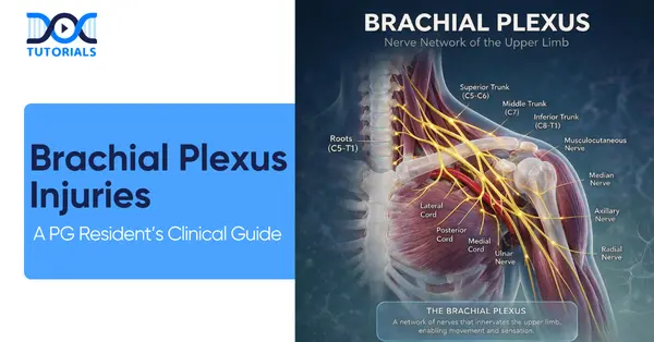
Mastering the intricate network of the brachial plexus is non-negotiable for clinical and examination success. This guide breaks down the essential anatomy, common exam traps, and the most vital diagnostic parameters surrounding a brachial plexus injury.
Get High Quality, High Yield PG Residency Orthopedics Notes for Free!
The Architectural Foundation of the Brachial Plexus
The biological wiring diagram of the brachial plexus follows a highly specific and logical sequence of formation. The sequence flows from Roots to Trunks to Divisions to Cords to Branches. Understanding this anatomical sequence is the first step in diagnosing and mapping any brachial plexus injury.
Roots, Trunks, and Divisions
The network originates from the ventral rami of spinal nerves C5, C6, C7, C8, and T1. These nerve roots quickly merge to form three distinct trunks.
- The Upper Trunk is formed by the union of the C5 and C6 roots.
- The Middle Trunk is formed exclusively by the continuation of the C7 root.
- The Lower Trunk is formed by the union of the C8 and T1 roots.
Once the trunks are formed, they traverse towards the clavicle and each splits into anterior and posterior divisions. This division step is crucial because it reorganizes the nerve fibers to eventually supply the anterior (flexor) and posterior (extensor) compartments of the upper extremity.
Cords and Terminal Branches
Following the divisions, the nerves reorganize into three cords. The cords of the brachial plexus utilize a strict nomenclature that is based entirely on their anatomical relationship to the axillary artery.
- The Lateral Cord is formed by the anterior divisions of the Upper and Middle trunks.
- The Medial Cord is formed solely by the anterior division of the Lower trunk.
- The Posterior Cord is uniquely formed by the posterior divisions from all three trunks (Upper, Middle, and Lower).
Mastering this RTDCB (Roots, Trunks, Divisions, Cords, Branches) sequence and the specific cord relations to the axillary artery will allow you to localize lesions perfectly in clinical practice.
Do all segments and connections within the brachial plexus give off peripheral nerve branches?
Answer: No. A defining anatomical rule of the brachial plexus is that divisions give off absolutely no branches. Assuming that all parts of the plexus give off branches is a very common exam trap designed to test your foundational anatomical knowledge.
Specific Nerve Branches and Clinical Correlates
The brachial plexus gives rise to multiple nerves, each with distinct motor and sensory responsibilities. Recognizing the function of these nerves helps quickly identify the level of a brachial plexus injury.
Root and Trunk Level Branches
Several nerves branch off before the formation of the cords.
- The Dorsal Scapular nerve and a contribution to the Phrenic nerve branch directly from the C5 root.
- The Long Thoracic nerve, which provides motor supply to the serratus anterior, originates from roots C5, C6, and C7.
- The Superior Trunk gives off the Suprascapular nerve and the Nerve to the subclavius.
A lone C5 root injury can drastically compromise respiratory function due to its critical contribution to the phrenic nerve and neck muscles. Furthermore, a brachial plexus injury to the upper roots (C5-C6) results in Erb’s palsy, while an injury to the lower roots (C8-T1) results in Klumpke’s palsy.
Cord Level Terminal Branches
The lateral, medial, and posterior cords give rise to the major terminal branches of the upper limb.
- The Lateral Cord branches into the Lateral Pectoral nerve (supplying the Pectoralis major), the Musculocutaneous nerve (supplying the Biceps, Brachialis, and Coracobrachialis), and the Lateral root of the Median nerve.
- The Medial Cord branches into the Medial Pectoral nerve, Medial Cutaneous nerves, the Ulnar nerve, and the Medial root of the Median nerve.
- The Posterior Cord gives rise to the Superior and Inferior Subscapular nerves, the Thoracodorsal nerve, the Axillary nerve, and the Radial nerve.
The Radial nerve is solely responsible for all extension mechanisms of the upper limb, including elbow, wrist, and digit extension. When evaluating hand innervation, be aware of ulnar motor exceptions: the thenar muscles and the lateral two lumbricals are median-innervated, not ulnar-innervated.
Mechanisms and Patterns of a Brachial Plexus Injury
When evaluating a patient with a brachial plexus injury, identifying the mechanism of trauma helps narrow down the damaged anatomy. Approximately 50% of these plexus injuries are caused by traction, which is predominantly seen in motor vehicle accidents (MVAs).
Common Etiologies and Vascular Associations
The anatomical location of the trauma often correlates strongly with specific vascular injuries. Distinguishing between supraclavicular and infraclavicular injury patterns clinically is a necessary skill for PG residents. Identifying the associated vascular injury will expertly help you localize the exact level of the brachial plexus injury.
| Injury Location | Anatomical Region | Mechanism of Injury | Associated Vascular Injury |
| Supraclavicular | Located above the clavicle | The neck and shoulder are violently forced apart | Subclavian artery |
| Infraclavicular | Located below the clavicle | Fractures or dislocations occurring around the shoulder joint | Axillary artery |
Nerve Microanatomy and Injury Classifications
To fully grasp the severity and prognosis of a brachial plexus injury, you must understand the microscopic layers of peripheral nerves. The severity of a nerve injury depends entirely on the depth of connective sheath disruption.
The Layers of a Peripheral Nerve
The nerve is composed of several protective layers surrounding the functional core.
- Epineurium: This is the outermost nerve covering.
- Perineurium: This layer covers individual fascicles, or bundles of axons.
- Endoneurium: This innermost connective layer covers individual axons.
- Axon: This is the functional unit capable of regeneration.
Overall prognosis is inversely proportional to connective tissue sheath involvement. Intact connective sheaths are critically required for directed axonal regeneration. Therefore, a lone axonal injury where the sheaths remain intact always carries a more favorable prognosis compared to covering-sheath disruptions.
Seddon and Sunderland Systems
Accurate grading of the injury using the Sunderland system is the most critical principle in clinical management, as it directly dictates the binary choice between clinical observation and surgical intervention.
| Sunderland Type | Seddon Equivalent | Histopathologic Changes | Recovery Potential |
| Type I | Neurapraxia | Myelin injury only (physiological conduction block) | Spontaneous auto-recovery within days to weeks |
| Type II | Axonotmesis | Axonal injury with an intact endoneurium | Auto-recovery expected due to intact sheath |
| Type III | Axonotmesis | Axonal and endoneurial injury | Spontaneous auto-recovery is still possible |
| Type IV | Neurotmesis (Mixed) | Perineurial injury and fascicular disruption | No spontaneous recovery; requires surgical repair |
| Type V | Neurotmesis | Complete nerve transection and epineurial disruption | No spontaneous recovery; requires surgical repair |
A common exam trap is equating Sunderland Type III with a guaranteed failure of recovery; however, spontaneous auto-recovery is still possible in Type III injuries because the axon remains capable of regeneration despite endoneurial damage. Sunderland Type IV is the critical threshold where spontaneous recovery becomes impossible due to perineurial disruption. The worst possible scenario is a complete root avulsion from the spinal origin, which represents the most severe, irreparable form of a primary brachial plexus injury.
Clinical Assessment and Prognosis
In managing a brachial plexus injury, the Tinel sign is your primary clinical monitor for tracking axonal regeneration.
The presence of a progressing Tinel sign fundamentally rules out Neurapraxia (Sunderland Type I), because Neurapraxia does not feature Wallerian degeneration. It also rules out complete Neurotmesis (Sunderland Type V/VI), where regeneration cannot naturally cross the transection.
- If a Tinel sign is progressing distally, axonal regeneration is actively occurring, and the resident should continue to observe the patient.
- If the Tinel sign is absent or stops progressing, you must reassess for stalled recovery or immediately plan for surgical repair.
Frequently Asked Questions (FAQs)
- What specific nerve roots form the brachial plexus?
The ventral rami origins of the brachial plexus are C5, C6, C7, C8, and T1.
- Which roots are involved in Erb’s palsy?
Erb’s palsy is an upper root brachial plexus injury that involves the C5 and C6 roots.
- Which roots are involved in Klumpke’s palsy?
Klumpke’s palsy is a lower root brachial plexus injury that involves the C8 and T1 roots.
- What is the motor function of the long thoracic nerve?
The long thoracic nerve, derived from roots C5, C6, and C7, supplies motor function to the serratus anterior muscle.
- How can a lone C5 root injury affect respiration?
A lone C5 root brachial plexus injury can compromise respiratory function due to its critical contribution to the phrenic nerve.
- Does the ulnar nerve innervate all intrinsic hand muscles?
No, always check the exceptions for ulnar nerve hand supply: the thenar muscles and lateral two lumbricals are spared, as they are median-innervated.
- What is the primary function of the radial nerve?
The radial nerve is solely responsible for all extension mechanisms of the upper limb, including elbow, wrist, and digit extension.
- What characterizes Neurapraxia?
Neurapraxia is a physiological conduction block involving myelin injury only, with the axon perfectly intact, allowing spontaneous recovery within days to weeks.
- What defines Axonotmesis?
Axonotmesis features axonal injury while the nerve sheath remains intact, and it always presents with Wallerian degeneration distal to the site of the brachial plexus injury.
- What is the clinical significance of a progressing Tinel sign?
A progressing Tinel sign fundamentally indicates that axonal regeneration is occurring in Sunderland Types II and III injuries, and it effectively rules out Neurapraxia and complete Neurotmesis.

Latest Blogs
-

NEET SS Exam 2024: Analysis, Key Dates, Counselling
The NEET SS 2024 exam kicked off on March 29, 2025. Over two days and two slots, candidates across 13…
-

INI SS Topper: 1 Dream, 1 Study Buddy, 1 Rank – The Journey of Dr. Prithivi Raaj, CML 1 & (AIIMS) AML 1 in DM Cardiology
Achieving the top rank in a competitive exam like the INI SS DM Cardiology requires tireless dedication, a structured approach,…
-

INI SS Results: Merit List, Exam Insights, and What Comes Next
The INI SS results for October 2024 are just around the corner. This is a life-changing moment for thousands of…