External Ear: Anatomy, Functions, and Disorders

Learning about the anatomy of the external ear enables you to understand how sound enters the ear and reaches the person’s hearing system. It also protects the inner components of the ear and prevents any kind of damage or infection.
Understanding its anatomical features, physiological roles, and related clinical conditions is essential, especially if you’re a NEET PG aspirant. Keep reading for a detailed understanding of ear anatomy and its associated disorders, which can significantly aid in clinical reasoning.
What is the External Ear?
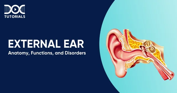
The external ear refers to the outermost section of the auditory system, which is visible on either side of the head. It collects sound and channels it through a tapered passageway towards the tympanic membrane, commonly known as the eardrum. There are specialised glands present in the skin of the external ear that can produce earwax, helping to protect the ear against dust and microbes.
What is the Anatomy of the External Ear?
The external ear anatomy consists of 3 parts:
- Pinna (Auricle)
It is one of the outer ear parts that can be seen and is positioned externally on the head. The exterior part, which is termed the pinna, is a cartilaginous flexible structure covered with skin, constructed in such a way that it forms a channel that accumulates and directs the wave of sound into the ear canal.
The pinna has numerous sebaceous glands within its skin that secrete sebum, which is an oily substance that keeps the skin soft and prevents it from drying out or becoming irritated. The following are the parts of pinna:
- Helix: This is simply the large outer edge of the ear, looking like a curve that starts at the point of attachment to the head and bends down to the earlobe.
- Antihelix: This is located immediately behind the helix, and it takes the shape of a Y apex that reflects the shape of the external ear.
- Crux of the Helix: Shaped like a curved band, this section bridges the upper helix to the lower antihelix.
- Tragus and Antitragus: These are small, cartilaginous projections located around the opening to the ear canal that could protect it.
- Concha: This is essentially the hollow space at the centre of the auricle, which directs the sound waves towards the auditory canal.
- Lobule: Commonly known as the earlobe, this soft, fleshy portion at the bottom of the ear lacks cartilage and is often used for wearing earrings.
- External Auditory Canal (External Auditory Meatus)
The external auditory meatus is commonly referred to as the ear canal. It forms a passage between the concha and the tympanic membrane. Structurally, it consists of a cartilaginous outer third and a bony inner two-thirds.
Rather than being a straight channel, the canal follows a natural S-shaped curve, which serves to shield the inner ear by limiting the entry of water, dust, and other foreign particles. Its lining contains numerous ceruminous glands, which secrete earwax. This waxy substance plays a protective role by trapping dirt and deterring small insects from progressing further into the ear.
- Tympanic Membrane
Also known as the eardrum, it is a delicate, thin structure that marks the boundary between the outer ear and the middle ear. It plays a crucial role in hearing by capturing sound vibrations that travel through the external auditory canal and conveying them to the tiny bones of the middle ear.
Its key anatomical features include:
- Umbo: This is the central depression where the handle (manubrium) of the malleus connects with the eardrum.
- Lateral Process of the Malleus: Positioned slightly above and in front of the umbo, this part of the malleus is visible during otoscopic examination.
- Anterior and Posterior Malleolar Folds: These are small folds of tissue that stretch forward and backwards from the lateral process, forming a boundary around the upper part of the eardrum.
- Pars Flaccida: Found above the malleolar folds, this loose, triangular region of the tympanic membrane appears less tense and more flexible than the rest.
- Pars Tensa: Comprising the majority of the eardrum, this section is thicker and tightly stretched, playing a crucial role in transmitting sound vibrations.
- Cone of Light: Seen during an ear exam as a bright, triangular reflection, it is located in the lower front quadrant of the eardrum, just below the umbo.
What are the Functions of the External Ear?
The external ear serves to gather sound vibrations and guide them toward the tympanic membrane. Its auricle plays a role in determining the direction of sound and in focusing acoustic energy. The blood supply of ear is provided by particular arteries and veins, and it receives sensory input from branches of the trigeminal and facial nerves.
The external auditory canal, which comprises both cartilaginous and bony segments, stretches from the auricle to the middle ear. This canal, along with the auricle, enhances sound transmission and shields deeper ear structures. While directional hearing is more accurate using both ears, some degree of sound localisation is achievable with one ear alone.
What are the Common Disorders of the External Ear?
A variety of conditions can affect the external ear, ranging from minor infections to more serious structural or functional issues. Below are some of the more frequently encountered disorders:
- Otitis Externa
This condition refers to inflammation within the ear canal, which may appear as a localised boil or present as widespread irritation that can sometimes involve the eardrum. It may be triggered by various factors, including bacterial or fungal infections, viral agents, or physical trauma.
Management typically includes careful removal of debris from the canal by using pain-relievers and anti-inflammatory medications.
- Ear Tumours
Benign growths such as keloids, cysts, and bony protrusions may develop in or around the external ear and often require surgical removal for comfort or cosmetic reasons. Malignant tumours, including squamous cell carcinoma, basal cell carcinoma, and melanoma, can also affect the ear.
Treatment depends on cancer type, stage, and extent of spread and may involve surgery, radiation, or chemotherapy.
- Tympanic Membrane Perforation
A tear or hole in the eardrum, known as a ruptured tympanic membrane, can result from infection, exposure to sudden loud noises, physical trauma, or the insertion of foreign objects into the ear. While such perforations often heal naturally, some cases may need surgical repair, such as a tympanoplasty.
- Perichondritis
It is an infection of the tissue layer covering the cartilage of the outer ear. It typically arises after some trauma, such as piercings or surgery. If left untreated, it may cause cartilage damage. Antibiotics are the standard treatment, though surgical drainage may be required if abscesses form.
- Trauma to the Ear
Blunt injury, fractures, or lacerations affecting the external ear can result in pain, deformity, or hearing complications. Treatment can entail basic or may involve a surgical procedure in the case of a serious injury.
FAQs About the External Ear
- What is the main function of the external ear?
The external ear collects sound waves and channels them towards the eardrum, aiding in hearing and sound localisation.
- Can earwax cause hearing problems?
Yes, excessive earwax can block the ear canal and temporarily reduce hearing, but it usually resolves with proper ear hygiene or medical removal.
- Are ear tumours always cancerous?
No, many ear tumours, like cysts or keloids, are benign. However, cancerous tumours can occur and require prompt diagnosis and treatment.
- What symptoms might suggest a problem with your external ear?
Several signs can point to an issue affecting the external ears. Common symptoms include:
- Discomfort or aching in the ear
- Swelling or redness in the ear
- A blocked or congested sensation
- Reduced clarity of hearing
- Persistent itching within the ear canal
- Episodes of dizziness or feeling sick
- A sensation of heaviness inside the ear
- Fluid or discharge leaking from the ear
- How can I prevent infections in the external ear?
To reduce your risk, avoid inserting objects into your ears, keep your ears dry after swimming or bathing, and refrain from using harsh chemicals or sprays near the ear canal.
Conclusion
A clear understanding of the anatomy, functions of the external ear, and types of disorders is necessary when learning theory, as well as when implementing it in practice. The external ear plays a crucial role in conducting sound and protecting the deeper ear structures, which makes it a high-yield topic in medical education.
As NEET PG aspirants, it is crucial for you to understand the function and disorders related to the external ear in order to diagnose the problem and take care of the patient. Strengthen your preparation by using the video lectures and the high-yield study materials offered by DocTutorials and the QRP (Quick Revision Programs) taught by experts in the medical field.
Ready to boost your NEET PG rank? Join our comprehensive NEET PG course today!
Latest Blogs
-

NEET SS Exam 2024: Analysis, Key Dates, Counselling
The NEET SS 2024 exam kicked off on March 29, 2025. Over two days and two slots, candidates across 13…
-

NEET PG Registration 2025: An Essential Guide For Exam Prep
The NEET PG registration, which is conducted online, is a crucial step in the exam process. Filling out the NEET…
-

NEET PG Syllabus 2026: A Must-Have Complete Guide for Exam Success
The NEET PG Syllabus acts as one of the foundation stones for aspiring postgraduate medical students like you who are…


