Pelvic Organ Prolapse: Causes, Symptoms, Diagnosis, and Treatment
Pelvic organ prolapse (POP) is often prevalent in women. It has been estimated that about 9.3% of all females were living with POP in 2010. Other more recent epidemiologic reviews state that the prevalence of pelvic examination is 30-50% in postmenopausal women, but surveys that rely on symptoms cite rates of 3-6%.
Smaller community research in India proposes a postpartum POP rate of nearly 1.6%, but the rates are underreported due to social stigma and unawareness. At the clinical level, POP is observed as bulging of the bladder, uterus, or rectum into or out of the vagina. It results in symptoms like pressure in the vagina, dysuria, defecation, and dyspareunia.
Pelvic organ prolapse is certainly a subject that should be understood well in the case you plan to take the NEET PG exam. Continue reading to get a detailed understanding of this condition.
What is Pelvic Organ Prolapse?
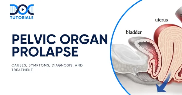
Pelvic organ prolapse (POP) is characterised by the inferior descent of pelvic organs, including the anterior/posterior vaginal walls, uterus, cervix, and vaginal apex, into the vaginal cavity. This happens because the support of pelvic floor muscles, fascia, and the ligaments is weakened, and the organs can bulge into the vagina opening, causing such disorders as cystocele, rectocele, enterocele, or uterine prolapse.
What are the Types of Pelvic Organ Prolapse?
Various types of pelvic organ prolapse in women exist according to the major organ involved and the point at which the vagina is involved.
Anatomical Classification of Pelvic Organ Prolapse
This system of classification depends on the main organ used in descent through the vaginal canal:
- Urethrocele: The urethra and the anterior vaginal wall move downwards towards or through the opening of the vagina.
- Cystocele: The bladder and the anterior vaginal wall drop down into the vagina.
- Cystourethrocele: Both the bladder and urethra prolapse along with the anterior vaginal wall.
- Uterovaginal Prolapse: Refers to the case when the primary part of the vaginal wall, the top part of the vagina (vaginal vault), and the cervix descend downwards.
- Enterocele: A projection of the single upper posterior wall of the vagina, usually protruding loops of small intestine.
- Rectocele: This is where there is protrusion and deformity of the lower posterior vagina by the rectum.
Classification Based on Vaginal Wall Involvement
Pelvic organ prolapse can also be organised according to the segment of the vaginal wall that is affected:
- Anterior Vaginal Wall Prolapse: It consists of cystocele and urethrocele. The possible problems that can be presented in cystoceles are that they can either be central and lateral, or a combination of both.
- Apical Prolapse: Comprises enteroceles, uterine prolapse, uterovaginal prolapse, and vaginal vault prolapse. These can be combined with front or back of the back types of prolapse.
- Posterior Vaginal Wall Prolapse: It includes mainly rectoceles and may be at the upper, middle or lower part of the posterior vaginal wall.
- Perineal Body Defects: Such defects affect the weakness or injury of the perineal body and may contribute to or worsen posterior wall prolapse.
What are the Causes of Pelvic Organ Prolapse?
Pelvic organ prolapse develops when the pelvic floor muscles and supportive tissues become weak or damaged. This condition is more frequently observed in postmenopausal women and with advancing age. The following are the pelvic organ prolapse causes:
- Pregnancy and vaginal delivery
- Obesity
- Chronic constipation and straining
- Persistent or chronic cough
- Previous pelvic surgery (e.g., Hysterectomy)
What are the Symptoms of Pelvic Organ Prolapse?
In some cases, pelvic organ prolapse (POP) may be asymptomatic. However, when symptoms do occur, they can vary depending on the type and severity of the prolapse. Common pelvic organ prolapse symptoms include:
- Visible or Palpable Vaginal Bulge: A sensation or observation of tissue protruding through or beyond the vaginal introitus.
- Pelvic Discomfort: A feeling of pelvic pressure, heaviness, or aching pain, particularly after prolonged standing or physical exertion.
- Lower Back Pain: Dull aching pain in the lumbosacral region, often associated with the sensation of dragging.
- Tampon Retention Issues: Difficulty in retaining or inserting tampons due to altered vaginal anatomy.
- Urinary Symptoms
- Increased urinary frequency
- Urgency with or without urge incontinence
- Incomplete bladder emptying
- Weak or intermittent urine stream
- Bowel Dysfunction: Constipation or the sensation of incomplete evacuation and the need to apply manual pressure (splinting) through the vagina to facilitate defecation.
- Sexual Dysfunction: Dyspareunia (painful intercourse) or reduced sexual satisfaction
- Multiorgan Involvement: Pelvic floor weakness often affects multiple compartments simultaneously. The presence of one type of organ prolapse (e.g., cystocele) increases the likelihood of coexisting prolapse of other pelvic structures (e.g., rectocele or uterine prolapse).
How to Diagnose Pelvic Organ Prolapse?
Pelvic organ pro
Pelvic organ prolapse (POP) is often prevalent in women. It has been estimated that about 9.3% of all females were living with POP in 2010. Other more recent epidemiologic reviews state that the prevalence of pelvic examination is 30-50% in postmenopausal women, but surveys that rely on symptoms cite rates of 3-6%.
Smaller community research in India proposes a postpartum POP rate of nearly 1.6%, but the rates are underreported due to social stigma and unawareness. At the clinical level, POP is observed as bulging of the bladder, uterus, or rectum into or out of the vagina. It results in symptoms like pressure in the vagina, dysuria, defecation, and dyspareunia.
Pelvic organ prolapse is certainly a subject that should be understood well in the case you plan to take the NEET PG exam. Continue reading to get a detailed understanding of this condition.
What is Pelvic Organ Prolapse?
Pelvic organ prolapse (POP) is characterised by the inferior descent of pelvic organs, including the anterior/posterior vaginal walls, uterus, cervix, and vaginal apex, into the vaginal cavity. This happens because the support of pelvic floor muscles, fascia, and the ligaments is weakened, and the organs can bulge into the vagina opening, causing such disorders as cystocele, rectocele, enterocele, or uterine prolapse.
What are the Types of Pelvic Organ Prolapse?
Various types of pelvic organ prolapse in women exist according to the major organ involved and the point at which the vagina is involved.
Anatomical Classification of Pelvic Organ Prolapse
This system of classification depends on the main organ used in descent through the vaginal canal:
- Urethrocele: The urethra and the anterior vaginal wall move downwards towards or through the opening of the vagina.
- Cystocele: The bladder and the anterior vaginal wall drop down into the vagina.
- Cystourethrocele: Both the bladder and urethra prolapse along with the anterior vaginal wall.
- Uterovaginal Prolapse: Refers to the case when the primary part of the vaginal wall, the top part of the vagina (vaginal vault), and the cervix descend downwards.
- Enterocele: A projection of the single upper posterior wall of the vagina, usually protruding loops of small intestine.
- Rectocele: This is where there is protrusion and deformity of the lower posterior vagina by the rectum.
Classification Based on Vaginal Wall Involvement
Pelvic organ prolapse can also be organised according to the segment of the vaginal wall that is affected:
- Anterior Vaginal Wall Prolapse: It consists of cystocele and urethrocele. The possible problems that can be presented in cystoceles are that they can either be central and lateral, or a combination of both.
- Apical Prolapse: Comprises enteroceles, uterine prolapse, uterovaginal prolapse, and vaginal vault prolapse. These can be combined with front or back of the back types of prolapse.
- Posterior Vaginal Wall Prolapse: It includes mainly rectoceles and may be at the upper, middle or lower part of the posterior vaginal wall.
- Perineal Body Defects: Such defects affect the weakness or injury of the perineal body and may contribute to or worsen posterior wall prolapse.
What are the Causes of Pelvic Organ Prolapse?
Pelvic organ prolapse develops when the pelvic floor muscles and supportive tissues become weak or damaged. This condition is more frequently observed in postmenopausal women and with advancing age. The following are the pelvic organ prolapse causes:
- Pregnancy and vaginal delivery
- Obesity
- Chronic constipation and straining
- Persistent or chronic cough
- Previous pelvic surgery (e.g., Hysterectomy)
What are the Symptoms of Pelvic Organ Prolapse?
In some cases, pelvic organ prolapse (POP) may be asymptomatic. However, when symptoms do occur, they can vary depending on the type and severity of the prolapse. Common pelvic organ prolapse symptoms include:
- Visible or Palpable Vaginal Bulge: A sensation or observation of tissue protruding through or beyond the vaginal introitus.
- Pelvic Discomfort: A feeling of pelvic pressure, heaviness, or aching pain, particularly after prolonged standing or physical exertion.
- Lower Back Pain: Dull aching pain in the lumbosacral region, often associated with the sensation of dragging.
- Tampon Retention Issues: Difficulty in retaining or inserting tampons due to altered vaginal anatomy.
- Urinary Symptoms
- Increased urinary frequency
- Urgency with or without urge incontinence
- Incomplete bladder emptying
- Weak or intermittent urine stream
- Bowel Dysfunction: Constipation or the sensation of incomplete evacuation and the need to apply manual pressure (splinting) through the vagina to facilitate defecation.
- Sexual Dysfunction: Dyspareunia (painful intercourse) or reduced sexual satisfaction
- Multiorgan Involvement: Pelvic floor weakness often affects multiple compartments simultaneously. The presence of one type of organ prolapse (e.g., cystocele) increases the likelihood of coexisting prolapse of other pelvic structures (e.g., rectocele or uterine prolapse).
How to Diagnose Pelvic Organ Prolapse?
Pelvic organ prolapse diagnosis involves a systematic approach that includes:
- Detailed Medical History
The diagnostic process begins with obtaining a thorough patient history. Important elements include obstetric history (number and mode of deliveries), history of menopause, previous pelvic surgeries, and symptoms such as:
- A sensation of vaginal bulge or pelvic pressure
- Urinary or faecal incontinence
- Difficulty with bowel movements
- Discomfort during sexual activity
- Pelvic Examination
To determine the extent and nature of prolapse, the clinical pelvic examination is conducted, whereby prolapse is visually and manually assessed. The patient is often observed in a supine and standing position and requested to perform the Valsalva manoeuvre (bearing down). This will help determine which pelvic compartment (anterior, posterior, or apical) has been involved.
- Pelvic Floor Strength Testing
During the pelvic exam, the examiner evaluates pelvic floor muscle tone and sphincter function. This involves asking the patient to contract pelvic muscles, as if trying to stop the flow of urine. It provides information about the muscular and ligamentous support of the:
- Vaginal walls
- Uterus
- Rectum
- Urethra
- Bladder
- Bladder Function Tests
In certain cases, additional urodynamic tests may be conducted to assess bladder function. These may include:
- Checking for urinary leakage when the bladder or prolapsed organ is manually repositioned.
- Measuring residual urine to evaluate how effectively the bladder empties.
- Identifying coexisting conditions like stress urinary incontinence.
- Imaging Studies
Imaging techniques are reserved for more complex or multi-compartment prolapse cases. Common imaging modalities include:
- Magnetic Resonance Imaging (MRI): Provides detailed views of pelvic floor anatomy.
- Transperineal or Transvaginal Ultrasound: Useful for visualising the position of pelvic organs during rest and strain.
What are the Treatment Options for Pelvic Organ Prolapse
Pelvic organ prolapse treatment depends on several factors, like the severity of the prolapse, its location, and how much it impacts your daily life. Both surgical and nonsurgical options are available. In mild cases where symptoms are minimal or absent, treatment might not be necessary.
- Nonsurgical Treatments
Non-surgical interventions are designed to relieve symptoms and improve quality of life, and not necessarily cure the problem, particularly in cases that are severe. Such conservative methods are favoured by many patients or initially used by them. Common nonsurgical options include:
- Vaginal Pessary
A removable silicone device inserted into the vagina by your provider to support and hold the prolapsed organ in place.
- Pelvic Floor Muscle Exercises (Kegel Exercises)
Weight-gain exercises aimed at pelvic floor muscle strengthening. Your doctor can put you in touch with a pelvic floor therapist or urogynecologist who can evaluate your muscle strength and instruct specific types of exercises.
- Surgical Treatments
Surgery is considered when conservative treatments fail to relieve symptoms or when surgery is deemed the best option to improve quality of life. Discuss the potential benefits and risks of surgery thoroughly with your healthcare provider.
Surgical interventions generally fall into 2 categories:
- Obliterative Surgery
This type narrows the vaginal opening to prevent organ prolapse but usually eliminates the possibility of penetrative intercourse.
- Colpocleisis: A common obliterative procedure that shortens the vagina and prevents organs from bulging externally. This is typically recommended for patients who are frail or do not desire future sexual activity.
- Reconstructive Surgery
This surgery repairs weakened pelvic floor structures and repositions the prolapsed organs. Common reconstructive procedures include:
- Sacrocolpopexy: Used to correct uterine or vaginal vault prolapse by attaching the vagina to a ligament near the tailbone with surgical mesh. Usually performed laparoscopically through small abdominal incisions.
- Sacrohysteropexy: Treats uterine prolapse by securing surgical mesh to the cervix and vagina, lifting the uterus back into its normal position by attaching it to the tailbone.
- Colporrhaphy: Corrects anterior and/or posterior vaginal wall prolapse by reinforcing vaginal walls via the vaginal route with dissolvable sutures to support the bladder and rectum.
- Uterosacral or Sacrospinous Ligament Fixation (Native Tissue Repair): In this procedure, your surgeon uses your own tissues to support the vagina by attaching it to pelvic ligaments or muscles through the vagina. Dissolvable sutures are used to secure the support, and they treat uterine or vaginal vault prolapse.
FAQs About Pelvic Organ Prolapse
- Is pelvic organ prolapse normal?
Pelvic organ prolapse is common, especially among women after childbirth, menopause, or ageing. While it’s not “normal,” mild prolapse occurs frequently and may not cause symptoms. Medical evaluation is essential to assess severity and guide management.
- How can I prevent pelvic organ prolapse?
Preventive measures include maintaining a healthy weight, avoiding heavy lifting, managing chronic cough or constipation, and regularly performing pelvic floor exercises (Kegels). These help strengthen pelvic muscles and reduce strain on pelvic organs, lowering the risk of prolapse development.
- Can I push back pelvic organ prolapse?
In some cases, a vaginal pessary device can be inserted to physically support and hold the prolapsed organs in place, effectively “pushing back” the prolapse. However, this is a temporary measure and requires medical supervision.
- What to avoid if I have pelvic organ prolapse?
Avoid heavy lifting, straining during bowel movements, prolonged standing, and high-impact exercises that increase intra-abdominal pressure. Managing chronic cough and constipation is vital to prevent worsening of the prolapse and related symptoms.
Conclusion
Pelvic organ prolapse may be of concern if you experience the signs of pelvic pressure, a feeling of vaginal bulge, urinary changes, or bowel changes. Proper and timely diagnosis followed by treatment is the key to avoiding any further progression of the patient and restoration of quality of life. With the persistence or aggravation of the symptoms, immediate medical assessment is needed.
Offering the best video lectures, expansive revision notes and focused study materials, DocTutorials is the first choice to prepare and study specifications important to pelvic organ prolapse. Join DocTutorials today and explore our NEET PG course to excel in your medical career.
lapse diagnosis involves a systematic approach that includes:
- Detailed Medical History
The diagnostic process begins with obtaining a thorough patient history. Important elements include obstetric history (number and mode of deliveries), history of menopause, previous pelvic surgeries, and symptoms such as:
- A sensation of vaginal bulge or pelvic pressure
- Urinary or faecal incontinence
- Difficulty with bowel movements
- Discomfort during sexual activity
- Pelvic Examination
To determine the extent and nature of prolapse, the clinical pelvic examination is conducted, whereby prolapse is visually and manually assessed. The patient is often observed in a supine and standing position and requested to perform the Valsalva manoeuvre (bearing down). This will help determine which pelvic compartment (anterior, posterior, or apical) has been involved.
- Pelvic Floor Strength Testing
During the pelvic exam, the examiner evaluates pelvic floor muscle tone and sphincter function. This involves asking the patient to contract pelvic muscles, as if trying to stop the flow of urine. It provides information about the muscular and ligamentous support of the:
- Vaginal walls
- Uterus
- Rectum
- Urethra
- Bladder
- Bladder Function Tests
In certain cases, additional urodynamic tests may be conducted to assess bladder function. These may include:
- Checking for urinary leakage when the bladder or prolapsed organ is manually repositioned.
- Measuring residual urine to evaluate how effectively the bladder empties.
- Identifying coexisting conditions like stress urinary incontinence.
- Imaging Studies
Imaging techniques are reserved for more complex or multi-compartment prolapse cases. Common imaging modalities include:
- Magnetic Resonance Imaging (MRI): Provides detailed views of pelvic floor anatomy.
- Transperineal or Transvaginal Ultrasound: Useful for visualising the position of pelvic organs during rest and strain.
What are the Treatment Options for Pelvic Organ Prolapse
Pelvic organ prolapse treatment depends on several factors, like the severity of the prolapse, its location, and how much it impacts your daily life. Both surgical and nonsurgical options are available. In mild cases where symptoms are minimal or absent, treatment might not be necessary.
- Nonsurgical Treatments
Non-surgical interventions are designed to relieve symptoms and improve quality of life, and not necessarily cure the problem, particularly in cases that are severe. Such conservative methods are favoured by many patients or initially used by them. Common nonsurgical options include:
- Vaginal Pessary
A removable silicone device inserted into the vagina by your provider to support and hold the prolapsed organ in place.
- Pelvic Floor Muscle Exercises (Kegel Exercises)
Weight-gain exercises aimed at pelvic floor muscle strengthening. Your doctor can put you in touch with a pelvic floor therapist or urogynecologist who can evaluate your muscle strength and instruct specific types of exercises.
- Surgical Treatments
Surgery is considered when conservative treatments fail to relieve symptoms or when surgery is deemed the best option to improve quality of life. Discuss the potential benefits and risks of surgery thoroughly with your healthcare provider.
Surgical interventions generally fall into 2 categories:
- Obliterative Surgery
This type narrows the vaginal opening to prevent organ prolapse but usually eliminates the possibility of penetrative intercourse.
- Colpocleisis: A common obliterative procedure that shortens the vagina and prevents organs from bulging externally. This is typically recommended for patients who are frail or do not desire future sexual activity.
- Reconstructive Surgery
This surgery repairs weakened pelvic floor structures and repositions the prolapsed organs. Common reconstructive procedures include:
- Sacrocolpopexy: Used to correct uterine or vaginal vault prolapse by attaching the vagina to a ligament near the tailbone with surgical mesh. Usually performed laparoscopically through small abdominal incisions.
- Sacrohysteropexy: Treats uterine prolapse by securing surgical mesh to the cervix and vagina, lifting the uterus back into its normal position by attaching it to the tailbone.
- Colporrhaphy: Corrects anterior and/or posterior vaginal wall prolapse by reinforcing vaginal walls via the vaginal route with dissolvable sutures to support the bladder and rectum.
- Uterosacral or Sacrospinous Ligament Fixation (Native Tissue Repair): In this procedure, your surgeon uses your own tissues to support the vagina by attaching it to pelvic ligaments or muscles through the vagina. Dissolvable sutures are used to secure the support, and they treat uterine or vaginal vault prolapse.
FAQs About Pelvic Organ Prolapse
- Is pelvic organ prolapse normal?
Pelvic organ prolapse is common, especially among women after childbirth, menopause, or ageing. While it’s not “normal,” mild prolapse occurs frequently and may not cause symptoms. Medical evaluation is essential to assess severity and guide management.
- How can I prevent pelvic organ prolapse?
Preventive measures include maintaining a healthy weight, avoiding heavy lifting, managing chronic cough or constipation, and regularly performing pelvic floor exercises (Kegels). These help strengthen pelvic muscles and reduce strain on pelvic organs, lowering the risk of prolapse development.
- Can I push back pelvic organ prolapse?
In some cases, a vaginal pessary device can be inserted to physically support and hold the prolapsed organs in place, effectively “pushing back” the prolapse. However, this is a temporary measure and requires medical supervision.
- What to avoid if I have pelvic organ prolapse?
Avoid heavy lifting, straining during bowel movements, prolonged standing, and high-impact exercises that increase intra-abdominal pressure. Managing chronic cough and constipation is vital to prevent worsening of the prolapse and related symptoms.
Conclusion
Pelvic organ prolapse may be of concern if you experience the signs of pelvic pressure, a feeling of vaginal bulge, urinary changes, or bowel changes. Proper and timely diagnosis followed by treatment is the key to avoiding any further progression of the patient and restoration of quality of life. With the persistence or aggravation of the symptoms, immediate medical assessment is needed.
Offering the best video lectures, expansive revision notes and focused study materials, DocTutorials is the first choice to prepare and study specifications important to pelvic organ prolapse.
Join DocTutorials today and explore our NEET PG course to excel in your medical career.


Latest Blogs
-

INI CET Exam 2026: Your Roadmap to Success – Key Topics, Strategies, and Lessons from Last Year’s Papers
The INI CET exam is more than just a test; it’s a significant milestone for many medical students aiming to…
-

INI CET Exam Success: Previous Year Question Papers & Ultimate Guide – INI CET PYQ
One can feel overwhelmed while preparing for the INI CET (Institute of National Importance Combined Entrance Test). A vast syllabus,…
-

NEET PG 2026 Preparation with Live Power Pack: Your Path to Success in 2026
Preparing for the NEET PG 2026 can be an overwhelming journey because of its vast syllabus and the pressure to…


