Mucormycosis: Types, Causes, Symptoms, and Treatment
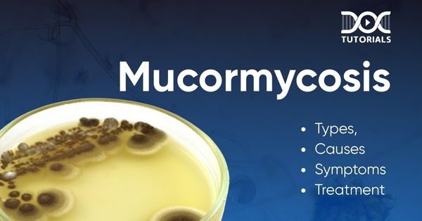
Mucormycosis is a fatal fungal infection that affected about 70% of the global population in March 2021. It occurs when fungal moulds from the Mucorales order infect human bodies. Mucormycosis affects individuals with weakened immune systems through a fast-moving infectious process that medical experts call the “black fungus.”
This article outlines all the essential details about mucormycosis, including its types, causes, risk factors, symptoms, and treatment approaches. It has gained worldwide attention because of the COVID-19 infection.
What is Mucormycosis?
Mucormycosis is a fungal condition caused by micromycetes, which reside unproblematically in environmental settings. These fungi remain harmless until they invade patients with weak immune systems. Mucormycosis infects different parts of the body, such as the sinuses, brain, lungs, digestive tract, and skin, and it shows distinct symptoms around each area of infection.
People who suffer from underlying illnesses that compromise their immune defences face the highest risk of developing this disease. Individuals with uncontrolled diabetes, cancer patients, patients with organ transplants, and those taking immunosuppressive drugs experience substantially elevated risk.
Medical intervention fails to diminish mucormycosis mortality rates because of its aggressive nature. Its spread happens rapidly, and the detection of the infection becomes necessary for patient survival.
Proficient and immediate medical intervention through medication and tissue removal procedures remains crucial for mucormycosis treatment since this condition typically results in death without such prompt intervention.
The immediate medical need for mucormycosis symptoms, with proper treatment of high-risk individuals, stresses the urgency of medical attention first thing when symptoms appear.
Types of Mucormycosis
The 5 different forms of mucormycosis affect distinct body systems through unique clinical pathways that vary in treatment results.
- Rhino-orbital-cerebral Mucormycosis
The most prevalent form is a sinus infection, particularly in diabetic patients and organ transplant recipients. This infection starts in the sinuses before it spreads quickly to the eyes and attacks the brain.
Patients usually experience pain, facial swelling, blocked nasal passages, and darkening around their noses or palates. This gradually results in vision disturbances, bulging eye appearance, and neurological complications.
- Pulmonary Mucormycosis
The disease targets people who have either hematologic malignancy or severe neutropenia. This infection spreads into lung tissue, producing general symptoms that duplicate bacterial pneumonia through fever and chest pain.
The primary characteristic of pulmonary mucormycosis is its ability to invade blood vessels through tissue necrosis, which leads to life-threatening lung cavity formation and bleeding during coughing.
- Cutaneous Mucormycosis
This infection appears in 2 different versions: primary (direct inoculation enters through skin damage or wounds) and secondary (infection spreads through the bloodstream from a different location). The beginning causes skin reddening, firm tissue swelling, and localised discomfort.
The infection follows a rapid course, ending in necrotic tissue development and distinctive black flank lesions. Primary cutaneous mucormycosis can develop in people with normal immune function after experiencing traumatic injuries.
- Gastrointestinal Mucormycosis
The highly fatal form of this condition occurs when patients consume fungal spores. The infection mainly affects patients who lack enough nutrition and premature newborns.
The digestive tract is infected in all its regions, although the stomach, colon, and ileum experience the highest incidence of involvement. Patients experience symptoms such as abdominal pain, distention, nausea, vomiting, and gastrointestinal bleeding.
- Disseminated Mucormycosis
The infection progresses to the worst stage when it advances throughout multiple organs simultaneously. Mucormycosis frequently develops from pulmonary infections in patients with low white blood cell counts, although it may start from various points throughout the body.
The brain remains the most frequently affected secondary area, whereas spread can also occur in the spleen, heart, skin, and other organs.
Causes and Risk Factors of Mucormycosis
The medical condition known as Mucormycosis primarily attacks patients who suffer from different illnesses. Understanding its origins and identifying vulnerable groups provides valuable information for halting its development before it needs medical intervention.
Causes of Mucormycosis
- Inhalation of Fungal Spores
Individuals usually get infected through inhalation of tiny fungal spores in the environment. These spores are plentiful and can be found in soil, decomposing vegetation, compost piles, and residential dust.
The respiratory defence mechanisms successfully these spores in healthy people. The fungal spores germinate in susceptible hosts and then invade lung tissue and blood vessels from the sinuses.
- Direct Tissue Inoculation
Mucormycetes penetrate the body through broken skin tissue. Any skin break, from burns to surgical wounds, traumatic injuries, or minor skin abrasions, can enable the fungus to enter the body. The pathogens multiply swiftly after establishing themselves in the tissues of vulnerable individuals and create localised infections that spread to additional areas.
- Ingestion of Contaminated Materials
Foodborne gastrointestinal mucormycosis manifests after eating foods containing fungal spores that cause infection. Such cases occur when food safety measures are inadequate and among individuals who lack sufficient nutrition.
Risk Factors of Mucormycosis
- Diabetes Mellitus
Individuals with uncontrolled diabetes and ketoacidosis face a very high risk of complications. These organisms experience an environment suitable for fungal growth because their bodies contain both high amounts of acid and sugar. Patients with diabetes account for the most significant proportion of mucormycosis cases globally.
- Hematologic Disorders and Malignancies
Any condition resulting in a decreased neutrophil count through neutropenia creates a significant opening for mucormycosis infection. Treatments such as chemotherapy and radiation therapy suppress immune cell production, which affects patients with leukaemias, lymphomas, and other hematologic malignancies.
- Organ and Stem Cell Transplantation
People who have received organ transplants need immunosuppressive drugs to protect their organs, but these medications make them more vulnerable to opportunistic infections. A combination of corticosteroids with other immunosuppressants creates condition for fungal infection to develop.
- Iron Overload Conditions
Patients with iron overload diseases like hemochromatosis and those undergoing deferoxamine therapy face increased fungal growth because micromycetes obtain iron from the patient’s metabolism to proliferate.
- COVID-19 Association
Medical professionals recognised a considerable rise in mucormycosis infections throughout the COVID-19 pandemic among diabetic patients who received corticosteroid therapy for COVID-19.
When the virus damages the immune system, and patients receive steroids and suffer from baseline medical conditions, it creates the required environment for this devastating secondary infection.
- Environmental and Occupational Exposures
People whose work or living conditions expose them to soil and decaying organic matter (construction work, farming, disaster-ridden areas) face a higher risk for mucormycosis, and such exposures need to be considered in combination with any predisposing medical conditions.
Symptoms of Mucormycosis
The symptoms appear distinctly depending on where the Mucormycosis infection occurs. Timely diagnosis via appropriate testing is essential since this infection rapidly progresses when patients do not receive treatment.
- Facial swelling and pain.
- Persistent head pain that develops into severe intensity.
- Nasal congestion or discharge, sometimes with blood
- Fever of varying intensity
- Black lesions on the nasal bridge or inside the mouth
- Double vision, blurry vision, and total loss of sight
- Toothache or loosening of teeth
- Facial numbness or tingling
- A persistent cough with blood.
- Shortness of breath or chest pain
- Fever and night perspiration
- General fatigue and weakness
- Rapidly increasing redness along with swelling.
- The appearance of blackened tissue cells (necrosis)
- Open sores or blisters which remain unhealed
- Pain or tenderness around the affected area
- Abdominal pain and distension
- Vomiting and nausea alongside potential bleeding incidents.
- Gastrointestinal bleeding
- Fever and general malaise
Diagnosis of Mucormycosis
- Medical History and Physical Examination
A medical assessment starts with testing for risk factors, including diabetes and cancer, alongside steroid use to identify black lesions at suspected infection sites. Mucormycosis can differentiate itself from other infections because it advances quickly along its course of symptoms.
- Imaging Studies
Computed Tomography scans (CT scans) show bone damage and tissue alterations in sinuses, lungs, and abdominal organs, while MRI provides clearer brain imaging. These medical image techniques provide doctors with information about infection progression.
- Laboratory Testing
Examining tissue samples through a microscope leads to a definitive diagnosis because mucormycosis displays distinctive broad ribbon-like structures. The identification of distinct fungal species through general methods requires lengthy processing, although these methods may produce incorrect negative results.
- Specialised Procedures
Doctors employ bronchoscopy for lung infections, endoscopic procedures for sinus involvement, and needle aspiration for infections that spread throughout the body. Speedy and precise diagnosis of the disease matters since early treatment determines the outcome of the resolving process.
Treatment Options for Mucormycosis
Patients with Mucormycosis need an urgent response consisting of surgery combined with antifungal drugs and proper treatment of underlying health conditions.
- Antifungal Medications
The initial treatment choice includes intravenous administration of high-dose lipid amphotericin B formulations during several weeks of therapy. Patients who experience amphotericin B intolerance or require step-down therapy can use Posaconazole and isavuconazole medications, available in intravenous and oral formulations.
The antifungal agents fluconazole, echinocandins, and voriconazole do not exhibit effective activity against Mucorales fungi.
- Surgical Management
The survival rate depends majorly upon the immediate aggressive surgical removal of infected tissue. Several surgical procedures might be required to eliminate infected tissue and prevent infection spread.
- Additional Approaches
Individuals with underlying medical conditions must carefully manage blood sugar and decrease immunosuppressive therapies whenever possible. Adjunctive therapy using hyperbaric oxygen demonstrates benefits through better tissue oxygenation and enhanced immune function.
FAQs about Mucormycosis
- Can mucormycosis be cured?
Yes, the fungal infection Mucormycosis, or black fungus, shows full curing potential when medical experts make early diagnoses and deliver prompt treatment.
- What healthcare objects trigger mucormycosis infections?
Healthcare-associated mucormycosis outbreaks have been associated with several sources, including adhesive bandages, wooden tongue depressors, hospital linens, negative pressure rooms, water leaks, poor air filtration, non-sterile medical devices, and building construction.
- How to test for mucormycosis?
Medical providers need both tissue samples from the bacterial site and pathologic investigations with test results to make a definitive diagnosis of mucormycosis. Medical experts use sterile specimens to confirm invasive mucormycosis infections since they demonstrate more clinical significance than samples from non-sterile areas.
- What medical treatment stands as the initial therapy for mucormycosis infections?
The initial FDA-approved drugs for treating mucormycosis infections are Amphotericin B medications, including their lipid formulations, which consist of liposomal preparations, lipid complexes, and colloidal dispersions. These drugs target fungal wall ergosterol to destabilise cell membranes.
- Can mucormycosis cause other conditions to develop?
Treatment of mucormycosis remains dangerous since the infection rapidly spreads across different body parts. According to medical experts the infection has the ability to spread through untreated cases into the lungs or brain. This can cause a brain infection, paralysis, pneumonia, seizures, and even death.
Conclusion
The medical attention needed for Mucormycosis treatment must be both immediate and intensive. For high-risk patients with diabetes or weakened immune systems, survival depends on immediate treatment through antifungal medications combined with surgical procedures after early detection and prompt diagnosis.
DocTutorials provides extensive educational courses that expand medical knowledge through a complete range of medical subjects. Master FMGE preparation with our specialised coaching, featuring expert faculty.


Latest Blogs
-

NEET SS Exam 2024: Analysis, Key Dates, Counselling
The NEET SS 2024 exam kicked off on March 29, 2025. Over two days and two slots, candidates across 13…
-

NEET PG Registration 2025: An Essential Guide For Exam Prep
The NEET PG registration, which is conducted online, is a crucial step in the exam process. Filling out the NEET…
-

NEET PG Exam 2025- Date, Pattern, Marking Scheme, Subject Wise Weightage, and Exam Mode
NEET PG Exam 2025 is the ultimate gateway for medical graduates aspiring to pursue postgraduate courses in medicine, including MD,…


