Thermal Injury: Causes, Symptoms, and Treatment
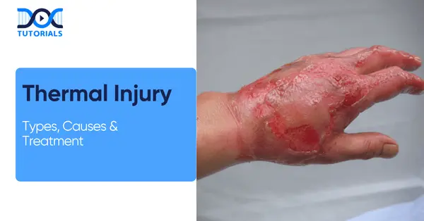
Thermal injuries (burns) are tissue damage caused by external heat sources, e.g., flame, hot liquids, steam, chemicals, electricity, or long-term radiation. These injuries are either superficial, first-degree burns, or deep full-thickness (third- or fourth-degree) burns, including those that affect more than one organ system when extensive.
In forensic medicine, the study of burn injuries is important in determining the cause and mode of injury or death. Keep reading to know more about its types, causes, diagnosis and treatment.
Classification of Thermal Injuries
There are 3 types of thermal injuries, classified by depth and mechanism. The depth of burns is superficial to full-thickness and deeper. Mechanisms include scalds, flame burns, contact burns, electrical burns, chemical burns, and radiation burns.
Thermal injuries are classified by the mechanism of injury and the depth (degree) of skin damage. The following table summarises the burn depth and features:
| Burn Degree | Skin Layers Involved | Key Features | Healing Time |
| First (Superficial) | Epidermis only | Red, dry, painful, no blisters; blanches on touch | ~5–10 days; heals without scarring |
| Second (Partial) | Epidermis + part of dermis | Red/moist with intact or ruptured blisters; very painful; blanches | 2–3 weeks; may scar |
| Third (Full) | Epidermis + dermis + subcutaneous | Leathery, white/brown/charred; painless (nerve loss); does not blanch | >8 weeks; requires surgery |
Fourth-degree burns (deep burns) penetrate all layers of the skin into muscle, bone or organs and result in charring. These are life-threatening and must always be surgically treated.
By mechanism, thermal injuries include:
- Scalds: Caused by hot liquids or steam (common in children).
- Flame Burns: Due to open-fire or clothing ignition.
- Contact Burns: Through contact with hot things (e.g., metal, irons).
- Electrical Burns: From AC (alternating current) or DC (direct current), producing serious thermal damage along the current path.
- Chemical Burns: Caused by acids or alkalis; they chemically generate heat on tissue contact.
- Radiation Burns: As a result of long UV (ultraviolet) exposure (sunburn) or ionising radiation.
Proper classification assists in estimating the total area burned and the treatment area. For example, the “Rule of Nines” divides the body into regions of 9% TBSA (total body surface area) each (e.g. head 9%, each arm 9%).
In children, the “Lund-Browder” chart adjusts for proportion changes. The severity of the burns is also graded (minor, moderate, major) according to TBSA, depth, age, and location.
Pathophysiology of Thermal Burns
Thermal injury causes protein denaturation and forms three zones: coagulation (necrosis), stasis (at risk), and hyperemia (reversible). Major burns cause fluid loss, shock, inflammation, and such complications as infection, respiratory damage, and organ failure.
Cellular damage from thermal energy includes protein denaturation and membrane destruction. Burns leave a characteristic three-zone pattern in the skin. They are:
- Zone of Coagulation: Central, irreversibly necrotic tissue.
- Zone of Stasis: Surrounding ischemic tissue that may survive or progress to necrosis.
- Zone of Hyperemia: Outer area with reversible injury.
Burns are dynamic, meaning the initial injury can worsen over a few hours or days (e.g., the stasis zone worsening). Larger size burns (>20% of body surface) result in a systemic inflammation response. It increases capillary permeability, resulting in excessive fluid loss (burn shock) and intravascular volume depletion.
Hypotension and hypoperfusion follow if not corrected. Exposure to superheated air or combustion products may also damage the airways and lungs, leading to respiratory failure. Tissue necrosis from burns also impairs the skin’s functions: infection barrier, temperature regulation, and fluid retention.
Loss of these functions leads to a high risk of infection, hypothermia, electrolyte imbalance, and organ dysfunction. Systemically, severe burns can cause hypermetabolism and release of inflammatory mediators (cytokines, prostaglandins), which, in turn, may result in the systemic inflammatory response syndrome (SIRS) and multi-organ failure.
In forensics, extensive burns may also result in poisonous exposures: carbon monoxide (in smoke) and cyanide (in plastics) intoxication are frequent causes of death.
Causes and Risk Factors of Thermal Injury
The burn definition medical professionals use describes the injury as damage to bodily tissues caused by hot liquids, flames, hot objects, electricity, lightning, chemicals and sunlight. The risk is high with the extremes of age, work-related exposures, and substance use, as well as unsafe homes. Burn patterns are used in forensic practice to determine the cause and manner of injury, such as possible abuse or homicide.
Thermal burns arise from many sources of excessive heat. Common causes include:
- Scalding liquids (hot water, steam, oil).
- Open flames (house fires, explosives).
- Hot objects (steam irons, stoves), and sunlight (ultraviolet).
Electrical injury or joule burn produces heat within the body when current passes through tissues, and there are usually entry and exit wounds. However, during a lightning strike, a filigree burns (also known as a Lichtenberg figure, arborescent burn, or feathering) is a distinctive, fern-like pattern on the skin
. Chemical burns (strong acids or alkalis) cause thermal-like injury via exothermic reactions.
Risk Factors
Certain conditions predispose to burns:
- Age Extremes: Young children and the elderly have thinner skin and slower reflexes to avoid hot sources.
- Occupational Hazards: Firefighters, industrial workers handling hot materials, and kitchen staff are at greater risk of exposure to flames.
- Substance Use: The effect of intoxication by alcohol or drugs compromises judgment near fire and hot materials.
- Domestic Risks: Lack of smoke detectors, unsafe stoves or heaters, and poor supervision of children.
The pattern and distribution of burns are also indicative of a cause in forensic practice. For example, “forced immersion” scalds (symmetrical burns on buttocks and extremities with distinct scald lines) raise suspicion of abuse in children or vulnerable adults.
Necklacing (homicidal burning method using a tyre) is a classic forensic burn scenario. The identification of these patterns aids forensic professionals in determining whether the injuries were deliberate, unintentional, or caused by others.
Clinical Features and Symptoms of Thermal Injury
Thermal burns are characterised by redness, blisters, swelling, charring, and eschar, whereas inhalation injury is characterised by facial burns, soot, and respiratory distress. Severe burns may result in oedema and renal failure.
Thermal burns have varying symptoms based on depth and extent. In partial-thickness (nerve endings are exposed) burns, pain is intense, but in full-thickness burns (nerve destruction), pain can be surprisingly weak. Common signs include:
- Redness (erythema)
- Blistering
- Swelling
- Charring
- Eschar formation
In inhalation injuries, one may see:
- Facial burns
- Singed nasal hairs
- Soot in the mouth or sputum
- Hoarseness
- Respiratory distress
Fluid shifts cause generalised oedema and often oliguric renal failure in severe cases. In forensic examination of a burned body, several findings distinguish antemortem from postmortem burns:
| Feature | Antemortem Burn | Postmortem Burn |
| Skin redness | Present (due to vasodilation) | Absent (no circulation) |
| Blisters | Present: fluid rich in albumin and chloride | May occur; contain air/thin fluid |
| Soot in airways | Carbon particles in the trachea/lungs | Absent |
| Carboxyhemoglobin (CO-Hb) | Elevated (>10%) from smoke inhalation | Normal |
| Haemorrhage at the injury site | Present (capillaries intact) | Absent (no vital reaction) |
| Curling’s ulcer (duodenum) | Common in fatal burns (stress ulcers) | Absent |
Other notable forensic findings include “pugilistic attitude” – a flexed posture of burned bodies due to muscle contraction at >60°C – and heat fractures of bones (bones split due to dehydration, often with a distinctive pattern that does not cross suture lines).
Burned bodies also exhibit thermolysis of soft tissues and “parchment” skin. The cause of death in fire victims is often inhalation injury (asphyxia from smoke, CO or cyanide poisoning) rather than cutaneous burns.
Findings in forensic reports can include lung weight, cherry-red tissue discolouration (CO poisoning), and cyanide atomic absorption.
Medical Diagnosis of Thermal Injury
Medical diagnosis of thermal injury involves both forensic and clinical evaluation. Forensic methods help determine the cause, severity, and identity, while clinicians assess burn size, depth, and related complications, such as inhalation injury.
Forensic-medical diagnosis of thermal injuries involves thorough external and internal examination. Investigations may include:
- Autopsy: Close dissection to determine depth of burns, internal organ injuries, and smoke inhalation.
- Toxicology: Determining the quantity of carboxyhemoglobin, cyanide, and other poisonous gases formed during combustion is essential.
- Histopathology: Skin biopsies can confirm the vitality of burns (e.g. neutrophils, red blood cells), extravasation in antemortem burns, and estimate the age of lesions.
- Imaging: Post-mortem CT (computed tomography) scans of the body could reveal heat fractures or retained foreign material.
- Forensic Odontology/Anthropology: Dental or skeletal analysis can help identify severely burned remains.
In clinical diagnosis, the healthcare practitioners measure the size and depth of the burns. The “Rule of Nines” or Lund-Browder chart quantifies TBSA. The depth is assessed by appearance, capillary refill (blanching), pain, and sensation. Patients with burns should be evaluated for associated injuries (inhalation, trauma) and for burn severity.
Management and Treatment of Thermal Injury
Thermal injuries are initially managed using the ABC principles (immediately after injury): airway protection, fluid resuscitation using the Parkland formula, and analgesics. Proper wound care, infection prevention, and tetanus prophylaxis are essential. Severe burns are surgically managed and strictly supported, with nutrition and observation of complications.
Initial management of thermal injuries follows trauma protocols (ABCs). Key steps include:
- Airway and Breathing: Check for inhalation injury. Early intubation in case of respiratory distress or facial burns.
- Circulation: Start large-bore IV access promptly. Remove constricting clothing/jewellery.
- Fluid Resuscitation: For major burns, aggressive IV fluid resuscitation is required. The Parkland formula is a standard guideline:
4 mL × body weight (kg) × % TBSA (second/third-degree only) of lactated Ringer’s over 24 hours, with half in the first 8 hours. Adjust fluid to maintain urine output (0.5–1 mL/kg/h in adults).
- Pain Control: Give analgesics (when necessary- opioids) as burns are painful.
- Burn Care: Cool the burn with lukewarm (not ice-cold) water to limit depth. Gently clean and debride wounds. Use topical antimicrobials (e.g. silver sulfadiazine) to avoid infection. Cover with a sterile dressing.
- Tetanus Prophylaxis: Update tetanus immunisation, as burns are prone to tetanus.
- Wound Management: Superficial burns heal spontaneously. Partially-thickness burns can need debridement and specialised dressings. Full-thickness burns require surgical treatment (excision and grafting).
- Supportive Care: Monitor for shock (IV fluids, hemodynamic monitoring), provide nutritional support (high-calorie diet), and prevent hypothermia (warm ambient temperature).
Admit to a burn centre if criteria are met (e.g. >20% TBSA, face/hands/genitals, electrical/chemical burns).
Rehabilitative therapy, scar management (pressure garments), and reconstructive surgery are treatment options that may be used on a long-term basis.
Prevention of Thermal Injury
Thermal injury prevention involves fire control, safe kitchen operations, controlled water temperature, and workplace safety precautions. Social consciousness and safety laws help reduce the incidence of burns, although forensic vigilance is important for identifying abuse or arson.
Preventing thermal injuries is critical, especially in high-risk settings. Safety measures include:
- Fire Safety: Install working smoke detectors and keep fire extinguishers accessible. Practice fire escape plans.
- Kitchen Safety: Use hot liquids with caution; handle pot handles carefully; be cautious around stoves. Never leave cooking unattended.
- Water Temperature: Set water heaters to less than 120 F (49 C) to avoid scalds; check bath water temperature.
- Workplace Precautions: Use protective equipment (gloves, face shields) when handling hot materials or chemicals. Follow electrical safety protocols.
- Education: Educate children on fire safety, do not play with matches, lighters or fireworks.
- Sun Protection: Wear sunscreen and protect yourself to avoid radiation burns (sunburn).
FAQs about Thermal Injuries
- How are burns classified by degree?
Burns can be categorised as deep: first-degree (epidermis, only, red, painful), second-degree (partial-thickness, blisters, very painful), and third-degree (full, charred, often painless). The fourth-degree burns extend to muscle or bone.
- What is the Rule of Nines?
The Rule of Nines is a quick method to estimate burn surface area. In adults: head/neck = 9%, each arm = 9%, anterior trunk = 18%, posterior trunk = 18%, each leg = 18%, perineum = 1%.
- How can you tell if burns occurred before death?
Antemortem burns show vital reactions – redness, blisters with protein fluid, haemorrhage at edges, and soot in airways. Blood tests reveal high carboxyhemoglobin and cyanide. Postmortem burns do not have these signs (there is no inflammation or inhaled soot).
- What is “pugilistic attitude”?
It is a typical flexed posture of a burned body due to muscle shrinkage in heat (> 60 °C). It resembles a boxing stance and results from heat-stiffened muscles.
- How are burns managed initially?
Follow trauma ABCs: secure airway (intubate if needed), support breathing, then start IV fluids. Cool the burn, provide analgesics, clean/debride wounds and place sterile dressings. Use formulas such as the Parkland formula (4 mL × kg × % TBSA) to guide fluid resuscitation.
- What is the Parkland formula?
A guideline for fluid resuscitation in major burns: 4 mL per kg of body weight per cent of TBSA burned (2nd/3rd-degree burns) of lactated Ringer’s in the first 24 hours. Half of that volume is given in the first 8 hours after the burn.
- Can electric injuries be considered thermal burns?
Electrical injuries cause serious thermal damage along current paths, but they also cause internal organ injury (arrhythmias, muscle damage). When providing a forensic classification, an electrical burn is frequently mentioned in contrast to a thermal burn.
- How do chemical burns differ from thermal burns?
Chemical burns occur due to an exothermic reaction with acid or alkali and can extend the damage to tissue until the agent is eliminated. They are treated by copious irrigation and neutralisation, unlike simple cooling for thermal burns.
- Why is burn depth painful initially but not in full-thickness burns?
Partial thickness burns reveal nerve ends, resulting in severe pain. Full-thickness burns destroy the nerves within the affected area; the victim might not experience any pain within the burnt area.
- What are some key forensic signs of burn-related death?
In addition to vital reactions, there is soot in the airways (from inhalation), thermal fractures of the bone, black lung (from inhaled carbon), and internal signs of systemic poisoning (e.g., cardiac carbonisation, cerebral infarcts due to hypoxia).
Conclusion
Burns can be thermal, chemical, radiation-induced, or electrical, each with distinct forensic and medical consequences. Medical students and forensic trainees must be able to understand burn classification, pathophysiology, and related factors.
In NEET PG forensic medicine, the Rule of Nines, burn depth features, and vital reactions in burns are high-yield topics. For additional guidance, DocTutorials can be your study companion. We offer crisp videos, clinical Qbank, exam-focused notes, flashcards, and mind maps.
Join DocTutorials today and explore our NEET PG course to excel in your medical career!


Latest Blogs
-
INI CET Exam Pattern 2027: A Complete Guide with Subject-Wise Weightage
INI CET Exam Pattern 2027: A Complete Guide with Subject-Wise Weightage The Institute of National Importance Combined Entrance Test (INI…
-

NEET PG Registration 2027: An Essential Guide For Exam Prep
The NEET PG registration, which is conducted online, is a crucial step in the exam process. Filling out the NEET…
-

NEET PG Exam 2025- Date, Pattern, Marking Scheme, Subject Wise Weightage, and Exam Mode
NEET PG Exam 2025 is the ultimate gateway for medical graduates aspiring to pursue postgraduate courses in medicine, including MD,…