Understanding the Biomechanics of the Pelvic Ring and Pelvic Ring Fractures
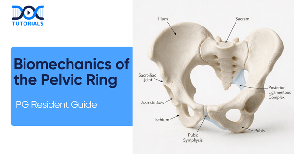
For postgraduate orthopedic residents, mastering the intricate biomechanics of the pelvic ring is an absolute necessity. The structural integrity of this anatomical region dictates patient survival and long-term functional outcomes following severe, high-velocity trauma.
When evaluating a pelvic ring fracture, one must immediately look beyond the broken bones and understand the profound, systemic failure of the soft-tissue envelope and ligamentous tension bands holding the pelvis together. This blog distills essential concepts regarding pelvic ring into actionable knowledge.
Get High Quality, High Yield Orthopedics PG Residency Notes for Free!
The Core Biomechanics of the Pelvic Ring
The fundamental principle underlying the biomechanics of the pelvic ring is that its stability relies entirely on ligamentous integrity. The sacroiliac (SI) joint lacks any inherent bony stability of its own. Therefore, understanding the ligamentous anatomy is directly tied to understanding stability.
When managing a pelvic ring fracture, residents must carefully distinguish between rotational support structures and vertical support structures.
Rotational vs. Vertical Support Structures
A critical clinical concept to remember is that a pelvis can be rotationally unstable yet remain vertically stable.
If rotational stability fails during a trauma event, the anterior side and the anterior SI ligaments typically rupture. Conversely, if vertical stability fails, it is an indication that the posterior interosseous and iliolumbar ligaments have completely ruptured.
Ligamentous Stabilizers of the Pelvic Ring
| Stability Type | Key Anatomical Structures Involved |
| Rotational Stability | Symphysis ligaments Sacrospinous ligament Anterior sacroiliac ligament |
| Vertical Stability | Posterior sacroiliac ligament Interosseous ligament Iliolumbar ligament |
Acute Resuscitation in a Pelvic Ring Fracture
A pelvic ring fracture typically results from high-velocity trauma, predominantly affecting younger patient populations. While most of these pelvic injuries are technically stable and non-surgical, catastrophic internal blood loss is the primary immediate threat to the patient’s life.
The primary source of lethal hemorrhage in a pelvic ring fracture is the superior gluteal artery and its associated venous plexus, as well as the internal iliac artery.
During acute trauma, the crisis metric to monitor is maximum blood loss, which can rapidly reach 1.5 to 2 liters. This volume of hemorrhage quickly leads to severe hypovolemic shock.
How should a pelvic binder be correctly applied during the acute resuscitation phase of a pelvic ring fracture?
A pelvic binder MUST be centered strictly over the greater trochanters, not the iliac crests, to effectively reduce the overall pelvic volume and provide mechanical tamponade.
The Trauma Bay Algorithm
When a patient arrives hemodynamically unstable with a suspected pelvic ring fracture, circumferential pelvic wrapping is the first step.
Following this, a FAST exam dictates the next moves: if the FAST is positive (indicating intraperitoneal bleeding), the patient requires an immediate laparotomy. If the FAST is negative, the patient should proceed to angiography to embolize the bleeding vessels. The primary goal in the first 24 hours of treating a hemodynamically unstable patient is hemorrhage control, not anatomical bony reduction.
Radiographic Signs and Classifications
To properly assess the altered biomechanics of the pelvic ring post-trauma, a standard radiographic trauma series is required. This series includes the standard AP View, the Inlet View (taken at 40° Caudal), and the Outlet View (taken at 40° Cephalad).
The Inlet view is utilized specifically to evaluate rotational displacement (front-to-back), whereas the Outlet view is essential for visualizing vertical displacement (up-and-down).
Critical Instability Thresholds and Classifications
Recognizing instability on an X-ray is paramount. For rotational instability, a symphysis diastasis (widening) of > 2.5 cm is the absolute clinical threshold that distinguishes a stable open-book injury (APC I) from an unstable one (APC II). For vertical instability, superior migration of one hemi-pelvis by 1.0 cm or more is the definitive sign.
The Tile Classification System (AO/OTA 61) breaks down a pelvic ring fracture into three categories:
- Type A: Stable (Note: Type A3 fractures involving the transverse sacrum or coccyx are technically considered spinal injuries rather than true pelvic ring disruptions).
- Type B: Rotationally Unstable, Vertically Stable (e.g., Open Book, Lateral Compression).
- Type C: Rotationally & Vertically Unstable.
Surgical Timing and Pelvic Ring Fracture Fixation
When transitioning from the trauma bay to the OR, the physiological timing of the surgery is just as important as understanding the biomechanics of the pelvic ring.
Surgical Timing Protocols for a Pelvic Ring Fracture
| Strategy | Clinical Indicator (Venous Lactate) | Protocol Summary |
| EAC (Early Appropriate Care) | < 4.0 mmol/L | Proceed with definitive fixation within 36 hours. |
| DCO (Damage Control Orthopedics) | > 4.0 mmol/L | Delay definitive fixation. Stabilize with external fixators, resuscitate in ICU until lactate drops. |
Operating for definitive fixation when a patient’s lactate is > 4.0 mmol/L significantly increases the mortality rate; DCO must be employed instead.
Fixation Strategies and Clinical Pearls
For anterior fixation, options include external fixation, Anterior Subcutaneous Internal Fixation (ASIF), and internal plating. However, because of continued physiological motion at the symphysis during healing, anterior plates will frequently break if posterior instability is not concurrently addressed.
For posterior fixation, Denis Zone 2 sacral fractures mandate Open Reduction Internal Fixation (ORIF) over percutaneous techniques due to a 30% risk of neurological injury that requires decompression. Conversely, an open posterior approach has absolute contraindications: it should never be performed if the patient has a Morel-Lavallée lesion or recent internal iliac artery embolization, as it creates an extreme risk of catastrophic soft tissue failure.
Frequently Asked Questions
1. What is the primary stabilizing factor of the pelvic ring?
The pelvic ring relies entirely on ligamentous integrity for its stability, as the sacroiliac joint lacks any inherent bony stability.
2. Which ligaments fail if a pelvis becomes vertically unstable?
If vertical stability fails, the posterior interosseous and iliolumbar ligaments have ruptured.
3. What is the primary source of lethal hemorrhage in a pelvic ring fracture?
The lethal hemorrhage source is typically the superior gluteal artery and venous plexus, or the internal iliac artery.
4. Where exactly should a pelvic binder be placed?
A pelvic binder must be centered strictly over the greater trochanters, rather than the iliac crests, to properly reduce pelvic volume.
5. What does the Inlet radiographic view demonstrate?
The Inlet view (taken at a 40° caudal angle) shows the inward or outward rotational displacement of the anterior pelvis.
6. What symphysis widening measurement indicates an unstable open-book fracture?
A symphysis diastasis of > 2.5 cm is the critical value that distinguishes a stable APC I from an unstable APC II injury.
7. Why are Tile A3 fractures unique?
Tile A3 fractures, which involve transverse fractures of the sacrum or coccyx, are technically classified as spinal injuries rather than true disruptions of the pelvic ring.
8. What is the danger of a Denis Zone 2 sacral fracture?
Denis Zone 2 (transforaminal) fractures carry a 30% risk of neurological compromise and mandate ORIF for neural decompression.
9. When should Damage Control Orthopedics (DCO) be utilized over early fixation?
DCO should be utilized when a patient’s venous lactate level is > 4.0 mmol/L or if they remain hemodynamically unstable.
10. Why might an anterior pubic symphysis plate break post-operatively?
Due to continued physiological motion at the symphysis during the healing process, anterior plates will frequently break if the associated posterior instability is not concurrently addressed surgically.

Latest Blogs
-

NEET SS Exam 2024: Analysis, Key Dates, Counselling
The NEET SS 2024 exam kicked off on March 29, 2025. Over two days and two slots, candidates across 13…
-

INI SS Topper: 1 Dream, 1 Study Buddy, 1 Rank – The Journey of Dr. Prithivi Raaj, CML 1 & (AIIMS) AML 1 in DM Cardiology
Achieving the top rank in a competitive exam like the INI SS DM Cardiology requires tireless dedication, a structured approach,…
-

INI SS Results: Merit List, Exam Insights, and What Comes Next
The INI SS results for October 2024 are just around the corner. This is a life-changing moment for thousands of…