Subtrochanteric Fractures: A Masterclass on Intramedullary Nail Fixation for PG Residents
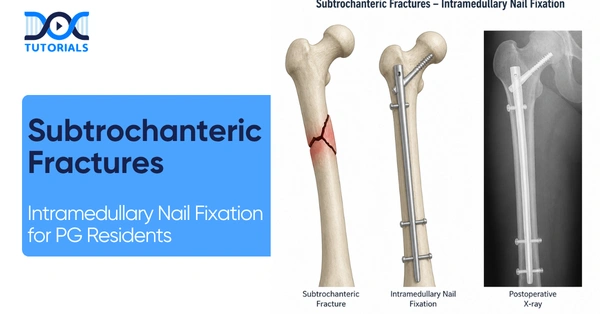
The subtrochanteric region represents a high-stress transition zone where profound biomechanical understanding is far more critical than isolated anatomical knowledge. For Orthopedics Post-Graduate (PG) Residents preparing for their exams and clinical practice, mastering these complex injuries requires a thorough grasp of the immense deforming forces at play and the meticulous application of an intramedullary nail.
Get High Quality, High Yield Orthopedics PG Residency Notes for Free!
Understanding the Anatomical Transition Zone
Before evaluating surgical implants like the intramedullary nail, it is imperative to strictly define the anatomical zone. A subtrochanteric fracture is defined as a fracture located entirely below the trochanters.
The proximal boundary of this region is the lesser trochanter, and the distal boundary is defined as exactly 5 cm distal to the lesser trochanter. This specific area acts as the transition zone to the femoral shaft. Any extension of the fracture line beyond this 5 cm mark changes the primary classification of the injury. Due to the biomechanical demands of this transition zone, the preferred implant is the intramedullary nail.
Biomechanics and Deforming Forces
The challenge of subtrochanteric fractures lies in the severe displacement caused by powerful regional musculature. A classic exam pearl dictates that the hallmark deformity is driven by the intact iliopsoas and abductors acting on the proximal fragment. To achieve a successful reduction before passing an intramedullary nail, surgeons must recognize and counteract these specific directional forces.
The proximal fragment is predictably flexed by the iliopsoas muscle, abducted by the abductor muscles, and externally rotated by the external rotators. Conversely, the distal fragment is adducted by the adductors and significantly shortened by the pull of the quadriceps and hamstrings. Correcting this multi-planar deformity is the primary prerequisite for accurate intramedullary nail insertion.
Biomechanical Deforming Forces in Subtrochanteric Fractures
| Bone Fragment | Acting Muscle Groups | Resulting Clinical Deformity |
| Proximal Fragment | Iliopsoas, Abductors, External Rotators | Flexed, Abducted, Externally Rotated |
| Distal Fragment | Adductors, Quadriceps, Hamstrings | Adducted, Shortened |
The Russell-Taylor Classification System
Surgical decision-making, particularly regarding the entry point of the intramedullary nail, relies heavily on accurate classification. The standard for these injuries is the Russell-Taylor Classification, which is primarily based on the fracture’s extension into the piriformis fossa.
Always assess the piriformis fossa first: if it is fractured, it is automatically a Type II injury. Type I fractures leave the piriformis fossa intact, while Type II fractures mean the fossa is involved.
Differentiating Subtypes
These main types are further subdivided based on the integrity of the lesser trochanter:
- Type IA and IIA: The lesser trochanter remains intact.
- Type IB and IIB: The lesser trochanter is fractured, which includes reverse oblique or oblique patterns.
Treatment Principles: The Role of the Intramedullary Nail
When managing a subtrochanteric fracture resulting from high-impact or high-velocity trauma, conservative treatment is generally not recommended. These injuries behave like femoral shaft injuries and require operative treatment.
The intramedullary nail (such as the PFN or PFNA) stands as the gold standard and the absolute treatment of choice. Plate fixation (using a Proximal Femoral Locking Plate or an Angled Blade Plate) is considered an alternative only if the intramedullary nail entry point is highly compromised or unsuitable.
Resident Focus Q&A: What is the most common surgical error encountered when inserting an intramedullary nail for a subtrochanteric fracture, and how is it corrected?
The most common surgical error is utilizing an excessively lateral entry point, which causes severe varus malalignment and delayed union. If the intramedullary nail drifts laterally, the bailout technique is to use an Anterior-to-Posterior (AP) Poller (Blocking) screw to force the intramedullary nail medially.
Surgical Techniques: Choosing the Right Entry Portal
The success of an intramedullary nail heavily depends on selecting the safest and most mechanically sound starting point.
The traditional piriformis fossa entry is technically demanding and carries a high risk of Avascular Necrosis (AVN) due to its proximity to the femoral head’s blood supply. It also carries a risk of an iatrogenic femoral neck fracture.
Consequently, the Greater Trochanter (GT) Tip is the commonly preferred approach. By utilizing a modified medial trochanter portal, surgeons gain a mechanically protective trajectory that ensures better alignment, an easier intramedullary nail technique, and improved reduction. A common exam trap is confusing varus and valgus errors; remember that a lateral entry point for the intramedullary nail causes varus malalignment, not valgus.
Intramedullary Nail Entry Portals and Associated Risks
| Intramedullary Nail Entry Portal | Benefits | Risks & Complications |
| Piriformis Fossa | Traditional alignment | High AVN risk, near blood supply, iatrogenic neck fracture |
| Greater Trochanter (GT) Tip | Preferred approach, avoids AVN risk | Requires a medial start to prevent varus deformity |
| Lateral Entry (Surgical Error) | None | Severe varus malalignment, nonunion, or delayed union |
Fixation Stability and Postoperative Rehabilitation
When utilizing an intramedullary nail, the integrity of the lesser trochanter is the critical stability factor. For highly unstable fractures (like R-T Type 1B, 2A, 2B), the proximal locking strategy requires a Reconstruction (Recon) mode. This configuration uses 2 or 3 proximal screws driven into the femoral head to provide superior fixation and control of the proximal fragment.
Postoperatively, the core principle is to avoid excessive force prematurely because the subtrochanteric region is subjected to massive bending moments. On Day 0, patients are restricted to touch-down weight bearing initially. Surgeons must wait a minimum of 6 weeks before allowing any advancement in weight-bearing, strictly relying on radiological evidence of callus formation.
Frequently Asked Questions (FAQs)
1. What defines the exact anatomical zone of a subtrochanteric fracture?
It is the zone starting from the lesser trochanter and extending exactly 5 cm distal to it.
2. What is the gold standard treatment for these injuries?
Operative treatment using an intramedullary nail is the standard of care.
3. In the Russell-Taylor classification, what dictates a Type II fracture?
Any involvement or extension of the fracture into the piriformis fossa automatically classifies it as a Type II.
4. What anatomical structure dictates the A vs. B sub-type in the Russell-Taylor system?
The integrity of the lesser trochanter dictates the sub-type; Type A is intact, and Type B is fractured.
5. Why is the Greater Trochanter tip preferred over the Piriformis Fossa for an intramedullary nail?
The Greater Trochanter tip avoids the high risk of Avascular Necrosis (AVN) associated with the piriformis fossa entry.
6. What is the consequence of an excessively lateral intramedullary nail entry point?
A lateral entry point causes severe varus malalignment and frequently leads to nonunion or delayed union.
7. How do you surgically correct a lateral drift of an intramedullary nail?
Surgeons should place an Anterior-to-Posterior (AP) Poller (blocking) screw to force the intramedullary nail medially.
8. What muscle forces cause the classic proximal fragment deformity?
The proximal fragment is flexed by the iliopsoas, abducted by the abductors, and externally rotated by the external rotators.
9. How many proximal screws are required for an intramedullary nail in Reconstruction (Recon) mode?
Recon mode requires 2 or 3 proximal locking screws placed into the femoral head.
10. When is the earliest a patient can advance beyond touch-down weight bearing after intramedullary nail fixation?
Weight-bearing should never be advanced before a strict minimum of 6 weeks, and only after an X-ray confirms radiological callus formation.

Latest Blogs
-

NEET SS Exam 2024: Analysis, Key Dates, Counselling
The NEET SS 2024 exam kicked off on March 29, 2025. Over two days and two slots, candidates across 13…
-

INI SS Topper: 1 Dream, 1 Study Buddy, 1 Rank – The Journey of Dr. Prithivi Raaj, CML 1 & (AIIMS) AML 1 in DM Cardiology
Achieving the top rank in a competitive exam like the INI SS DM Cardiology requires tireless dedication, a structured approach,…
-

INI SS Results: Merit List, Exam Insights, and What Comes Next
The INI SS results for October 2024 are just around the corner. This is a life-changing moment for thousands of…