
Mastering Trauma Surgery: A Comprehensive Guide for PG Residents
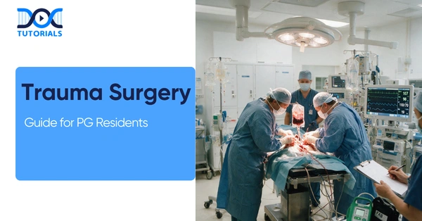
This essential guide on trauma surgery, tailored specifically for postgraduate (PG) surgical residents. Navigating the high-stakes environment of trauma surgery requires a structured, evidence-based approach to patient assessment, operative intervention, and postoperative care. This article provides a comprehensive overview of critical trauma surgery protocols, emphasizing the management of abdominal trauma, the application of damage control surgery, and high-yield clinical pearls to enhance your surgical residency training.
Get High Quality, High Yield Surgery PG Residency Notes for Free!
Mechanisms and Classification of Abdominal Trauma
In trauma surgery, understanding the mechanism of injury is the critical first step in predicting organ damage and guiding your diagnostic approach. Abdominal trauma is broadly categorized into blunt and penetrating injuries, each with a distinct pattern of visceral involvement.
When managing blunt abdominal trauma, the spleen is the most frequently injured organ, followed by the liver and the small intestine. Interestingly, in the pediatric population, the spleen remains the most commonly injured organ, but the kidney takes the second spot. Conversely, penetrating abdominal trauma most commonly affects the liver, followed by the small intestine and the diaphragm.
Patient classification based on hemodynamic status is a cornerstone of trauma surgery triage. Patients are classified as:
- Responders: Maintain stable hemodynamics.
- Transient Responders: Initially improve but then crash, indicating ongoing pathology.
- Non-Responders: Require immediate intervention due to persistent instability.
Common Mechanisms of Abdominal Trauma
| Trauma Type | Primary Mechanism | Most Commonly Injured Organs (in order of frequency) |
| Blunt | Motor vehicle accidents, falls, compression | 1. Spleen, 2. Liver, 3. Small Intestine |
| Penetrating | Stab wounds, gunshot wounds | 1. Liver, 2. Small Intestine, 3. Diaphragm |
| Pediatric Blunt | Falls, sports injuries, vehicle accidents | 1. Spleen, 2. Kidney |
Diagnostic Modalities in Trauma Surgery
Rapid and accurate diagnosis is vital in trauma surgery. For a stable patient with blunt abdominal trauma, a Contrast-Enhanced Computed Tomography (CECT) of the abdomen is the gold standard. However, if the patient is unstable, exhibits peritonitis (guarding/rigidity), or has a positive eFAST exam, an immediate exploratory laparotomy is indicated.
The Role of eFAST and CT Scans
The Extended Focused Assessment with Sonography for Trauma (eFAST) is the most sensitive initial test for unstable hemorrhage. The eFAST exam adds a pericardial view to the standard abdominal and pelvic windows (Morison’s Pouch, Splenorenal, and Pouch of Douglas).
Q: What are the primary blind spots and limitations of the eFAST exam in trauma surgery?
A: While rapid, the eFAST exam has significant blind spots. It cannot rule out hollow viscus perforation, cannot assess the retroperitoneum, and is often obscured by obesity and bowel gas. Furthermore, it cannot identify the exact source of bleeding and is unreliable for fluid volumes less than 100 ml.
For penetrating abdominal trauma, stable patients require a targeted approach. Flank or back injuries necessitate a triple contrast CT (Oral/IV/Rectal) to rule out retroperitoneal or colon injuries. Anterior abdominal injuries may require local wound exploration; if the peritoneum is breached, diagnostic laparoscopy is mandatory.
Organ-Specific Trauma Surgery Protocols
Effective trauma surgery requires tailored protocols for specific organ injuries, balancing the need to stop hemorrhage with organ preservation whenever possible.
Splenic and Liver Injuries
The spleen is highly susceptible to deceleration forces in blunt abdominal trauma. The AAST scale grades splenic injuries from Grade 1 (minor subcapsular hematoma) to Grade 5 (a “shattered” spleen). Stable patients with Grade 1-3 injuries are typically managed conservatively with hematocrit monitoring, utilizing angioembolization if their grade deteriorates. If a splenectomy is performed, residents must be vigilant about Overwhelming Post-Splenectomy Infection (OPSI), ensuring patients receive Pneumococcal, Meningococcal, and Hib vaccines.
For liver injuries, trauma surgery relies on the “4 P’s” for hemorrhage control: Pressure, Plug, Pringle’s Maneuver, and Pack. The Pringle’s maneuver occludes the portal vein and hepatic artery; if bleeding persists despite this, the source is likely the hepatic veins or inferior vena cava due to retrograde flow.
Damage Control Surgery in Bowel Trauma
In trauma surgery, the decision to perform Damage Control Surgery (DCS) versus Early Total Care is often dictated by the patient’s physiological state and lactate levels. A lactate level below 2 in a physiologically stable patient allows for Early Total Care, including resection and primary anastomosis of bowel injuries.
However, if a patient has a lactate level of 3-5+ or is a transient responder, Damage Control Surgery is imperative. The surgical principle here is to avoid anastomosis in unstable patients due to a high risk of leakage.
Damage Control Surgery vs. Early Total Care
| Clinical Parameter | Early Total Care | Damage Control Surgery (DCS) |
| Patient Status | Physiologically Stable | Physiologically Unstable / Transient Responder |
| Lactate Level | Lactate < 2 | Lactate 3-5+ / Acidosis |
| Surgical Action | Resection + Anastomosis | Resect, staple ends (discontinuity) or Stoma |
| Next Steps | Standard post-op recovery | ICU Stabilization followed by planned re-operation |
Retroperitoneal and Genitourinary Trauma
The retroperitoneum is famously known as the surgeon’s minefield in trauma surgery. Management depends heavily on the zone of injury. Zone 1 (Central) injuries must always be explored in both blunt and penetrating abdominal trauma. In contrast, Zone 3 (Pelvic) hematomas should generally not be explored due to the high tamponade effect; opening this zone can lead to torrential bleeding, so it is better managed with angioembolization and packing.
Lower genitourinary trauma presents uniquely. Urethral injuries present with blood at the meatus, retention, and a high-riding bladder; crucially, these patients must not be catheterized. Instead, trauma surgery protocol dictates a suprapubic cystostomy followed by delayed urethroplasty.
Frequently Asked Questions (FAQs) in Trauma Surgery
1. What is the most common organ injured in blunt abdominal trauma?
The spleen is the most frequently injured organ in blunt abdominal trauma, followed by the liver and small intestine.
2. When is a CECT abdomen indicated in trauma surgery?
A CECT abdomen is the gold standard for stable patients with blunt trauma. It is triggered by evidence of a hollow viscus injury or free fluid without solid organ injury.
3. What are the indications for an immediate exploratory laparotomy?
Immediate laparotomy is indicated for hemodynamic instability, peritonitis (guarding/rigidity), or a positive eFAST in an unstable patient.
4. What is the threshold for destructive colon injuries requiring a stoma?
In trauma surgery, a destructive colon injury is defined as having greater than 50% circumference loss or devascularization, necessitating resection, and a stoma.
5. How are rectal injuries assessed and managed?
A rectal injury is considered destructive if there is greater than 25% circumference loss, which requires a Hartmann’s Procedure or loop colostomy.
6. What defines Damage Control Surgery in bowel trauma?
Damage Control Surgery involves resecting the injured bowel, stapling the ends or creating a stoma, followed by ICU stabilization and a planned re-operation, avoiding primary anastomosis.
7. What is Quincy’s Triad in liver trauma?
Quincy’s Triad indicates hemobilia and presents as jaundice, colicky pain, and melena.
8. Which pathogens are targeted for OPSI prevention post-splenectomy?
Prevention targets encapsulated organisms, primarily Streptococcus pneumoniae, by administering Pneumococcal, Meningococcal, and Hib vaccines.
9. How should a Zone 3 pelvic hematoma be managed?
A Zone 3 pelvic hematoma should not be surgically explored to preserve the tamponade effect; it is managed with angioembolization and packing.
10. What is the diagnostic protocol for a penetrating flank injury?
A penetrating injury to the flank or back in a stable patient requires a triple contrast CT (Oral/IV/Rectal) to rule out retroperitoneal or colon injuries.

Latest Blogs
-

NEET SS Exam 2024: Analysis, Key Dates, Counselling
The NEET SS 2024 exam kicked off on March 29, 2025. Over two days and two slots, candidates across 13…
-

INI SS Topper: 1 Dream, 1 Study Buddy, 1 Rank – The Journey of Dr. Prithivi Raaj, CML 1 & (AIIMS) AML 1 in DM Cardiology
Achieving the top rank in a competitive exam like the INI SS DM Cardiology requires tireless dedication, a structured approach,…
-

INI SS Results: Merit List, Exam Insights, and What Comes Next
The INI SS results for October 2024 are just around the corner. This is a life-changing moment for thousands of…