pg residency Archives -

Vascular Surgery: High-Yield Concepts for PG Residents 
Enter Mobile Number

Enter your mobile number to access study materials
Enter valid phone number
📱 OTP via SMS
Zz%!P)
Enter the code exactly as shown (case sensitive)
We'll send you a verification code

Verify OTP

OTP sent to

Complete Registration

Academic Details

🎓 Please provide your academic information

Processing...
No internet connection. Please check your network.
✓ Session Active
vascular surgery

Mastering vascular surgery requires a deep understanding of complex hemodynamic cycles, precise diagnostic criteria, and rapid surgical decision-making. This guide breaks down the core concepts of arterial and mesenteric ischemia to help you elevate your clinical practice and score better on your board exams. 

Get High Quality, High Yield PG Residency Notes for Free! 

Peripheral Arterial Disease (PAD) and Limb Ischemia 

Peripheral Arterial Disease (PAD) represents a disease of the arterial system presenting as acute, acute on chronic, or chronic conditions. For PG residents, understanding the exact progression of this vascular surgery staple is crucial. 

Chronic Limb Ischemia and the Claudication Triad 

Chronic limb ischemia is characterized by a significant reduction in oxygen supply to the muscles and skin needed for activity. This manifests through a specific ischemic cycle: exertion leads to anaerobic metabolism, which in turn increases Substance P, causing local pain that limits further exertion. 

The diagnosis relies heavily on the Claudication Triad: 

  • Exertional: Pain is produced only by activity (e.g., walking) and is localized to specific muscle groups, such as the calf. 
  • Cramp-like: The sensation has a specific quality; it is not sharp, shooting, or burning. 
  • Reproducible: Walking a specific distance brings on the pain, rest allows it to subside, and walking that exact same distance reproduces the pain. 

Disease Progression: Boyd Classification 

The primary etiology of PAD is atherosclerosis, and the key metric for tracking its severity is claudication distance, which decreases as the stage increases. The Boyd Classification outlines this progression: 

  • Stage 1: Pain occurs after a distance, but the patient continues walking. 
  • Stage 2: The patient perceives pain and walks with effort. 
  • Stage 3: Severe pain forces the patient to stop or rest. 
  • Stage 4: The patient develops rest pain, famously described as the “crying of the dying nerves”. 

Critical limb ischemia is established when rest pain occurs at the most distal part (the foot), disturbs sleep, is not relieved by two weeks of analgesia (including opioids), and ultimately requires surgical or endovascular intervention. Gravity plays a role here; gravity assists arterial flow, so pain decreases when the limb is dependent, whereas arterial pressure facing gravity resistance leads to increased pain when the limb is elevated. 

Clinical Examination and Diagnostic Modalities 

Physical signs of ischemia include thin or shiny skin, hair loss, brittle nails, and muscle wasting. The Ankle-Brachial Index (ABI) is a critical bedside tool. It is calculated by dividing the higher ankle pressure (Dorsalis Pedis or Posterior Tibial) by the higher brachial pressure. An ABI of 1.0 is considered normal, <0.8 is pathological, and <0.5 indicates critical ischemia. 

Selecting the Right Imaging Tool 

When evaluating vascular surgery patients, choosing the appropriate imaging modality is vital. 

Comparison of Vascular Investigation Modalities 

Modality Key Feature Pros Cons 
Duplex Ultrasound Measures Peak Systolic Velocity (PSV). Non-invasive, no radiation, low cost. Operator dependent, limited by obesity/edema. 
DSA (Digital Subtraction Angiography) Intra-luminal & Dynamic (Live) imaging. Gold Standard visuals, allows moving limb acquisition. Invasive, nephrotoxic contrast, radiation risk. 
CT Angiogram (CTA) 3D Reconstruction. Visualizes calcifications, aneurysms, and surrounding anatomy. Higher contrast dose, radiation exposure. 

Q: How do you differentiate between the early and late signs of Acute Mesenteric Ischemia during a clinical examination?  

A: In the early stages, the abdomen is typically soft, non-tender, and lacks guarding; the hallmark is pain out of proportion to the examination. In the late stages (necrosis), the patient will present with fever, distension, melena, and peritonitis. 

Mesenteric Ischemia: Anatomy and Pathology 

The vascular supply to the gut is highly collateralized. The Celiac Trunk supplies the foregut, the Superior Mesenteric Artery (SMA) supplies the midgut, and the Inferior Mesenteric Artery (IMA) supplies the hindgut. Extensive anastomoses, such as the Arc of Buhler and the Arc of Riolan (Marginal Artery of Drummond), allow for flow compensation between these territories. 

Acute Mesenteric Ischemia (AMI) 

AMI is a severe vascular surgery emergency. The most common vessel involved is the SMA, and the etiology is typically an embolic shower (often cardiac) or a thrombotic event. 

Angiogram Findings in AMI 

Pathology Angiogram Characteristics 
Embolus The initial segment remains free, and the embolus lodges distally (e.g., Middle Colic Artery), creating a “Meniscus Sign”. 
Thrombosis Occlusion occurs at the origin or proximal segment, resulting in an abrupt cut-off. 

Management begins with immediate resuscitation, utilizing antibiotics and full-dose heparin to prevent clot propagation. From there, the pathway splits: 

  • Open Surgery: The primary goal is to assess bowel viability. Interventions include embolectomy for an embolus or bypass for thrombosis. 
  • Endovascular Intervention: Catheter-Directed Thrombolysis (CDT) dissolves the clot to reveal the underlying lesion. However, a major warning is thNon-Occlusive and Chronic Mesenteric Ischemia 
  • at CDT cannot assess bowel viability. 

Non-Occlusive Mesenteric Ischemia (NOMI): This condition occurs typically in critically ill, elderly, ventilated, post-MI, or SIRS patients. The pathophysiology involves diffuse narrowing or segmental collapse. The treatment requires vasodilator infusions (like Papaverine), and thrombolysis is strictly contraindicated. 

Chronic Mesenteric Ischemia (Intestinal Angina): This is driven by the “Food Fear Cycle.” Meal ingestion leads to increased oxygen demand. Due to insufficient flow from stenosis, the patient experiences postprandial pain, leading to a fear of eating, which results in weight loss and malaise. Diagnosis requires significant stenosis or occlusion in at least 2 of the 3 visceral arteries (Celiac, SMA, IMA). 

Recent Advances in Vascular Surgery 

Vascular surgery relies heavily on mechanical interventions to clear plaque and thrombi: 

  • Atherectomy: Uses a drill or blade to remove calcified lesions. 
  • Filter Devices: Designed to capture distal debris and atheroemboli. 
  • Penumbra: Utilizes suction thrombectomy. 
  • AngioJet: A rheolytic device where a saline jet creates a Venturi effect to break up clots. 

Future technologies in stent and imaging include Intravascular Ultrasound (IVUS) for cross-sectional luminal images, Virtual Histology to color-code tissue density (differentiating calcium from necrotic core or fibro-fatty tissue), and advanced stents like Paclitaxel drug-eluting stents and bioabsorbable scaffolds. Furthermore, MRI Fusion overlays a pre-op 3D MRI model onto live fluoroscopy for precise navigation, while CO2 Angiography provides a safe alternative for patients with renal compromise. 

Frequently Asked Questions (FAQs) 

1. What is the classic triad of arterial claudication?  

The triad includes exertional pain, a cramp-like sensation, and reproducible symptoms based on walking distances. 

2. At what ABI level is limb ischemia considered critical?  

An Ankle-Brachial Index (ABI) of less than 0.5 indicates critical ischemia. 

3. What is the gold standard imaging for Peripheral Arterial Disease?  

Digital Subtraction Angiography (DSA) remains the gold standard because it provides dynamic, live intra-luminal imaging. 

4. How does Stage 4 of the Boyd Classification present?  

Stage 4 presents as severe rest pain, which is clinically referred to as the “crying of the dying nerves”. 

5. What is the most common artery affected in Acute Mesenteric Ischemia?  

The Superior Mesenteric Artery (SMA) is the most commonly affected vessel. 

6. What is the “Clinical Paradox” in acute mesenteric ischemia?  

In early stages, the abdomen is soft and non-tender, but the patient experiences severe pain that is entirely out of proportion to the physical examination. 

7. How do you distinguish an embolus from a thrombus on an angiogram?  

An embolus lodges distally and shows a meniscus sign, while a thrombus causes an abrupt cut-off at the proximal segment or origin. 

8. What is the primary limitation of Catheter-Directed Thrombolysis (CDT) in mesenteric ischemia?  

While CDT can dissolve the clot, it cannot assess the viability of the bowel, which is critical in preventing necrotic complications. 

9. What is the typical patient profile for Non-Occlusive Mesenteric Ischemia (NOMI)? 

NOMI typically affects critically ill patients, including the elderly, those on ventilators, post-MI patients, and those with SIRS. 

10. What drives the “Food Fear Cycle” in Chronic Mesenteric Ischemia?  

Eating increases intestinal oxygen demand, but arterial stenosis prevents sufficient blood flow, causing severe postprandial pain. This makes the patient afraid to eat, leading to weight loss. 

DocTutorials App Download Now

Latest Blogs

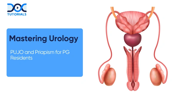
Comprehensive Urology Guidelines: PUJO and Priapism 

 Mastering Urology

For postgraduate residents specializing in reconstructive surgeries and clinical emergencies, mastering complex genitourinary pathologies is essential. A nuanced understanding of conditions like Pelviureteric Junction Obstruction (PUJO) and the acute management of priapism defines a competent specialist.  

This comprehensive guide breaks down these high-yield topics, offering a highly structured approach to pathogenesis, diagnostic modalities, and stepwise surgical interventions tailored specifically for the rigorous demands of your residency training.  

Get High Quality, High Yield PG Residency Notes for Free!

Pelviureteric Junction Obstruction (PUJO) in Urology 

Pathogenesis and Mechanisms of PUJO 

In clinical urology, PUJO is defined as a functional impairment of urinary transport directly at the kidney-ureter junction, which ultimately leads to severe obstruction. The accepted diagnostic standard for identifying hydronephrosis in this context is an antero-posterior (AP) diameter exceeding 12 mm of the intrarenal pelvis on a standard ultrasound.  

The etiology of this condition is typically divided into congenital and acquired causes. Intrinsic congenital defects frequently involve abnormal smooth muscle peristalsis, obstructive mucosal valves, or a pathological decrease in the interstitial cells of Cajal. Alternatively, anatomical obstruction due to crossing vessels is a major factor, present in 63% of PUJO cases, compared to merely 20% in normal kidneys. Acquired causes routinely encountered include ureteric stones, strictures, neoplasms, and post-operative fibrosis. Furthermore, when evaluating duplex systems, residents must remember that the lower moiety is the one most commonly obstructed.  

Diagnostic Paradigm and Functional Confirmation 

The diagnostic workup follows a strict stepwise funnel. Ultrasound (USG) serves as the first-line screening modality, highly capable of detecting the classic prenatal flank mass. Following screening, a non-contrast Computed Tomography (CT) scan is deployed to precisely define the anatomy, evaluate the extent of parenchymal thinning, and reliably identify the presence of crossing vessels.  

Functional confirmation remains a cornerstone of modern practice, achieved via MAG-3 Diuretic Renography. MAG-3 is preferred over DTPA because it accurately assesses split renal function and drainage dynamics, yielding a classic obstructive pattern on the renogram curve. Finally, retrograde pyelography (RGP) is routinely performed intraoperatively to dynamically visualize the relevant anatomy immediately prior to making the surgical incision.  

Q: What is the defining clinical timeline and presentation for PUJO across different age groups in urology?  

A: According to the clinical timeline, neonates and infants typically present asymptomatically with a flank mass initially detected as a prenatal ultrasound finding. In stark contrast, older children and adults classically present with Dietl’s crisis (intermittent, severe flank pain), accompanied by nausea, vomiting, and occasionally rare signs like hematuria or hypertension.  

Surgical Management of PUJO in Urology 

Endourological management, specifically endopyelotomy, is frequently favored for short-segment PUJO. The technique can be approached percutaneously (antegrade) or via the urethra (retrograde). The procedure involves inserting a flexible scope and creating a full-thickness lateral incision using a hook knife or a 200µ Holmium laser fiber. Surgical patency is validated intraoperatively via contrast extravasation, immediately followed by the placement of a 14 x 7 Fr stent. However, this technique is strictly contraindicated in patients presenting with active infection, underlying coagulopathy, or long strictures exceeding 2 cm.  

The absolute gold standard intervention remains the Anderson-Hynes Dismembered Pyeloplasty. This meticulously executed four-step procedure involves: excision of the stricture, spatulation of the ureter, reduction of the renal pelvis, and a watertight anastomosis. It is highly indicated for cases featuring a redundant pelvis, a tortuous ureter, or a high ureteral insertion. Residents should note it is unsuitable for long strictures or a predominantly intrarenal pelvis. Laparoscopic and robotic approaches are now preferred via the transperitoneal route due to familiar anatomy and superior working space, offering high success rates (>95%). Post-operative care mandates a stent placement, which is generally removed after 4 to 6 weeks.  

Advanced Flap Reconstruction Techniques in Urology 

Flap Technique Primary Clinical Indication Reconstructive Mechanism 
Foley’s Y-V Plasty High ureteral insertion.  Reconstructs the junction by converting a Y-shaped incision into a wide V.  
Spiral Flap (Scardino-Prince) Long proximal ureteric strictures.  Wraps a spiral flap of the redundant renal pelvis downward to bridge long defects.  
Vertical Flap Box-shaped renal pelvis.  Utilizes a vertical pelvic tissue flap to recreate a natural, dependent junction.  

Priapism: A Critical Emergency in Urology 

Ischemic vs. Non-Ischemic Classifications 

Priapism represents a profound clinical emergency, strictly defined as a full or partial erection lasting longer than 4 hours without sexual stimulation, or persisting beyond climax. It is broadly classified into three primary types: ischemic (low-flow or veno-occlusive), non-ischemic (high-flow or arterial), and stuttering (recurrent ischemic).  

Ischemic priapism presents as a severely painful, rigid erection that characteristically features a soft, unaffected glans (glans sparing). The pathophysiology relies on decreased cavernous arterial inflow, leading to stagnant blood stasis, severe hypoxia, hypercapnia, and local acidosis. Diagnostic markers on a cavernous blood gas reveal a low pO2 (< 30 mmHg), high pCO2 (> 60 mmHg), and an acidic pH (< 7.25). Doppler ultrasound will demonstrate a complete absence of arterial inflow. The critical urgency window is less than 36 hours; delays beyond this risk irreversible corporal fibrosis and permanent loss of potency.  

Conversely, non-ischemic priapism is generally painless and presents as a non-rigid tumescence. It is most frequently caused by perineal trauma (e.g., a straddle injury) that creates an arterial-sinusoidal fistula. A diagnostic workup reveals a completely normal cavernous blood gas profile.  

Stuttering Priapism Mechanisms 

Stuttering priapism consists of intermittent, repetitive ischemic episodes and is heavily associated with Sickle Cell Disease (SCD). The molecular mechanism involves the rupture of sickled RBCs, releasing free hemoglobin. This free hemoglobin acts by scavenging nitric oxide (NO), while arginase depletes L-arginine. The subsequent drop in NO availability severely impairs smooth muscle relaxation, triggering venous outflow obstruction. Triggers for stuttering episodes include nocturnal sleep, cold exposure, dehydration, and fever.  

Stepwise Management of Priapism in Urology 

Pharmacological triggers for priapism include alpha-blockers (prazosin, tamsulosin), psychotropic medications (sertraline, bupropion, phenothiazines), and recreational drugs like cocaine and marijuana. Intracavernosal injection therapies carry variable risks: papaverine carries a high risk (~35%) of prolonged erection, trimix carries a moderate risk, and PDE-5 inhibitors carry a low risk unless combined with recreational drugs. The structured management ladder for ischemic priapism is an essential protocol to master.  

The Stepwise Ischemic Management Algorithm in Urology 

Intervention Step Clinical Action & Surgical Details 
Step 1: Aspiration & Irrigation Decompress the corpora cavernosa using a needle and confirm the diagnosis via a cavernous blood gas test.  
Step 2: Pharmacologic Injection Administer Intracavernosal Phenylephrine at 0.5-1 mL every 5-10 minutes, up to a strict maximum of 1 mg, while monitoring patient vitals.  
Step 3: Distal Shunts Establish distal drainage via Winter’s (needle technique), Ebbehoj (blade technique), Al-Ghorab (excision), or Corporal Snake (dilator).  
Step 4: Proximal Shunts Execute Quackel’s (Corpora-Spongiosum shunt) or Grayhack (Vein-to-Corpora shunt) procedures.  
Step 5: Penile Prosthesis A refractory indication if the priapism duration exceeds 36 hours, due to the high risk of irreversible fibrosis.  

For non-ischemic cases, management is highly conservative (observation or site-specific compression) because many resolve spontaneously, though selective arterial embolization may be utilized. Stuttering priapism management focuses on prevention using hormonal therapies (GnRH agonists, ketoconazole), non-hormonal agents (pseudoephedrine, baclofen), or paradoxical low-dose PDE-5 inhibitors to downregulate the NO pathway and nocturnal cycles.  

Frequently Asked Questions (FAQs) in Urology 

1. What defines PUJO radiologically?  

It is defined as functional impairment causing obstruction, definitively marked by an antero-posterior (AP) diameter >12 mm of the intrarenal pelvis on a renal ultrasound.  

2. What is the classic clinical presentation of PUJO in adults?  

Adults typically present with Dietl’s crisis, which is characterized by intermittent flank pain, nausea, and severe vomiting.  

3. Which specific renal moiety is obstructed in a duplex system?  

A fundamental anatomical rule is that the lower moiety is the one most commonly obstructed in duplex collecting systems.  

4. What is the preferred diuretic renography agent for PUJO?  

MAG-3 is strictly preferred over DTPA because it allows for a highly accurate assessment of both split renal function and dynamic urinary drainage.  

5. When is an Anderson-Hynes pyeloplasty clinically contraindicated?  

This gold standard reconstructive procedure is deemed unsuitable for patients presenting with long strictures, multiple strictures, or a predominantly intrarenal pelvis.  

6. How is ischemic priapism diagnosed using a cavernous blood gas?  

Diagnostics will reveal severe local acidosis and hypoxia, specifically: a pH < 7.25, a pO2 < 30 mmHg, and a pCO2 > 60 mmHg.  

7. Why does the glans remain remarkably soft during ischemic priapism?  

The ischemic pathology specifically targets and engorges the corpora cavernosa but classically spares the corpus spongiosum, resulting in a distinctively soft glans during physical examinations.  

8. What is the precise mechanism of stuttering priapism in Sickle Cell Disease?  

Sickled RBC hemolysis releases free hemoglobin that binds available nitric oxide, deeply impairing smooth muscle relaxation and causing persistent venous stasis.  

9. What is the absolute maximum dose of intracavernosal phenylephrine?  

Phenylephrine must be carefully injected at 0.5-1 mL every 5-10 minutes, capped strictly at a maximum dose of 1 mg while maintaining continuous hemodynamic monitoring.  

10. What is the critical time window for resolving ischemic priapism?  

Guidelines heavily emphasize a critical intervention window of less than 36 hours; surgical delays beyond this dramatically increase the risk of irreversible corporal fibrosis and permanent erectile dysfunction. 

DocTutorials App Download Now

Latest Blogs

Mastering Trauma Surgery: A Comprehensive Guide for PG Residents 

Trauma Surgery

This essential guide on trauma surgery, tailored specifically for postgraduate (PG) surgical residents. Navigating the high-stakes environment of trauma surgery requires a structured, evidence-based approach to patient assessment, operative intervention, and postoperative care. This article provides a comprehensive overview of critical trauma surgery protocols, emphasizing the management of abdominal trauma, the application of damage control surgery, and high-yield clinical pearls to enhance your surgical residency training. 

Get High Quality, High Yield Surgery PG Residency Notes for Free! 

Mechanisms and Classification of Abdominal Trauma 

In trauma surgery, understanding the mechanism of injury is the critical first step in predicting organ damage and guiding your diagnostic approach. Abdominal trauma is broadly categorized into blunt and penetrating injuries, each with a distinct pattern of visceral involvement.  

When managing blunt abdominal trauma, the spleen is the most frequently injured organ, followed by the liver and the small intestine. Interestingly, in the pediatric population, the spleen remains the most commonly injured organ, but the kidney takes the second spot. Conversely, penetrating abdominal trauma most commonly affects the liver, followed by the small intestine and the diaphragm.  

Patient classification based on hemodynamic status is a cornerstone of trauma surgery triage. Patients are classified as: 

  • Responders: Maintain stable hemodynamics.  
  • Transient Responders: Initially improve but then crash, indicating ongoing pathology.  
  • Non-Responders: Require immediate intervention due to persistent instability.  

Common Mechanisms of Abdominal Trauma 

Trauma Type Primary Mechanism Most Commonly Injured Organs (in order of frequency) 
Blunt Motor vehicle accidents, falls, compression 1. Spleen, 2. Liver, 3. Small Intestine  
Penetrating Stab wounds, gunshot wounds 1. Liver, 2. Small Intestine, 3. Diaphragm  
Pediatric Blunt Falls, sports injuries, vehicle accidents 1. Spleen, 2. Kidney  

Diagnostic Modalities in Trauma Surgery 

Rapid and accurate diagnosis is vital in trauma surgery. For a stable patient with blunt abdominal trauma, a Contrast-Enhanced Computed Tomography (CECT) of the abdomen is the gold standard. However, if the patient is unstable, exhibits peritonitis (guarding/rigidity), or has a positive eFAST exam, an immediate exploratory laparotomy is indicated.  

The Role of eFAST and CT Scans 

The Extended Focused Assessment with Sonography for Trauma (eFAST) is the most sensitive initial test for unstable hemorrhage. The eFAST exam adds a pericardial view to the standard abdominal and pelvic windows (Morison’s Pouch, Splenorenal, and Pouch of Douglas).  

Q: What are the primary blind spots and limitations of the eFAST exam in trauma surgery?  

A: While rapid, the eFAST exam has significant blind spots. It cannot rule out hollow viscus perforation, cannot assess the retroperitoneum, and is often obscured by obesity and bowel gas. Furthermore, it cannot identify the exact source of bleeding and is unreliable for fluid volumes less than 100 ml.  

For penetrating abdominal trauma, stable patients require a targeted approach. Flank or back injuries necessitate a triple contrast CT (Oral/IV/Rectal) to rule out retroperitoneal or colon injuries. Anterior abdominal injuries may require local wound exploration; if the peritoneum is breached, diagnostic laparoscopy is mandatory.  

Organ-Specific Trauma Surgery Protocols 

Effective trauma surgery requires tailored protocols for specific organ injuries, balancing the need to stop hemorrhage with organ preservation whenever possible. 

Splenic and Liver Injuries 

The spleen is highly susceptible to deceleration forces in blunt abdominal trauma. The AAST scale grades splenic injuries from Grade 1 (minor subcapsular hematoma) to Grade 5 (a “shattered” spleen). Stable patients with Grade 1-3 injuries are typically managed conservatively with hematocrit monitoring, utilizing angioembolization if their grade deteriorates. If a splenectomy is performed, residents must be vigilant about Overwhelming Post-Splenectomy Infection (OPSI), ensuring patients receive Pneumococcal, Meningococcal, and Hib vaccines.  

For liver injuries, trauma surgery relies on the “4 P’s” for hemorrhage control: Pressure, Plug, Pringle’s Maneuver, and Pack. The Pringle’s maneuver occludes the portal vein and hepatic artery; if bleeding persists despite this, the source is likely the hepatic veins or inferior vena cava due to retrograde flow.  

Damage Control Surgery in Bowel Trauma 

In trauma surgery, the decision to perform Damage Control Surgery (DCS) versus Early Total Care is often dictated by the patient’s physiological state and lactate levels. A lactate level below 2 in a physiologically stable patient allows for Early Total Care, including resection and primary anastomosis of bowel injuries.  

However, if a patient has a lactate level of 3-5+ or is a transient responder, Damage Control Surgery is imperative. The surgical principle here is to avoid anastomosis in unstable patients due to a high risk of leakage.  

Damage Control Surgery vs. Early Total Care 

Clinical Parameter Early Total Care Damage Control Surgery (DCS) 
Patient Status Physiologically Stable  Physiologically Unstable / Transient Responder  
Lactate Level Lactate < 2  Lactate 3-5+ / Acidosis  
Surgical Action Resection + Anastomosis  Resect, staple ends (discontinuity) or Stoma  
Next Steps Standard post-op recovery ICU Stabilization followed by planned re-operation  

Retroperitoneal and Genitourinary Trauma 

The retroperitoneum is famously known as the surgeon’s minefield in trauma surgery. Management depends heavily on the zone of injury. Zone 1 (Central) injuries must always be explored in both blunt and penetrating abdominal trauma. In contrast, Zone 3 (Pelvic) hematomas should generally not be explored due to the high tamponade effect; opening this zone can lead to torrential bleeding, so it is better managed with angioembolization and packing.  

Lower genitourinary trauma presents uniquely. Urethral injuries present with blood at the meatus, retention, and a high-riding bladder; crucially, these patients must not be catheterized. Instead, trauma surgery protocol dictates a suprapubic cystostomy followed by delayed urethroplasty.  

Frequently Asked Questions (FAQs) in Trauma Surgery 

1. What is the most common organ injured in blunt abdominal trauma?  

The spleen is the most frequently injured organ in blunt abdominal trauma, followed by the liver and small intestine.  

2. When is a CECT abdomen indicated in trauma surgery?  

A CECT abdomen is the gold standard for stable patients with blunt trauma. It is triggered by evidence of a hollow viscus injury or free fluid without solid organ injury.  

3. What are the indications for an immediate exploratory laparotomy?  

Immediate laparotomy is indicated for hemodynamic instability, peritonitis (guarding/rigidity), or a positive eFAST in an unstable patient.  

4. What is the threshold for destructive colon injuries requiring a stoma?  

In trauma surgery, a destructive colon injury is defined as having greater than 50% circumference loss or devascularization, necessitating resection, and a stoma.  

5. How are rectal injuries assessed and managed?  

A rectal injury is considered destructive if there is greater than 25% circumference loss, which requires a Hartmann’s Procedure or loop colostomy.  

6. What defines Damage Control Surgery in bowel trauma?  

Damage Control Surgery involves resecting the injured bowel, stapling the ends or creating a stoma, followed by ICU stabilization and a planned re-operation, avoiding primary anastomosis.  

7. What is Quincy’s Triad in liver trauma?  

Quincy’s Triad indicates hemobilia and presents as jaundice, colicky pain, and melena.  

8. Which pathogens are targeted for OPSI prevention post-splenectomy?  

Prevention targets encapsulated organisms, primarily Streptococcus pneumoniae, by administering Pneumococcal, Meningococcal, and Hib vaccines.  

9. How should a Zone 3 pelvic hematoma be managed?  

A Zone 3 pelvic hematoma should not be surgically explored to preserve the tamponade effect; it is managed with angioembolization and packing.  

10. What is the diagnostic protocol for a penetrating flank injury?  

A penetrating injury to the flank or back in a stable patient requires a triple contrast CT (Oral/IV/Rectal) to rule out retroperitoneal or colon injuries. 

DocTutorials App Download Now

Latest Blogs

A Comprehensive Guide to Surgical Gastroenterology for PG Residents 

surgical Gastroenterology

Mastering clinical genetics, dysmorphic assessments, and advanced molecular treatments like CAR T Cell Therapy is no longer optional for today’s post-graduate (PG) medical residents. This comprehensive guide connects foundational pediatric bedside examinations with the cutting-edge frontiers of gene modification and CAR T Cell Therapy. 

Get High Quality, High Yield PG Residency Notes for Free!

Endogenous Mechanisms and Pathophysiology in Surgical Gastroenterology 

A profound understanding of Surgical Gastroenterology begins with the endogenous anti-reflux mechanisms that protect the esophagus. The body relies on a delicate balance of anatomical structures. The Lower Oesophageal Sphincter (LOS) creates a critical zone of high pressure via intrinsic distal esophageal muscle. This sphincter remains tonically contracted and only relaxes for approximately 500 milliseconds during swallowing.  

Working in tandem with the LOS is the crural diaphragm. During inspiration, the anteroposterior diameter decreases, which increases pressure and mechanically augments the sphincter through a “pinch-cock” action. Furthermore, Surgical Gastroenterology highlights the importance of the diagonal sling fibres of the cardia and the Angle of His, which acts as a flap valve to prevent reflux. The intra-abdominal pressure is transmitted to the gastroesophageal (GE) junction to counteract gastric pressure.  

The Pathophysiology of the Barrier Breach 

A core concept in Surgical Gastroenterology is understanding how this barrier fails. Pathological reflux occurs when intra-gastric pressure exceeds 12 mmHg, overpowering the distal oesophageal high-pressure zone, which drops below 8 mmHg. This mechanical imbalance overrides the body’s protective barriers.  

This breach can be caused by a hypotensive LOS, which is a decreased pressure in the normal position or associated with a hiatal hernia (where the GE junction migrates into the posterior mediastinum). Alternatively, it can occur via spontaneous relaxation, where normal LOS pressure inappropriately relaxes in the absence of peristalsis, often triggered by gastric distention or vagal stimulation. While physiological reflux is normal for belching and bloating relief, it becomes a pathological concern in Surgical Gastroenterology based on total acid exposure, symptom severity, and mucosal damage like esophagitis or Barrett’s esophagus.   

Question: In the context of Surgical Gastroenterology, how does the management of GERD in Idiopathic Pulmonary Fibrosis (IPF) patients differ between chemical and mechanical treatments?  

Answer: Chemical Proton Pump Inhibitor (PPI) therapy only neutralizes acid; it is effective for acid reflux but completely ineffective against non-acid or bile reflux. In contrast, a mechanical fundoplication restores the physical barrier, stopping the micro-aspiration of bile salts and pepsin, and offering a ~70% likelihood of improvement for extra-oesophageal symptoms.  

Clinical Presentation and Pre-Operative Workup in Surgical Gastroenterology 

Patients in a Surgical Gastroenterology clinic present with both typical and atypical symptoms. Typical symptoms include heartburn (an epigastric/retrosternal caustic sensation), water brash (a sour taste from acid/bile reflux into the oropharynx), and regurgitation (the return of digested food). Atypical or extra-oesophageal symptoms are caused by micro-aspiration leading to laryngeal/respiratory injury, or vagal nerve reflexes causing bronchospasm.  

During physical examination, Surgical Gastroenterology residents should observe for dental erosion (loss of dentine), an injected oropharynx (redness), chronic sinusitis, and behavioral signs like leaning forward to flatten the diaphragm or constant water drinking to buffer symptoms. Crucially, residents must look for red flags: dysphagia requires mandatory evaluation, and practitioners must rule out malignancy (checking for supraclavicular lymphadenopathy) and musculoskeletal pain like Tietze syndrome.  

The Pulmonary Link 

Surgical Gastroenterology frequently intersects with pulmonology. Proximal reflux leads to micro-aspiration, causing alveolar injury and subsequent pulmonary decline. Strikingly, there is a 94% incidence of GERD in Idiopathic Pulmonary Fibrosis (IPF) patients. For conditions like asthma, anti-reflux surgery is more effective than medical therapy, yielding 90% symptom improvement in children and 70% in adults. PPIs may slow decline, but Laparoscopic anti-reflux surgery provides a mechanical barrier to mitigate progression, as evidenced by the Wrap-IPF trial.  

The Mandatory Diagnostic Workup 

Before any Surgical Gastroenterology intervention, a rigorous pre-operative workup is mandatory.  

Essential Diagnostic Modalities in Surgical Gastroenterology 

Diagnostic Test Primary Function Key Clinical Insights in Surgical Gastroenterology 
Ambulatory pH & Impedance The Quantifier The gold standard for quantifying acid exposure. Impedance monitoring detects non-acid reflux and flow direction.  
Oesophageal Manometry The Mechanic Assesses motility and sphincter function; critical to exclude Achalasia prior to surgery.  
Oesophagoscopy (OGD) The Visual Evaluates mucosa and valve grade in retroflexion. Checks for hiatal hernia size and Barrett’s oesophagus.  
Barium Oesophagogram The Map Provides detailed anatomical mapping. Preferred over CT for GERD symptoms to rule out diverticulosis and tumors.  

In Surgical Gastroenterology, the DeMeester Score is pivotal during pH monitoring. A score > 14.72 confirms GERD based on pH < 4 exposure (upright, supine, and total), episode frequency, and episode duration greater than 5 minutes. Furthermore, the Los Angeles Classification during OGD dictates that Grade A/B is mild and requires a pH test, while Grade C/D is severe and pathognomonic for GERD, requiring no pH test.  

Esophageal Motility Disorders in Surgical Gastroenterology 

Esophageal motility disorders form a complex and highly tested chapter in Surgical Gastroenterology. Manometry helps identify these conditions, noting that acid exposure time is the best predictor of treatment response. High-resolution catheter manometry takes about 15 minutes and involves ten swallows to identify defective LOS and ineffective motility (present in 25-48% of GERD patients).  

Comparing Motility Disorders in Surgical Gastroenterology 

Disorder Clinical & Manometry Criteria Surgical Gastroenterology Management 
Diffuse Esophageal Spasm (DES) Chest pain mimicking angina; paradoxical dysphagia. Manometry shows a “corkscrew esophagus” with simultaneous contractions, DCI > 450, and normal LOS relaxation (IRP < 15 mmHg).  Step 1: Lifestyle/Psych eval. Step 2: Nitrates, CCBs, Botox. Step 3: Long Esophageal Myotomy + Fundoplication or POEM.  
Jackhammer (Nutcracker) Esophagus A hypermotility disorder characterized by excessive contractility. Two successive swallows with DCI > 8000; LES relaxation is normal and regurgitation is uncommon.  Avoid caffeine and temperature triggers. POEM is the ideal surgical procedure if refractory to medical therapy.  
Hypertensive LES / Achalasia Part of an evolutionary spectrum of EGJ outflow obstruction. IRP > 15 mmHg (failed relaxation) with a tight ring, but often preserved peristalsis in the body initially.  Temporary: Botox or Hydrostatic balloon dilatation. Surgical: Laparoscopic Heller Myotomy augmented with Dor/Toupet fundoplication.  

When treating motility disorders in Surgical Gastroenterology, if peristalsis is impaired, a partial fundoplication is preferred to prevent post-op dysphagia.  

Frequently Asked Questions (FAQs) 

1. What is the gold standard diagnostic test for GERD in Surgical Gastroenterology? 

Ambulatory pH & Impedance monitoring is the gold standard, as it quantifies acid exposure and differentiates liquid versus gas reflux.  

2. What DeMeester score confirms a GERD diagnosis?  

A DeMeester score > 14.72 is the diagnostic threshold for GERD in Surgical Gastroenterology.  

3. Why is Oesophageal Manometry essential before Surgical Gastroenterology procedures?  

It is critical to evaluate motility and explicitly exclude Achalasia, which is a contraindication for standard anti-reflux surgery.  

4. What is the incidence of GERD in patients with Idiopathic Pulmonary Fibrosis?  

There is a 94% incidence of GERD in patients suffering from Idiopathic Pulmonary Fibrosis (IPF).  

5. Which OGD findings are pathognomonic for GERD in Surgical Gastroenterology?  

According to the Los Angeles Classification, Grade C and Grade D esophagitis are severe and pathognomonic, requiring no further pH testing.  

6. What are the clinical red flags during a Surgical Gastroenterology evaluation for reflux?  

Dysphagia is a major red flag requiring mandatory evaluation to rule out malignancy (such as checking for supraclavicular lymphadenopathy).  

7. How is Jackhammer Esophagus diagnosed on manometry?  

Jackhammer Esophagus is diagnosed when there are two successive swallows with a Distal Contractile Integral (DCI) > 8000.  

8. What is the definitive surgical management for Diffuse Esophageal Spasm (DES)?  

If medical management fails, the preferred Surgical Gastroenterology intervention is a Long Esophageal Myotomy (distal 2/3) combined with a Fundoplication, or a POEM procedure.  

9. Why might a surgeon choose a partial fundoplication over a full wrap?  

In Surgical Gastroenterology, a partial fundoplication (like Dor or Toupet) is preferred if esophageal peristalsis is impaired, in order to prevent post-operative dysphagia.  

10. How does a normal Lower Oesophageal Sphincter function during swallowing?  

The normal LOS creates a zone of high pressure that is tonically contracted, and it relaxes for approximately 500ms to allow food to pass during swallowing. 

DocTutorials App Download Now

Latest Blogs

Plastic Surgery: High-Yield Principles for PG Residents 

Plastic Surgery

Mastering the vast domain of Plastic Surgery requires an in-depth understanding of anatomy, surgical principles, and patient management. This comprehensive guide distills critical concepts from fundamental facial aesthetics to complex burn resuscitation to enhance your clinical acumen in Plastic Surgery.  

Get High Quality, High Yield Plastic surgery PG Residency Notes for Free! 

Principles of Upper Face Rejuvenation 

Patient selection is the cornerstone of successful Plastic Surgery. Success from a patient’s perspective means managing expectations and addressing specific complaints. However, surgeons must watch for contraindications, or “red flags,” such as Body Dysmorphic Disorder (BDD), substance abuse, or unrealistic expectations (e.g., hoping surgery solves relationship issues).  

Brow Lifts and Anatomical Planes 

Upper face rejuvenation requires precise anatomical knowledge. Dissection often occurs in the subgaleal plane, a potential space between the galea aponeurotica and the pericranium. Brow lifts address lateral eyebrow ptosis, forehead wrinkles, and upper eyelid crowding.  

  • Open Lift: Utilizes a bicoronal or hairline incision, elevating the tissue in the subgaleal plane, and trimming excess skin.  
  • Endoscopic Lift: A minimally invasive approach using 5 portals. It involves subgaleal dissection, resection of the corrugator/procerus muscles, and fixation via screws.  

Periorbital Surgery (Blepharoplasty) 

Blepharoplasty is a staple in aesthetic Plastic Surgery. Upper blepharoplasty targets ptosis, excess skin, and fat prolapse via a supra-crease incision, which includes the excision of an orbicularis oculi slip and resection of excess fat pads.  

Lower blepharoplasty presents the challenge of assessing skin laxity and globe adherence. Mild laxity is corrected with Canthopexy (plication of the lateral canthal tendon), whereas severe laxity requires Canthoplasty (incision and fixation of the eyelid to Whitnall’s Tubercle).  

Surgeons must remain vigilant for complications like dry eye, scleral show, and ectropion (outward turning of the eyelid causing exposure risk).  

Advanced Facial Rejuvenation and Hair Transplants 

Mastering the SMAS Concept 

Modern rhytidectomy (facelifts) in Plastic Surgery must move beyond simple skin excision. True longevity relies on manipulating the Superficial Musculo-Aponeurotic System (SMAS)—the “power layer” that carries facial muscles.  

Techniques include: 

  • SMAS Plication: Folding and suturing the sagging SMAS.  
  • SMAS Ectomy: Excising the excess muscle layer before suturing.  
  • Deep Plane: Lifting the entire muscle complex with deep structures, yielding the longest-lasting results.  

Additionally, platysmaplasty tightens neck muscles to correct vertical banding. Common complications include hematoma (the most frequent), hypertrophic scarring, and greater auricular nerve injury.  

Q: What is the core biological principle governing hair transplantation success?  

A: The core principle is “Donor Dominance.” Graft survival depends on the characteristics of the donor site (e.g., occipital hair is permanent), not the recipient’s site.  

Hair Transplantation and Rhinoplasty 

In restorative Plastic Surgery, Follicular Unit Grafting (FUG) is the gold standard for hair transplants. It involves transplanting natural groupings of 1, 2, or 3 hairs to achieve natural density and avoid a “cobblestone” appearance. Implantation strategies vary: the forehead requires a forward vector, while the vertex needs a spiral pattern.  

For nasal refinement, Rhinoplasty approaches are divided into open (using a columellar inverted V incision for full visualization of osteotomy/grafts) and closed (no external scar, but limited manipulation). Key maneuvers include osteotomies to narrow bones, spreader grafts for valve collapse, and strut grafts for tip support.  

Skin Resurfacing Techniques in Plastic Surgery 

Feature Ablative Lasers (e.g., CO2) Fractional Photothermolysis 
Mechanism Thermal coagulation of the dermis  Coagulative injury columns  
Recovery Time 7-10 Days  24-48 Hours  
Risk / Benefit Risk of Hyperpigmentation  Faster healing, spares surrounding tissue  

Body Contouring and Breast Aesthetics 

Breast Surgery Fundamentals 

In aesthetic Plastic Surgery of the breast, augmentation safety is paramount. Placing implants in the submuscular plane reduces the risk of capsular contracture. Surgeons must also be aware of BIA-ALCL (Breast Implant-Associated Anaplastic Large Cell Lymphoma), a CD30+ lymphoma linked to textured implants and biofilms, typically presenting as a peri-implant seroma.  

For breast reduction, hypertrophy is removed while preserving the Nipple-Areolar Complex (NAC) on a pedicle. Mastopexy corrects ptosis caused by attenuated Cooper’s ligaments. Gynecomastia management depends on the type: florid (ductal hyperplasia treated with liposuction) versus fibrous (stromal fibrosis treated with excision).  

Abdominal and Post-Bariatric Contouring 

Abdominoplasty in Plastic Surgery is a contouring procedure, not a weight-loss tool. A standard abdominoplasty involves a transverse incision, diastasis plication, and umbilical transposition. For massive weight loss patients with horizontal excess, the Fleur-de-lis technique adds a vertical incision.  

Post-bariatric Plastic Surgery requires strict criteria: stable weight for 4 months, a BMI under 30, and waiting 1.5 years post-surgery. Procedures like belt lipectomy and brachioplasty (medial arm skin excision) carry high risks of skin necrosis (especially in smokers), seromas, and DVT/PE.  

Critical Burn Management in Plastic Surgery 

Pathophysiology and Classification 

Burns are a critical component of reconstructive Plastic Surgery. Jackson’s Zones of injury define the pathophysiology: 

  1. Zone of Coagulation: Central area of irreversible necrosis.  
  1. Zone of Stasis: Area of impaired perfusion at risk of dying; this is the critical target for fluid resuscitation.  
  1. Zone of Hyperemia: Inflamed but viable tissue.  

Burn size is calculated using the Rule of Nines (Head 9%, each Arm 9%, each Leg 18%, Torso 36%), the Palm Method (patient’s palm + fingers = 1% TBSA), or the Lund-Browder chart for objective age adjustments.  

Burn Depth Assessment and Management 

Classification Depth & Appearance Healing & Treatment 
Superficial 2nd Degree (Group A) Papillary dermis; blisters, pink/moist, blanches  Heals <14 days; conservative care (moisture/antibiotics)  
Deep 2nd Degree (Group B) Reticular dermis; pale/mottled, no blanching  Will not heal without scars (2-5 weeks); requires surgery  
3rd Degree Full thickness; leathery eschar, insensate  Will not heal; requires surgical excision and grafting  

Resuscitation and Metabolic Response 

The metabolic response to severe burns happens in two phases: the Ebb phase (0-48h) characterized by shock and low cardiac output, followed by the Flow phase (>5 days) featuring a massive catecholamine surge, hypermetabolic state, and high cardiac output. Losing over 40% of muscle mass yields high mortality. Systemic impacts include acute tubular necrosis in the kidneys and bacterial translocation in the gut.  

Acute management starts with cooling the burn with water (10-15 mins), dry dressings, and IV Morphine. Fluid resuscitation uses Ringer’s Lactate and is guided by the Parkland Formula (4 mL x Body Weight (kg) x % TBSA), giving 50% in the first 8 hours and 50% in the next 16 hours. 

Surgical Excision and Pharmacology 

In reconstructive Plastic Surgery, early tangential excision of the eschar down to punctate bleeding within 48 hours decreases mortality. For coverage, the reconstruction ladder starts with temporary coverage (allograft/xenograft), moves to dermal substitutes (Integra/BTM) if the dermis is lost, and finishes with a Split Thickness Skin Graft (STSG) for definitive coverage.  

Pharmacological support relies on topical agents like Silver Sulfadiazine (which has limited penetration) or Mafenide Acetate (which penetrates eschar but can cause metabolic acidosis). Systemically, Propranolol inhibits heart rate and catabolism, while Oxandrolone preserves muscle mass. Nutrition must meet high protein needs (1-2 g/kg/day) according to the Curreri Formula.  

Frequently Asked Questions (FAQs) 

  1. What is the subgaleal plane in Plastic Surgery?  

It is a potential space between the galea aponeurotica and pericranium used in brow lifts.  

  1. What are the absolute contraindications for aesthetic Plastic Surgery?  

Body Dysmorphic Disorder (BDD), substance abuse, and unrealistic expectations. 

  1. How is severe lower eyelid laxity corrected in Plastic Surgery?  

It is corrected using Canthoplasty, fixing the eyelid to Whitnall’s Tubercle.  

  1. Which rhytidectomy technique offers the longest-lasting results?  

The deep plane facelift, which lifts the entire muscle complex.  

  1. What is the gold standard for hair transplants in Plastic Surgery?  

Follicular Unit Grafting (FUG), utilizing natural groupings of 1-3 hairs.  

  1. How does submuscular implant placement benefit Plastic Surgery patients?  

It significantly reduces the risk of capsular contracture.  

  1. What defines Jackson’s Zone of Stasis in burn Plastic Surgery?  

It is an area of impaired perfusion that is the primary target for resuscitation.  

  1. What is the Parkland Formula used in burn Plastic Surgery?  

4 mL x Body Weight (kg) x % TBSA, with 50% given in the first 8 hours.  

  1. When is an escharotomy indicated in Plastic Surgery?  

When abdominal compartment syndrome pressure exceeds 30mmHg or limb ischemia pressure exceeds 40mmHg.  

  1. What is the purpose of Propranolol in burn-related Plastic Surgery?  

It acts as a beta-blocker to decrease heart rate, catabolic activity, and fatty liver risks. 

DocTutorials App Download Now

Latest Blogs

Comprehensive Neurosurgery Guidelines: Cervical Trauma and SAH Management 

Cervical Trauma

In the highly demanding and rigorous field of neurosurgery, mastering the acute management of spinal trauma and vascular emergencies is paramount for postgraduate residents. This educational and informative guide provides a factually robust overview of critical conditions, ranging from the complex Jefferson Fracture to life-threatening subarachnoid hemorrhages.  

By closely adhering to established diagnostic and therapeutic algorithms, neurosurgery residents can ensure they are fully equipped with the high-yield clinical knowledge required for optimal patient outcomes. 

Get High Quality, High Yield Neurosurgery PG Residency Notes for Free! 

Understanding Cervical Spine Trauma in Neurosurgery 

The upper cervical spine, structurally spanning from the skull base down to C2, is a frequent site of traumatic injury that demands precise neurosurgery evaluation. 

Upper Cervical Spine Injuries 

One of the hallmark injuries encountered in trauma neurosurgery is the Atlas or Jefferson Fracture. The primary mechanism responsible for a Jefferson Fracture is axial loading. Clinical diagnosis of a Jefferson Fracture is often aided by an Open Mouth View X-ray, which prominently reveals C1 lateral mass displacement. 

The overall stability of a Jefferson Fracture is highly dependent on the integrity of the transverse ligament. A Jefferson Fracture is explicitly considered unstable if the combined lateral mass deviation is greater than 6.9 mm, a finding that implies a complete transverse ligament rupture. The standard neurosurgery treatment for a Jefferson Fracture is mostly non-operative, typically utilizing a protective collar or halo. 

Other critical upper cervical injuries assessed in neurosurgery include Occipital Condyle Fractures, which require conservative treatment utilizing a collar for 6 to 8 weeks. Additionally, Occipital Atlanto Dislocations are classified as high-energy or fatal injuries. In neurosurgery practice, the assessment for this dislocation is conducted via the Power Ratio on a Triple Junction View. In cases of isolated traumatic transverse ligament rupture, the indicated neurosurgery procedure is a posterior C1-C2 fusion, which utilizes a C1 lateral mass screw alongside a C2 pedicle screw. 

Axis (C2) Fractures and Spondylolisthesis 

Axis (C2) Dens fractures are meticulously classified into three distinct types within neurosurgery. Type 1 involves the tip, Type 2 involves the base or waist, and Type 3 involves the body. Displaced Type 2 fractures specifically require neurosurgery intervention utilizing an anterior decompression screw. 

Another condition is the Hangman’s Fracture, which is defined as the traumatic spondylolisthesis of the Axis (C2) on C3. Imaging signs for a Hangman’s Fracture show minimal forward translation or C2-C3 subluxation. The neurosurgery treatment dictates posterior stabilization for significant displacement. 

Target Patient Group Recommended Management Protocol 
Acute/Majority Non-operative (Hard collar/Halo x 3 months) 
Displaced Internal fixation 
Nonunion Posterior C1-C2 fusion 
Elderly/Unfit Conservative (Soft collar) 

Sub-Axial Injury Mechanisms 

For the sub-axial cervical spine, encompassing C3 to C7, neurosurgery teams must directly correlate the mechanism of injury with the specific fracture type. Hyperflexion directly leads to compression fractures. Axial compression results in burst fractures. Distraction causes facet subluxation, while hyperextension leads to teardrop fractures. The primary surgical goal in these neurosurgery cases is decompression and stabilization, such as an Anterior Cervical Discectomy and Fusion (ACDF), which is used for cervical burst fractures presenting with cord contusion. 

Question: Are cervical collars universally applied in all suspected cervical spine traumas by the neurosurgery team? 

Answer: No. In patients with Ankylosing Spondylitis (Bamboo Spine), a collar is completely contraindicated. The pathology of this disease involves auto-fusion and a rigid spine, resulting in a high susceptibility to fractures; therefore, the neurosurgery management protocol strictly dictates avoiding a collar and managing the patient in a position of comfort. 

Subarachnoid Hemorrhage (SAH): A Neurosurgery Emergency 

Subarachnoid Hemorrhage (SAH) is a critical neurosurgery emergency requiring immediate and aggressive care. 

Pathogenesis and SAH Origins 

The pathogenesis of SAH typically involves the spontaneous rupture of an aneurysm or an arteriovenous malformation (AVM). High-risk zones for these aneurysms include crucial branch points, specifically the Anterior Communicating Artery (A Comm) and the ACA/MCA bifurcation. Major risk factors that precipitate this neurosurgery emergency include female sex, hypertension, smoking, and a family history involving two or more relatives. 

Feature Perimesencephalic SAH Aneurysmal SAH 
Origin Venous origin Arterial origin 
Location Basal cistern (anterior to pons) Diffuse / specific sites 
Prognosis/Risk Good prognosis High risk 

Clinical Presentation and Diagnostic Algorithm 

Clinically, a neurosurgery patient with SAH will present with a thunderclap headache, yet statistically, one-third of these patients are incorrectly diagnosed at initial presentation. Clinical grading is strictly measured after resuscitation. Grade 1 denotes a GCS of 15 with no focal deficit, whereas Grade 5 denotes a GCS of 3-9 with or without a focal deficit. 

The diagnostic algorithm for SAH is a fundamental pillar of neurosurgery practice. The primary modality is a CT Scan; however, its sensitivity drops significantly if performed more than one week post-incident. If the CT is negative, a Lumbar Puncture must be performed. During the lumbar puncture, spectrophotometry is utilized to detect oxyhemoglobin and bilirubin, which is best achieved between 6 to 12 hours. The absolute gold standard diagnostic tool remains Digital Subtraction Angiography (DSA). For intracerebral or ventricular involvement, the Fisher Grading on CT is designated as Grade 4. 

Neurosurgery Treatment and ICU Interventions 

For definitive aneurysm treatment, the primary neurosurgery options include surgical clipping or endovascular coiling, noting that early intervention is crucial. The risk of a fatal re-bleed is heavily monitored; it spikes to 4% at 24 hours, and continues at a rate of 1.5% per day. A dire prognosis warning within neurosurgery dictates that 80% of patients who experience a re-bleed will have a poor outcome. 

ICU Care Bundles are essential in post-operative neurosurgery care. These bundles include strict DVT prophylaxis, careful fluid management with strict intake/output (I/O) monitoring, and aggressive symptom control utilizing analgesics and antiemetics. To prevent vasospasm and Delayed Ischemic Neurologic Deficit (DIND), a prescription of Nimodipine at 60mg PO every 4 hours is administered. 

Despite optimal neurosurgery interventions, complications frequently arise. Neurological complications include vasospasm (seen in 15-20% of cases), DIND, and hydrocephalus. Systemic complications involve hyponatremia (which is very common), severe hypertension, cardiac arrhythmia, and neurogenic pulmonary edema. 

Frequently Asked Questions 

1. What is the primary mechanism of a Jefferson Fracture in neurosurgery? 

The mechanism that causes a Jefferson Fracture is axial loading. 

2. At what measurement is a Jefferson Fracture considered unstable? 

A Jefferson Fracture is considered unstable if the combined lateral mass deviation is greater than 6.9 mm. 

3. What neurosurgery treatment is recommended for an Occipital Condyle Fracture? 

Conservative treatment utilizing a collar for 6-8 weeks is recommended. 

4. How is Occipital Atlanto Dislocation radiographically assessed? 

It is assessed using the Power Ratio via a Triple Junction View. 

5. What defines a Hangman’s Fracture? 

A Hangman’s Fracture is the traumatic spondylolisthesis of the Axis (C2) on C3. 

6. Why is a cervical collar contraindicated for patients with Ankylosing Spondylitis? 

These patients have auto-fusion and a rigid spine, making them highly susceptible to fractures; they must be managed in a position of comfort. 

7. What is the gold standard diagnostic test for SAH? 

The gold standard is Digital Subtraction Angiography (DSA). 

8. What medication prevents Delayed Ischemic Neurologic Deficit (DIND) after an SAH? 

Nimodipine, given at 60mg PO every 4 hours, prevents DIND and vasospasm. 

9. What is the risk of re-bleeding at 24 hours for an SAH patient? 

The risk of re-bleed at 24 hours is 4%. 

10. How often is Subarachnoid Hemorrhage misdiagnosed upon initial presentation? 

One-third (1/3) of patients are incorrectly diagnosed at presentation. 

DocTutorials App Download Now

Latest Blogs

Mastering Laparoscopy Surgery: Essential Guide for PG Residents 

Mastering Laparoscopy Surgery

The transition from open procedures to minimally invasive techniques demands a high level of technical precision and unwavering focus. Laparoscopy surgery presents a uniquely steep learning curve, primarily due to the distinct lack of tactile feedback compared to traditional open surgery.  

For a PG resident, mastering laparoscopy is not just about understanding the steps of a procedure, but developing an intimate familiarity with the equipment, the physics of the instruments, and the rigid safety protocols required to navigate this complex environment. Crafting high-yield knowledge for surgical residents, this guide serves as a foundational resource to elevate your competency in the operating theater.  

Get High Quality, High Yield Surgery PG Residency Notes for Free! 

The Fundamentals of Setup and Patient Positioning 

Before making a single incision, the foundation for a successful laparoscopy surgery is laid out in the operating theater setup. Proper patient positioning is paramount and is dictated strictly by the intended surgical site and the patient’s specific pathology. For non-pelvic pathologies, particularly in male patients, a flat position is universally standard. This allows for straightforward access to the upper abdominal quadrants and generalized exploration.  

Conversely, when addressing pelvic pathology, particularly in female patients, or when the preoperative diagnosis remains unclear, the modified lithotomy position is the standard of care. This positioning provides the necessary access to the lower pelvis while allowing the surgeon and assistant to maneuver comfortably around the primary port at the umbilicus and alternative ports at Palmer’s point (left subcostal/LUQ) or the left lower quadrant (9th intercostal space).  

The Veress Needle: Mechanics and Mandatory Safety Tests 

Gaining safe initial access to the peritoneal cavity is often the most critical portion of laparoscopy surgery. The Veress needle is designed specifically to mitigate the risks associated with blind entry. Understanding its mechanics is crucial: during Stage A (traversing tissues like the fascia and muscle layer), the needle faces resistance, causing the inner blunt shield to retract and exposing the sharp edge for penetration. Once it enters the gas-filled peritoneal cavity (Stage B), the lack of resistance allows the blunt tip to spring forward, safely covering the sharp edge and protecting underlying viscera.  

However, relying solely on the mechanical “click” of the Veress needle is insufficient. PG residents must rigorously perform mandatory safety tests pre-insufflation. The Click Test should yield exactly two clicks at the umbilicus, indicating successful traversal of the fascia and the peritoneum. The Aspiration Test utilizes a syringe to ensure no blood or bowel content is drawn up. The Drop Test confirms that saline flows freely down the needle without resistance. Finally, the VIP (Volume, Initial Pressure) Test must register an initial pressure of less than 10 mmHg before insufflation can safely commence.  

Q: What are the absolute contraindications for performing laparoscopy surgery?  

A: The absolute contraindications for laparoscopy surgery are severely restrictive and include a ruptured diaphragm combined with a tension pneumothorax, as well as overarching hemodynamic instability.  

Navigating Trocars and The Hasson Technique 

Following successful insufflation via the Veress needle, selecting the appropriate trocar is the next critical step. Trocars act as the working channels for all subsequent laparoscopy instruments.  

For patients presenting with a history of previous abdominal surgeries or suspected severe adhesions, blind entry using a Veress needle carries an unacceptably high risk. In these scenarios, the Hasson Technique (open entry) is the absolute safest method. This technique involves direct surgical dissection down to the peritoneum, the insertion of a blunt obturator, and securing the port with fascial sutures to create an airtight seal.  

Laparoscopic Trocar Selection Guide 

Trocar Type Primary Characteristics Best Use Case / Notes 
Disposable Sharp, expensive, shield mechanism.  Standard initial entry when budget allows. 
Reusable Stainless steel, cost-effective, maintenance heavy.  Ideal for high-volume surgical centers. 
Optical Direct vision entry.  Provides visual confirmation during tissue traversal. 
Bladeless Radial dilation (separates fibers).  Reduces hernia risks by separating rather than cutting. 

Instrument Anatomy and Ergonomic Precision 

Laparoscopy surgery requires instruments designed to act as extensions of the surgeon’s hands. A standard laparoscopic instrument ranges from 33 to 36 cm in length, while bariatric instruments extend up to 45 cm to accommodate thicker abdominal walls. The anatomy of these tools includes a ring handle for tactile feedback, a rotation knob allowing 360-degree orientation, a ratchet mechanism for locking tissue, and an insulated shaft—which is essential for electrosafety.  

Ergonomics play a massive role in reducing surgeon fatigue during prolonged procedures. The ideal grip angle for laparoscopic handles is between 140 and 143 degrees. Handle variants include the Pistol Grip (often used for high force applications or staplers) and the Axial grip (ideal for precise hooks or suction tools).  

Distal Effectors: The Working Ends 

The distal effectors (tips) of laparoscopic instruments dictate their specific function. Graspers must be selected carefully based on tissue fragility, while scissors are frequently connected to electrosurgery units and feature serrated edges to prevent tissue slip during transection.  

Distal Effectors and Their Surgical Applications 

Effector Name Design Classification Primary Surgical Function 
Maryland Dissector Atraumatic (Manipulation)  Gentle tissue manipulation and fine dissection.  
Cobra / Alligator Traumatic (Resection/Removal)  Aggressive grasping for resection or tissue removal.  
Metzenbaum Scissors Cutting  Precise tissue transection; often electrified.  
Needle Holder Suturing (Tungsten Carbide)  Secure grip on needles for intracorporeal suturing.  

Advanced Energy Devices and Complication Management 

Modern laparoscopy surgery relies heavily on advanced energy devices for simultaneous exposure and hemostasis. The Ultrasonic (Harmonic) scalpel utilizes high-frequency vibration for cutting with exceptionally low thermal spread. Alternatively, Advanced Bipolar devices (like Ligasure) provide reliable vessel sealing jaws for larger vasculature. For exposure, instruments like the Fan Retractor expand inside the cavity for broad organ retraction.  

With advanced energy comes the profound risk of insulation failure. PG residents must adhere to strict maintenance protocols, including disassembling and enzymatically cleaning instruments to remove bio-burden. Checking the insulation on shafts is mandatory; an invisible break can cause capacitive coupling, leading to severe, unnoticed bowel burns outside the visual field. Mechanical failures, such as a broken jaw fragment, or a lost instrument inside the abdomen, require immediate retrieval, potentially forcing a conversion to open surgery.  

Recognizing Complications and Indications for Conversion 

Recognizing when to convert a laparoscopy surgery to an open procedure is a hallmark of a mature surgeon. Indications include massive bleeding, iatrogenic injury, a bloody visual field, severe adhesions (“frozen abdomen”), or obesity limitations resulting in critically poor angles. If visualization is the primary issue, troubleshooting should be systematic: change the scope (swapping a 30-degree for a 45-degree), adjust the patient position (adding tilt or Trendelenburg), or add a suprapubic port to change the instrument.  

Looking ahead, future trends in laparoscopy surgery heavily feature robotic integration equipped with haptic feedback, closing the gap on the lack of tactile sensation, alongside 3D-printed patient-specific instrument designs.  

Frequently Asked Questions (FAQs) 

1. What is the main disadvantage of laparoscopy surgery for beginners?  

It possesses a steep learning curve and severely lacks the tactile feedback present in open surgery.  

2. When is the modified lithotomy position required in laparoscopy?  

It is strictly required for females with pelvic pathology or whenever the preoperative diagnosis remains unclear.  

3. How does a Veress needle prevent visceral injury during entry?  

Once it passes through the fascia and enters the gas-filled peritoneal cavity, lack of resistance allows a blunt shield to spring forward and cover the sharp edge.  

4. What constitutes a successful Click Test for the Veress needle?  

Hearing exactly 2 clicks at the umbilicus, signaling traversal of both the fascia and the peritoneum.  

5. What is the target pressure for the VIP test before insufflation?  

The initial pressure must register at less than 10 mmHg before insufflation can safely commence.  

6. Who is the ideal candidate for the Hasson Technique?  

Patients with a history of previous abdominal surgeries or those suspected of having severe intra-abdominal adhesions.  

7. Why is checking the insulated shaft of a laparoscopic instrument critical?  

It is absolutely mandatory for electrosafety; invisible breaks or compromised insulation can lead to capacitive coupling and catastrophic bowel burns.  

8. What is the standard length of a laparoscopy surgery instrument?  

Standard instruments range from 33-36 cm, while bariatric variations extend up to 45 cm. 

9. How does an Ultrasonic (Harmonic) device safely cut tissue?  

It cuts using high-frequency vibration, which has the distinct benefit of creating very low lateral thermal spread.  

10. What are the primary indications for converting to open surgery?  

Massive bleeding, unmanageable iatrogenic injury, severe adhesions, or insurmountable equipment limitations and poor angles due to obesity. 

DocTutorials App Download Now

Latest Blogs

The Ultimate Guide to General Surgery: Principles, Patient Flow, and Shock Management 

Master General Surgery

Welcome to your comprehensive primer on general surgery. Designed specifically for PG residents, this guide breaks down the essential principles of operative care, patient selection, and critical emergency management. Whether you are managing ward admissions or handling critical trauma cases, mastering these core general surgery concepts is vital for improving patient outcomes and excelling in your residency. 

 Get High Quality, High Yield PG Residency Notes for Free! 

Day Case Surgery: Optimizing Patient Flow and Selection 

In modern general surgery, day case surgery has become a cornerstone of efficient patient management. Day case surgery is defined as the admission of a selected patient for a planned procedure with discharge on the same day, typically completing the entire process in under 12 hours.  

To maximize indoor bed availability and reduce hospital-acquired infections, general surgery departments classify these stays into three distinct categories:  

  • Outpatient: No ward admission required, usually reserved for minor procedures.  
  • Overnight stay: Admission lasting up to 23 hours.  
  • Short stay: Admission lasting up to 72 hours.  

Successful outcomes in general surgery depend heavily on the filtering process. Poor patient selection inevitably leads to increased morbidity, and inadequate information transfers the post-operative care burden to the family.  

Medical and Surgical Selection Criteria for Day Case Surgery 

Category Criteria for Inclusion in Day Case General Surgery Red Flag Signs (Require Optimization & ICU) 
Surgical Low risk of catastrophic bleeding; Airway safety; Procedures lasting 3-4 hours (e.g., Laparoscopic cholecystectomy).  N/A 
Medical Fit patients: ASA I, ASA II, Stable ASA III; Controlled Diabetes (HbA1c < 8.5%); Controlled Epilepsy; Morbid obesity (included only in expert hands).  Unstable ASA III, IV, V; Functional capacity issues; Uncontrolled BP/CHF; SpO2 < 94%; STOP-BANG score > 5; Obesity Surgery Mortality Risk Score > 3.  

Preoperative Optimization and The Fasting Clock 

Every successful general surgery procedure begins long before the first incision. Preoperative optimization ensures that patients are physically prepared for anesthesia and surgical stress. For general surgery patients, adhering to the “Fasting Clock” is non-negotiable to prevent complications. 

The standard preoperative fasting guidelines dictate counting back from the scheduled time of surgery:  

  • 2 hours: Clear liquids only.  
  • 6 hours: Light meals or milk.  
  • 8 hours: Fried food or meat.  

Furthermore, general surgery residents must understand that discharge is contingent upon strict criteria rather than an arbitrary time restriction (with the exception of tonsillectomies, which require a minimum of 6 hours). A general surgery patient can only be discharged when vitals have been stable for over 1 hour, they are oriented and able to dress/walk, have passed urine (voiding), initiated oral intake, have their pain controlled orally, and have a responsible escort present.  

Q: What are the primary factors contributing to Post-Operative Nausea and Vomiting (PONV) in general surgery, and how is it scored? 

A: Post-operative nausea and vomiting (PONV) is the most common complication following general surgery procedures. It is clinically assessed using the Apfel Score for PONV risk, which evaluates four key risk factors: female gender, a history of PONV or motion sickness, being a non-smoker, and the intraoperative use of opioids. The probability of experiencing PONV increases significantly with the number of risk factors present. For instance, a patient with 0 factors has a roughly 10% risk, while a patient with 3 factors has approximately a 61% risk. If all 4 factors are present, the probability of PONV surges to about 79%.  

Understanding Shock in General Surgery 

A fundamental pillar of general surgery training is the rapid identification and management of shock. Shock is defined as a systemic state of low tissue perfusion that is inadequate for normal cellular respiration.  

General surgery residents must be adept at recognizing the four main classifications of shock: 

  1. Hemorrhagic / Hypovolemic: Caused by volume loss. Characterized by a low Jugular Venous Pressure (JVP), cold periphery, low cardiac output, and high Systemic Vascular Resistance (SVR).  
  1. Cardiogenic: Caused by pump failure, such as from myocardial infarction or arrhythmias. Presents with high JVP.  
  1. Distributive: Caused by vasodilation or pooling, seen in sepsis or neurogenic conditions. This is a “warm shock” marked by low SVR and a hyperdynamic high cardiac output.  
  1. Obstructive: Caused by extracardiac obstruction, like cardiac tamponade or pulmonary embolism, presenting with a raised JVP.  

Hemorrhagic Shock Staging 

In acute trauma scenarios within general surgery, hemorrhagic shock requires immediate staging to guide blood product transfusion and surgical intervention. Tachycardia is often the earliest sign of shock, followed by a narrowing of the pulse pressure (where systolic drops but diastolic maintains) before frank hypotension occurs.  

Stages of Hemorrhagic Shock and Clinical Indicators 

Class Blood Loss Volume Blood Loss % Key Clinical Signs in General Surgery Patients 
Class I < 750 mL  < 15%  Minimal clinical signs.  
Class II 750 – 1500 mL  15% – 30%  Tachycardia; Pulse pressure narrows.  
Class III 1.5 – 2 Liters  31% – 40%  Hypotension; Confusion; Needs Blood Products.  
Class IV > 2 Liters  > 40%  Severe hypotension; Oliguria/Anuria; Lethal risk.  

The Lethal Triad and Resuscitation Strategies 

When dealing with severe bleeding in general surgery, the pathophysiology of peripheral pooling leads to ischemia, while tachypnea causes respiratory alkalosis. More critically, massive trauma can trigger the “Lethal Triad”—the vicious cycle of trauma death.  

This cycle consists of: 

  • Acidosis: Ischemia causes lactate buildup, leading to decreased ATP production and acting as a cardiac depressant.  
  • Hypothermia: Cold environments lead to metabolic failure. This slows down coagulation and causes platelet inactivity, which is considered severe if temperatures drop below 32°C.  
  • Coagulopathy: Resulting from microvascular thrombi and leading to uncontained bleeding.  

The core management principle in general surgery is that you must treat all three conditions simultaneously; treating the pH alone is entirely insufficient.  

During resuscitation, a fluid response test utilizing a 1L rapid infusion determines the next steps. Responders whose bleeding has stopped proceed to Perfusion-Targeted Resuscitation to normalize base deficit and lactate while optimizing preload/afterload. Non-responders or those with transient responses exhibiting active bleeding require Damage Control Resuscitation (DCR). DCR protocols include a 1:1:1 ratio of RBC, FFP, and Platelets, permissive hypotension to avoid “popping the clot”, and administration of Tranexamic Acid (1g loading + 1g infusion as per CRASH-2).  

Frequently Asked Questions (FAQs) 

1. What is the most common complication in general surgery?  

The most common complication is Post-operative nausea and vomiting (PONV).  

2. What is the primary cause for hospital readmission in general surgery?  

Secondary haemorrhage is documented as the most common cause for readmission following general surgery procedures.  

3. What constitutes a high STOP-BANG score in general surgery preoperative assessments?  

A STOP-BANG score greater than 5 is considered a red flag, indicating the need for preoperative optimization and required ICU booking with an experienced anaesthesiologist.  

4. How is the Shock Index calculated in general surgery?  

The Shock Index (SI) is calculated as Heart Rate divided by Systolic Blood Pressure and serves as an indicator of stability.  

5. What is the rule for fasting from clear liquids before a general surgery procedure?  

Patients must abstain from clear liquids for a minimum of 2 hours prior to the general surgery procedure according to the Fasting Clock.  

6. At what stage of hemorrhagic shock is blood product transfusion mandatory?  

Blood products are typically required starting at Class III hemorrhagic shock, which involves a 31-40% blood volume loss (1.5 – 2 Liters).  

7. How is hypothermia managed effectively in general surgery trauma cases?  

Conduction methods, like body cavity lavage or cardiopulmonary bypass, are highly efficient. Convection methods, such as a Bair Hugger, are inefficient, delivering only ~10 kcal/hr.  

8. What is the significance of the Base Deficit in general surgery shock management? 

A base deficit is a key measure of metabolic acidosis. A level greater than 6 mmol/L is associated with increased patient mortality.  

9. Why is permissive hypotension used in general surgery for trauma resuscitation?  

Permissive hypotension is utilized during Damage Control Resuscitation (DCR) to prevent the dislodgment of newly formed clots (“don’t pop the clot”) while active bleeding is present.  

10. What dictates the discharge of a day case general surgery patient?  

Discharge requires stable vitals for over an hour, being oriented and able to dress/walk, passing urine, initiated oral intake, pain controlled orally, and having a responsible escort present. There are no mandatory time restrictions, except for procedures like a tonsillectomy which require a minimum of 6 hours. 

DocTutorials App Download Now

Latest Blogs

A PG Resident’s Guide to Pediatric Endocrine Challenges: Obesity & Suprasellar Masses 

Pediatric Endocrine

This guide breaks down high-yield concepts—ranging from the assessment and management of childhood obesity to dissecting complex suprasellar masses—equipping PG residents with the factual accuracy and clinical algorithms needed for everyday practice and exams. 

Get High Quality, High Yield Pediatrics PG Residency Notes for Free! 

Understanding Pediatric Obesity in the Pediatric Endocrine Sphere 

India currently ranks third globally in the prevalence of pediatric obesity, following only the USA and China. This staggering statistic places a heavy responsibility on those studying pediatric endocrine conditions. Obesity is fundamentally defined as a metabolic disorder in which energy intake exceeds energy expenditure. 

Assessment and the Barker Hypothesis 

When evaluating a child in a pediatric endocrine clinic, the primary tool is the Body Mass Index (BMI), calculated as Weight (kg) / Height² (m²). However, BMI has limitations because it cannot differentiate muscle from fat, often overestimating adiposity in muscular children and underestimating it in tall or lean children. For a more nuanced pediatric endocrine assessment, clinicians use surrogate markers like the Waist-to-Hip Ratio (WHR)—where high cardiovascular risk is noted if >1.0 for men or >0.85 for women—and skinfold thickness measured via Vernier Calipers at the triceps and subscapular sites. 

Fat distribution morphology is vital in assessing metabolic risk. The “Apple Shape” represents central or visceral adiposity, carrying a high metabolic risk for Syndrome X and Type 2 Diabetes Mellitus (T2DM). Conversely, the “Pear Shape” represents gluteofemoral or subcutaneous fat, which carries a lower metabolic risk. 

The pathogenesis is often traced back to the Barker Hypothesis, emphasizing “The First 1000 Days” from conception to two years, where poor intrauterine nutrition alters metabolic programming. This creates a neuro-hormonal imbalance, increasing orexigenic (hunger) signals and compromising anorexigenic (satiety) signals, particularly driven by leptin resistance. Sustained positive energy balance leads to adipose saturation and subsequent visceral spillover into the liver, heart, and pancreas. 

Etiological Classification & Syndromic Obesity 

The majority of obesity cases (90%) are exogenous, driven by lifestyle factors such as diet, physical inactivity, and excessive screen time. The remaining 10% represent endogenous etiologies, a major focus for pediatric endocrine specialists. Endogenous causes include hormonal imbalances like hypothyroidism (low Basal Metabolic Rate), Cushing’s Syndrome (hypercortisolism leading to central obesity), hypothalamic damage (affecting the satiety center), and Growth Hormone (GH) deficiency (resulting in high fat mass and low lean mass). 

A key pediatric endocrine diagnostic skill is differentiating exogenous from endogenous obesity using growth velocity. An obese child with tall or normal height typically has exogenous/lifestyle obesity, as excessive nutrition drives growth. Conversely, a child presenting with short stature or growth failure points toward an endocrine or syndromic cause, because conditions like hypercortisolism or hypothyroidism suppress growth. In such cases, the standard pediatric endocrine workup includes checking TSH, cortisol, karyotype, and occasionally an MRI. 

Some pediatric endocrine cases involve syndromic obesity: 

  • Prader-Willi Syndrome: Features include hypotonia, almond-shaped eyes, hyperphagia, and small hands and feet. 
  • Bardet-Biedl Syndrome: Presents with retinal dystrophy, polydactyly, and obesity. 
  • Alström Syndrome: Characterized by vision and hearing loss alongside cardiomyopathy. 

Management of Pediatric Obesity 

Treating obesity requires a multi-disciplinary pediatric endocrine approach involving pediatricians, dieticians, psychologists, and physiotherapists. The intervention strategy depends heavily on the child’s age and BMI. 

Treatment Need Matrix 

Age Group Action Required 
2-7 yrs Weight maintenance (unless BMI >95th percentile) 
>7 yrs Weight loss advised if BMI >95th percentile or complications present 

Dietary Guidelines and the 0-1-2-5-10 Rule 

Dietary composition should consist of 45-60% carbohydrates, 10-20% protein, and less than 30% fat, keeping saturated fats under 10%. Pediatric endocrine experts advocate the “Rainbow Diet,” heavy breakfasts with light dinners, and strictly no screens during meals. The food guidelines are categorized by a traffic light system: Green Light for unlimited consumption (vegetables, whole fruits, whole grains), Yellow Light for moderation (potatoes, rice, mango, grapes), and Red Light for avoidance (bakery items, sugary soda, fried foods). 

For physical activity, the pediatric endocrine recommendation targets at least 60 minutes per day of aerobic physical activity like walking, running, or school sports. Screen time limits are strictly set: zero for children under 2 years, less than 1 hour a day for those aged 2-6 years, and less than 2 hours a day for children over 6 years. Should these fail, pediatric endocrine teams may escalate to pharmacotherapy or bariatric surgery for severe and morbid obesity. 

Q: How do pediatric endocrine specialists distinguish between the onset of lifestyle obesity and pathological (endocrine) obesity in children based purely on growth patterns? 

A: Pediatric endocrine specialists rely heavily on growth velocity. In exogenous (lifestyle) obesity, excessive nutrition drives linear growth, resulting in a child who is tall or of normal height. In contrast, endogenous or syndromic obesity—such as that caused by hypercortisolism or hypothyroidism—suppresses growth, presenting as obesity accompanied by short stature or growth failure. 

Cracking Pediatric Endocrine Cases: The Suprasellar Mass 

A classic pediatric endocrine presentation involves pituitary mass lesions. A notable case study features a 7-year-old boy with a history of consanguinity, who was initially misdiagnosed and treated for seizures without an endocrine workup at a primary center. 

Symptom Progression and Physical Examination 

The symptom timeline for a pediatric endocrine sellar mass progresses through distinct stages: 

  • Onset: Polyuria and polydipsia, suggesting dilute urine and Diabetes Insipidus. 
  • Progression: Virilization and precocious puberty, presenting as facial hair, pubic hair, and increased phallus size at age 7. 
  • Late Stage: Neurological and systemic symptoms such as dizziness, seizures, lethargy, and cold intolerance. 

Physical examination revealed normal fundus but asymmetric vision loss, indicating retrobulbar or chiasmal pathology. Notably, the absence of cafe-au-lait macules ruled out Neurofibromatosis 1 and McCune-Albright Syndrome. 

Endocrine Workup & “The Twist” 

The core pediatric endocrine diagnosis was panhypopituitarism combined with peripheral precocious puberty. This happens due to a three-part “twist” in the pathophysiology: 

  1. Stalk Compression: Blocks dopamine inhibition, leading to elevated prolactin. 
  1. Gland Compression: Causes a loss of function, leading to central hypothyroidism (decreased T3/T4) and adrenal insufficiency (decreased ACTH/Cortisol). 
  1. The Twist: The tumor mass secretes β-hCG, which stimulates Leydig cells mimicking Luteinizing Hormone (LH), thus driving precocious puberty.  

Lab results confirm this via high β-hCG and low endogenous LH/FSH. High AFP and β-hCG are specific for a Non-germinomatous Germ Cell Tumor (NGGCT), which is aggressive and requires chemo and surgery, whereas a pure germinoma is radio-sensitive. 

Differential Diagnosis of Suprasellar Masses 

Feature Craniopharyngioma Germinoma (Case Diagnosis) 
Age / Location Common in children (5-14y) Midline/Suprasellar 
Calcification Present (Common) Absent 
Puberty Impact Usually Delayed Puberty Precocious Puberty (if β-hCG secreting) 
Visual Field Inferior field loss Variable 
Tumor Markers None Elevated AFP / Elevated β-hCG 

The Steroid Safety Protocol 

In pediatric endocrine emergencies involving panhypopituitarism, residents must memorize the “Steroid Safety” Protocol. The golden pediatric endocrine rule is: Steroids First, Then Thyroxine. Administering thyroxine first increases cortisol clearance and can precipitate a fatal adrenal crisis. 

The pediatric endocrine sequence is strictly defined: 

  1. Diagnose Panhypopituitarism. 
  1. Administer Glucocorticoids (Critical): Give a stress dose of Hydrocortisone at induction. 
  1. Monitor for Diabetes Insipidus (DI) Unmasking: Steroids restore free water clearance, causing polyuria spikes; start Desmopressin. 
  1. Administer Thyroxine: Never before step 2. 

Frequently Asked Questions (FAQs) 

1. What is the primary pediatric endocrine tool for assessing childhood obesity? 

The primary tool is the Body Mass Index (BMI), though it cannot differentiate muscle from fat. 

2. Which fat distribution indicates a higher metabolic risk? 

The “Apple Shape,” representing central or visceral adiposity, indicates a high metabolic risk for Syndrome X and T2DM. 

3. How does the Barker Hypothesis relate to pediatric endocrine obesity? 

It emphasizes that poor intrauterine nutrition during the “First 1000 Days” (conception to 2 years) alters long-term metabolic programming. 

4. What are the red flags for syndromic obesity? 

Signs like short stature, hypotonia, almond-shaped eyes (Prader-Willi), retinal dystrophy, or polydactyly (Bardet-Biedl) signal a pathological pediatric endocrine cause. 

5. How is exogenous obesity clinically differentiated from endogenous obesity? 

Exogenous obesity presents with tall or normal height due to over-nutrition driving growth, while endogenous obesity suppresses growth, presenting with short stature. 

6. What is the pediatric endocrine rule for screen time? 

Zero screen time under 2 years, less than 1 hour for 2-6 years, and less than 2 hours for children older than 6 years. 

7. Why is a high $\beta$-hCG relevant in a pediatric endocrine sellar mass? 

The tumor secretes β-hCG which mimics LH, stimulating Leydig cells and causing peripheral precocious puberty. 

8. What tumor markers indicate a Non-germinomatous Germ Cell Tumor (NGGCT)? 

Elevated Alpha-fetoprotein (AFP) and elevated β-hCG. 

9. Why must glucocorticoids be given before thyroxine in panhypopituitarism? 

Thyroxine increases cortisol clearance, so giving it before steroids can precipitate a life-threatening adrenal crisis. 

10. What pediatric endocrine mechanism unmasks Diabetes Insipidus post-steroid administration? 

Glucocorticoids restore free water clearance, which leads to a sudden spike in polyuria, requiring Desmopressin. 

DocTutorials App Download Now

Latest Blogs

Pediatric Dermatology for PG Residents: Decoding Skin Lesions and Vesicles 

Pediatric Dermatology

Mastering clinical genetics, dysmorphic assessments, and advanced molecular treatments like CAR T Cell Therapy is no longer optional for today’s post-graduate (PG) medical residents. This comprehensive guide connects foundational pediatric bedside examinations with the cutting-edge frontiers of gene modification and CAR T Cell Therapy. 

Get High Quality, High Yield PG Residency Notes for Free!

The Foundation of Dysmorphic Examination 

Evaluating a child for genetic syndromes begins with a meticulous general assessment and anthropometry. Understanding foundational metrics helps clinicians identify underlying genetic abnormalities before moving to advanced treatments like CAR T Cell Therapy. 

Anthropometry and Crucial Metrics 

A non-negotiable metric for any syndromic assessment is the Upper Segment to Lower Segment (US:LS) ratio. This ratio must always be correlated with age-specific norms. As a rule for all dysmorphic children, clinicians must document the US:LS ratio even if it falls within normal limits. Alongside this, the clinical checklist should include assessing consciousness, vital signs, and malnutrition staging (acute, chronic, or acute-on-chronic). A rapid screen for general signs like pallor, icterus, cyanosis, and clubbing is also essential. 

Head, Skull, and Facial Features 

Plotting the occipitofrontal circumference (OFC) on a growth chart determines whether a patient has microcephaly, macrocephaly, or normal head growth. Furthermore, analyzing the anterior fontanel for size, shape, and bulging (if open) is critical. Skull shapes vary significantly in craniosynostosis: scaphocephaly presents with increased AP diameter, while brachycephaly shows increased biparietal diameter. 

Facial features provide immense diagnostic clues. For instance, an upward lateral canthus (mongoloid slant) is associated with Down Syndrome, whereas a downward lateral canthus (antimongoloid slant) points to Noonan Syndrome. 

Below is a table summarizing key facies types and their associated conditions. 

Common Facies and Associated Syndromes 

Facies Type Key Clinical Features Associated Conditions 
Coarse Facies Thick lips, flat bridge, large tongue MPS, Cretinism 
Elfin Facies Wide mouth, smooth philtrum, pointed teeth Williams Syndrome 
Hemolytic Facies Frontal bossing, crowded teeth Thalassemia 
Other Facies Moon, Hepatic, Mask-like Cushing’s, CLD, Moebius 

The Evolution of Gene Therapy Strategies 

While physical examinations identify the phenotypic expressions of genetic disorders, the treatment landscape is rapidly shifting toward molecular interventions, laying the groundwork for complex modalities like CAR T Cell Therapy. Gene therapy strategies are broadly categorized into In Vivo and Ex Vivo mechanisms. 

In Vivo Therapies 

In Vivo gene therapy involves direct administration inside the body, where gene modification occurs internally. The primary methods for delivery include viral vectors (like AAV), nanoparticles, and RNA/Ribosomes. 

How do Antisense Oligonucleotides (ASO) function in the treatment of Duchenne Muscular Dystrophy (DMD)? 

In DMD, which is an X-linked recessive disorder characterized by deficient dystrophin, ASO therapy works via exon skipping. The ASO binds to the pre-mRNA and masks specific exons—such as skipping Exon 51 with Eteplirsen—to bypass the mutation stop codon, ultimately allowing translation into a shorter but functional dystrophin protein. 

In Vivo delivery systems heavily rely on advanced transport mechanisms, such as GalNAc conjugates which are receptor-dependent and liver-specific (administered subcutaneously), and Lipid Nanoparticles (LNP) which utilize non-receptor dependent entry via membrane fusion or endocytosis. 

In Vivo ASO Therapeutics Overview 

Disease Drug Name Gene Target Route of Administration 
Spinal Muscular Atrophy Nusinersen (Spinraza) SMN2 Gene Intrathecal 
TTR-Amyloidosis Inotersen Transthyretin (TTR) Subcutaneous 
Hyperlipidaemia Mipomersen Apolipoprotein B100 Subcutaneous 
Familial Chylomicronaemia Volanesorsen Apolipoprotein C3 Subcutaneous 

The Breakthrough of CAR T Cell Therapy (Ex Vivo) 

Ex Vivo gene therapy takes cells harvested from the patient, modifies them in a laboratory setting, and returns them to the patient. This framework is exactly how CAR T Cell Therapy operates. 

CAR T Cell Therapy is a revolutionary immunotherapy primarily utilized in pediatrics for the treatment of Acute Lymphoblastic Leukaemia (ALL). The process of CAR T Cell Therapy begins with the isolation of the patient’s own T-cells. Once isolated, these cells undergo modification ex vivo to express the Chimeric Antigen Receptor (CAR). 

After being successfully modified to become CAR T-cells, they are expanded in the laboratory and subsequently re-infused into the patient’s bloodstream. The core action of CAR T Cell Therapy is to grant specific cytotoxic activity against target cancer cells. By leveraging the body’s own immune system, CAR T Cell Therapy ensures the direct and targeted destruction of leukemia cells.  

The integration of lentiviral vectors is frequently utilized in these Ex Vivo additions to ensure functional gene expression. Thus, CAR T Cell Therapy represents a paradigm shift in how hematological malignancies are treated in pediatric patients. 

Advanced Ex Vivo Gene Editing: CRISPR/Cas9 

Beyond CAR T Cell Therapy, Ex Vivo strategies also utilize CRISPR/Cas9 technology, functioning as “molecular scissors.” A prominent example is the treatment for Sickle Cell Anaemia using Exagamglogene Autotemcel (Exa-cel/Casgevy). This mechanism involves the inactivation of the BCL11A gene, which serves as the switch that normally stops fetal hemoglobin (HbF) production. The resulting increase in HbF successfully prevents red blood cell sickling and subsequent crises. 

However, much like the stringent monitoring required after CAR T Cell Therapy, CRISPR therapies demand careful safety oversight. Clinicians must monitor for off-target effects and potential malignant transformations, as these represent permanent genetic changes compared to treatments like Hydroxyurea. 

Frequently Asked Questions (FAQs) 

1. What is the primary pediatric indication for CAR T Cell Therapy? 

The primary pediatric use for CAR T Cell Therapy is the treatment of Acute Lymphoblastic Leukaemia (ALL). 

2. How are cells modified in CAR T Cell Therapy? 

In CAR T Cell Therapy, a patient’s T-cells are isolated, modified ex vivo to express Chimeric Antigen Receptors (CAR), expanded, and then re-infused. 

3. What is the mechanism of action for CAR T Cell Therapy? 

CAR T Cell Therapy grants the modified T-cells specific cytotoxic activity directed against target cancer cells, leading to cancer cell destruction. 

4. How does CAR T Cell Therapy differ from In Vivo gene therapy? 

CAR T Cell Therapy is an Ex Vivo method, meaning cells are harvested, modified outside the body in a lab, and then returned. In Vivo therapy involves direct administration and internal modification. 

5. What is the US:LS ratio in dysmorphic examinations? 

The Upper Segment to Lower Segment (US:LS) ratio is a non-negotiable metric essential for all dysmorphic children and must correlate with age-specific norms. 

6. Which condition is indicated by an upward lateral canthus? 

An upward lateral canthus, also known as a mongoloid slant, is a feature associated with Down Syndrome. 

7. How is Spinal Muscular Atrophy (SMA) treated using gene therapy? 

SMA is treated using Nusinersen (Spinraza), an In Vivo therapy administered intrathecally that activates the inactive SMN2 gene to compensate for the missing SMN1. 

8. What vectors are used in Ex Vivo therapies like CAR T Cell Therapy? 

Ex Vivo therapies often utilize Lentiviral vectors for the laboratory integration of functional genes. 

9. How does CRISPR/Cas9 help Sickle Cell Anaemia patients? 

It inactivates the BCL11A gene, increasing Fetal Hemoglobin (HbF) production, which prevents sickling and crises. 

10. What dermatological marker indicates Tuberous Sclerosis? 

The presence of an Ash Leaf Macule (a hypopigmented spot) is a hidden dermatological marker for Tuberous Sclerosis. 

DocTutorials App Download Now

Latest Blogs

NEET PG Online Live Power Pack
DocTutorials App Download Now