Comprehensive Urology Guidelines: PUJO and Priapism
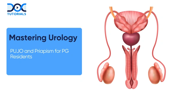
For postgraduate residents specializing in reconstructive surgeries and clinical emergencies, mastering complex genitourinary pathologies is essential. A nuanced understanding of conditions like Pelviureteric Junction Obstruction (PUJO) and the acute management of priapism defines a competent specialist.
This comprehensive guide breaks down these high-yield topics, offering a highly structured approach to pathogenesis, diagnostic modalities, and stepwise surgical interventions tailored specifically for the rigorous demands of your residency training.
Get High Quality, High Yield PG Residency Notes for Free!
Pelviureteric Junction Obstruction (PUJO) in Urology
Pathogenesis and Mechanisms of PUJO
In clinical urology, PUJO is defined as a functional impairment of urinary transport directly at the kidney-ureter junction, which ultimately leads to severe obstruction. The accepted diagnostic standard for identifying hydronephrosis in this context is an antero-posterior (AP) diameter exceeding 12 mm of the intrarenal pelvis on a standard ultrasound.
The etiology of this condition is typically divided into congenital and acquired causes. Intrinsic congenital defects frequently involve abnormal smooth muscle peristalsis, obstructive mucosal valves, or a pathological decrease in the interstitial cells of Cajal. Alternatively, anatomical obstruction due to crossing vessels is a major factor, present in 63% of PUJO cases, compared to merely 20% in normal kidneys. Acquired causes routinely encountered include ureteric stones, strictures, neoplasms, and post-operative fibrosis. Furthermore, when evaluating duplex systems, residents must remember that the lower moiety is the one most commonly obstructed.
Diagnostic Paradigm and Functional Confirmation
The diagnostic workup follows a strict stepwise funnel. Ultrasound (USG) serves as the first-line screening modality, highly capable of detecting the classic prenatal flank mass. Following screening, a non-contrast Computed Tomography (CT) scan is deployed to precisely define the anatomy, evaluate the extent of parenchymal thinning, and reliably identify the presence of crossing vessels.
Functional confirmation remains a cornerstone of modern practice, achieved via MAG-3 Diuretic Renography. MAG-3 is preferred over DTPA because it accurately assesses split renal function and drainage dynamics, yielding a classic obstructive pattern on the renogram curve. Finally, retrograde pyelography (RGP) is routinely performed intraoperatively to dynamically visualize the relevant anatomy immediately prior to making the surgical incision.
Q: What is the defining clinical timeline and presentation for PUJO across different age groups in urology?
A: According to the clinical timeline, neonates and infants typically present asymptomatically with a flank mass initially detected as a prenatal ultrasound finding. In stark contrast, older children and adults classically present with Dietl’s crisis (intermittent, severe flank pain), accompanied by nausea, vomiting, and occasionally rare signs like hematuria or hypertension.
Surgical Management of PUJO in Urology
Endourological management, specifically endopyelotomy, is frequently favored for short-segment PUJO. The technique can be approached percutaneously (antegrade) or via the urethra (retrograde). The procedure involves inserting a flexible scope and creating a full-thickness lateral incision using a hook knife or a 200µ Holmium laser fiber. Surgical patency is validated intraoperatively via contrast extravasation, immediately followed by the placement of a 14 x 7 Fr stent. However, this technique is strictly contraindicated in patients presenting with active infection, underlying coagulopathy, or long strictures exceeding 2 cm.
The absolute gold standard intervention remains the Anderson-Hynes Dismembered Pyeloplasty. This meticulously executed four-step procedure involves: excision of the stricture, spatulation of the ureter, reduction of the renal pelvis, and a watertight anastomosis. It is highly indicated for cases featuring a redundant pelvis, a tortuous ureter, or a high ureteral insertion. Residents should note it is unsuitable for long strictures or a predominantly intrarenal pelvis. Laparoscopic and robotic approaches are now preferred via the transperitoneal route due to familiar anatomy and superior working space, offering high success rates (>95%). Post-operative care mandates a stent placement, which is generally removed after 4 to 6 weeks.
Advanced Flap Reconstruction Techniques in Urology
| Flap Technique | Primary Clinical Indication | Reconstructive Mechanism |
| Foley’s Y-V Plasty | High ureteral insertion. | Reconstructs the junction by converting a Y-shaped incision into a wide V. |
| Spiral Flap (Scardino-Prince) | Long proximal ureteric strictures. | Wraps a spiral flap of the redundant renal pelvis downward to bridge long defects. |
| Vertical Flap | Box-shaped renal pelvis. | Utilizes a vertical pelvic tissue flap to recreate a natural, dependent junction. |
Priapism: A Critical Emergency in Urology
Ischemic vs. Non-Ischemic Classifications
Priapism represents a profound clinical emergency, strictly defined as a full or partial erection lasting longer than 4 hours without sexual stimulation, or persisting beyond climax. It is broadly classified into three primary types: ischemic (low-flow or veno-occlusive), non-ischemic (high-flow or arterial), and stuttering (recurrent ischemic).
Ischemic priapism presents as a severely painful, rigid erection that characteristically features a soft, unaffected glans (glans sparing). The pathophysiology relies on decreased cavernous arterial inflow, leading to stagnant blood stasis, severe hypoxia, hypercapnia, and local acidosis. Diagnostic markers on a cavernous blood gas reveal a low pO2 (< 30 mmHg), high pCO2 (> 60 mmHg), and an acidic pH (< 7.25). Doppler ultrasound will demonstrate a complete absence of arterial inflow. The critical urgency window is less than 36 hours; delays beyond this risk irreversible corporal fibrosis and permanent loss of potency.
Conversely, non-ischemic priapism is generally painless and presents as a non-rigid tumescence. It is most frequently caused by perineal trauma (e.g., a straddle injury) that creates an arterial-sinusoidal fistula. A diagnostic workup reveals a completely normal cavernous blood gas profile.
Stuttering Priapism Mechanisms
Stuttering priapism consists of intermittent, repetitive ischemic episodes and is heavily associated with Sickle Cell Disease (SCD). The molecular mechanism involves the rupture of sickled RBCs, releasing free hemoglobin. This free hemoglobin acts by scavenging nitric oxide (NO), while arginase depletes L-arginine. The subsequent drop in NO availability severely impairs smooth muscle relaxation, triggering venous outflow obstruction. Triggers for stuttering episodes include nocturnal sleep, cold exposure, dehydration, and fever.
Stepwise Management of Priapism in Urology
Pharmacological triggers for priapism include alpha-blockers (prazosin, tamsulosin), psychotropic medications (sertraline, bupropion, phenothiazines), and recreational drugs like cocaine and marijuana. Intracavernosal injection therapies carry variable risks: papaverine carries a high risk (~35%) of prolonged erection, trimix carries a moderate risk, and PDE-5 inhibitors carry a low risk unless combined with recreational drugs. The structured management ladder for ischemic priapism is an essential protocol to master.
The Stepwise Ischemic Management Algorithm in Urology
| Intervention Step | Clinical Action & Surgical Details |
| Step 1: Aspiration & Irrigation | Decompress the corpora cavernosa using a needle and confirm the diagnosis via a cavernous blood gas test. |
| Step 2: Pharmacologic Injection | Administer Intracavernosal Phenylephrine at 0.5-1 mL every 5-10 minutes, up to a strict maximum of 1 mg, while monitoring patient vitals. |
| Step 3: Distal Shunts | Establish distal drainage via Winter’s (needle technique), Ebbehoj (blade technique), Al-Ghorab (excision), or Corporal Snake (dilator). |
| Step 4: Proximal Shunts | Execute Quackel’s (Corpora-Spongiosum shunt) or Grayhack (Vein-to-Corpora shunt) procedures. |
| Step 5: Penile Prosthesis | A refractory indication if the priapism duration exceeds 36 hours, due to the high risk of irreversible fibrosis. |
For non-ischemic cases, management is highly conservative (observation or site-specific compression) because many resolve spontaneously, though selective arterial embolization may be utilized. Stuttering priapism management focuses on prevention using hormonal therapies (GnRH agonists, ketoconazole), non-hormonal agents (pseudoephedrine, baclofen), or paradoxical low-dose PDE-5 inhibitors to downregulate the NO pathway and nocturnal cycles.
Frequently Asked Questions (FAQs) in Urology
1. What defines PUJO radiologically?
It is defined as functional impairment causing obstruction, definitively marked by an antero-posterior (AP) diameter >12 mm of the intrarenal pelvis on a renal ultrasound.
2. What is the classic clinical presentation of PUJO in adults?
Adults typically present with Dietl’s crisis, which is characterized by intermittent flank pain, nausea, and severe vomiting.
3. Which specific renal moiety is obstructed in a duplex system?
A fundamental anatomical rule is that the lower moiety is the one most commonly obstructed in duplex collecting systems.
4. What is the preferred diuretic renography agent for PUJO?
MAG-3 is strictly preferred over DTPA because it allows for a highly accurate assessment of both split renal function and dynamic urinary drainage.
5. When is an Anderson-Hynes pyeloplasty clinically contraindicated?
This gold standard reconstructive procedure is deemed unsuitable for patients presenting with long strictures, multiple strictures, or a predominantly intrarenal pelvis.
6. How is ischemic priapism diagnosed using a cavernous blood gas?
Diagnostics will reveal severe local acidosis and hypoxia, specifically: a pH < 7.25, a pO2 < 30 mmHg, and a pCO2 > 60 mmHg.
7. Why does the glans remain remarkably soft during ischemic priapism?
The ischemic pathology specifically targets and engorges the corpora cavernosa but classically spares the corpus spongiosum, resulting in a distinctively soft glans during physical examinations.
8. What is the precise mechanism of stuttering priapism in Sickle Cell Disease?
Sickled RBC hemolysis releases free hemoglobin that binds available nitric oxide, deeply impairing smooth muscle relaxation and causing persistent venous stasis.
9. What is the absolute maximum dose of intracavernosal phenylephrine?
Phenylephrine must be carefully injected at 0.5-1 mL every 5-10 minutes, capped strictly at a maximum dose of 1 mg while maintaining continuous hemodynamic monitoring.
10. What is the critical time window for resolving ischemic priapism?
Guidelines heavily emphasize a critical intervention window of less than 36 hours; surgical delays beyond this dramatically increase the risk of irreversible corporal fibrosis and permanent erectile dysfunction.

Latest Blogs
-

NEET SS Exam 2024: Analysis, Key Dates, Counselling
The NEET SS 2024 exam kicked off on March 29, 2025. Over two days and two slots, candidates across 13…
-

INI SS Topper: 1 Dream, 1 Study Buddy, 1 Rank – The Journey of Dr. Prithivi Raaj, CML 1 & (AIIMS) AML 1 in DM Cardiology
Achieving the top rank in a competitive exam like the INI SS DM Cardiology requires tireless dedication, a structured approach,…
-

INI SS Results: Merit List, Exam Insights, and What Comes Next
The INI SS results for October 2024 are just around the corner. This is a life-changing moment for thousands of…