Comprehensive Neurosurgery Guidelines: Cervical Trauma and SAH Management
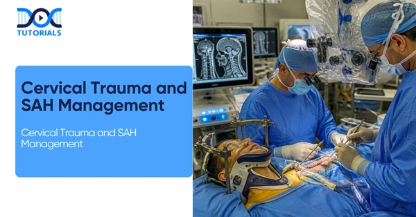
In the highly demanding and rigorous field of neurosurgery, mastering the acute management of spinal trauma and vascular emergencies is paramount for postgraduate residents. This educational and informative guide provides a factually robust overview of critical conditions, ranging from the complex Jefferson Fracture to life-threatening subarachnoid hemorrhages.
By closely adhering to established diagnostic and therapeutic algorithms, neurosurgery residents can ensure they are fully equipped with the high-yield clinical knowledge required for optimal patient outcomes.
Get High Quality, High Yield Neurosurgery PG Residency Notes for Free!
Understanding Cervical Spine Trauma in Neurosurgery
The upper cervical spine, structurally spanning from the skull base down to C2, is a frequent site of traumatic injury that demands precise neurosurgery evaluation.
Upper Cervical Spine Injuries
One of the hallmark injuries encountered in trauma neurosurgery is the Atlas or Jefferson Fracture. The primary mechanism responsible for a Jefferson Fracture is axial loading. Clinical diagnosis of a Jefferson Fracture is often aided by an Open Mouth View X-ray, which prominently reveals C1 lateral mass displacement.
The overall stability of a Jefferson Fracture is highly dependent on the integrity of the transverse ligament. A Jefferson Fracture is explicitly considered unstable if the combined lateral mass deviation is greater than 6.9 mm, a finding that implies a complete transverse ligament rupture. The standard neurosurgery treatment for a Jefferson Fracture is mostly non-operative, typically utilizing a protective collar or halo.
Other critical upper cervical injuries assessed in neurosurgery include Occipital Condyle Fractures, which require conservative treatment utilizing a collar for 6 to 8 weeks. Additionally, Occipital Atlanto Dislocations are classified as high-energy or fatal injuries. In neurosurgery practice, the assessment for this dislocation is conducted via the Power Ratio on a Triple Junction View. In cases of isolated traumatic transverse ligament rupture, the indicated neurosurgery procedure is a posterior C1-C2 fusion, which utilizes a C1 lateral mass screw alongside a C2 pedicle screw.
Axis (C2) Fractures and Spondylolisthesis
Axis (C2) Dens fractures are meticulously classified into three distinct types within neurosurgery. Type 1 involves the tip, Type 2 involves the base or waist, and Type 3 involves the body. Displaced Type 2 fractures specifically require neurosurgery intervention utilizing an anterior decompression screw.
Another condition is the Hangman’s Fracture, which is defined as the traumatic spondylolisthesis of the Axis (C2) on C3. Imaging signs for a Hangman’s Fracture show minimal forward translation or C2-C3 subluxation. The neurosurgery treatment dictates posterior stabilization for significant displacement.
| Target Patient Group | Recommended Management Protocol |
| Acute/Majority | Non-operative (Hard collar/Halo x 3 months) |
| Displaced | Internal fixation |
| Nonunion | Posterior C1-C2 fusion |
| Elderly/Unfit | Conservative (Soft collar) |
Sub-Axial Injury Mechanisms
For the sub-axial cervical spine, encompassing C3 to C7, neurosurgery teams must directly correlate the mechanism of injury with the specific fracture type. Hyperflexion directly leads to compression fractures. Axial compression results in burst fractures. Distraction causes facet subluxation, while hyperextension leads to teardrop fractures. The primary surgical goal in these neurosurgery cases is decompression and stabilization, such as an Anterior Cervical Discectomy and Fusion (ACDF), which is used for cervical burst fractures presenting with cord contusion.
Question: Are cervical collars universally applied in all suspected cervical spine traumas by the neurosurgery team?
Answer: No. In patients with Ankylosing Spondylitis (Bamboo Spine), a collar is completely contraindicated. The pathology of this disease involves auto-fusion and a rigid spine, resulting in a high susceptibility to fractures; therefore, the neurosurgery management protocol strictly dictates avoiding a collar and managing the patient in a position of comfort.
Subarachnoid Hemorrhage (SAH): A Neurosurgery Emergency
Subarachnoid Hemorrhage (SAH) is a critical neurosurgery emergency requiring immediate and aggressive care.
Pathogenesis and SAH Origins
The pathogenesis of SAH typically involves the spontaneous rupture of an aneurysm or an arteriovenous malformation (AVM). High-risk zones for these aneurysms include crucial branch points, specifically the Anterior Communicating Artery (A Comm) and the ACA/MCA bifurcation. Major risk factors that precipitate this neurosurgery emergency include female sex, hypertension, smoking, and a family history involving two or more relatives.
| Feature | Perimesencephalic SAH | Aneurysmal SAH |
| Origin | Venous origin | Arterial origin |
| Location | Basal cistern (anterior to pons) | Diffuse / specific sites |
| Prognosis/Risk | Good prognosis | High risk |
Clinical Presentation and Diagnostic Algorithm
Clinically, a neurosurgery patient with SAH will present with a thunderclap headache, yet statistically, one-third of these patients are incorrectly diagnosed at initial presentation. Clinical grading is strictly measured after resuscitation. Grade 1 denotes a GCS of 15 with no focal deficit, whereas Grade 5 denotes a GCS of 3-9 with or without a focal deficit.
The diagnostic algorithm for SAH is a fundamental pillar of neurosurgery practice. The primary modality is a CT Scan; however, its sensitivity drops significantly if performed more than one week post-incident. If the CT is negative, a Lumbar Puncture must be performed. During the lumbar puncture, spectrophotometry is utilized to detect oxyhemoglobin and bilirubin, which is best achieved between 6 to 12 hours. The absolute gold standard diagnostic tool remains Digital Subtraction Angiography (DSA). For intracerebral or ventricular involvement, the Fisher Grading on CT is designated as Grade 4.
Neurosurgery Treatment and ICU Interventions
For definitive aneurysm treatment, the primary neurosurgery options include surgical clipping or endovascular coiling, noting that early intervention is crucial. The risk of a fatal re-bleed is heavily monitored; it spikes to 4% at 24 hours, and continues at a rate of 1.5% per day. A dire prognosis warning within neurosurgery dictates that 80% of patients who experience a re-bleed will have a poor outcome.
ICU Care Bundles are essential in post-operative neurosurgery care. These bundles include strict DVT prophylaxis, careful fluid management with strict intake/output (I/O) monitoring, and aggressive symptom control utilizing analgesics and antiemetics. To prevent vasospasm and Delayed Ischemic Neurologic Deficit (DIND), a prescription of Nimodipine at 60mg PO every 4 hours is administered.
Despite optimal neurosurgery interventions, complications frequently arise. Neurological complications include vasospasm (seen in 15-20% of cases), DIND, and hydrocephalus. Systemic complications involve hyponatremia (which is very common), severe hypertension, cardiac arrhythmia, and neurogenic pulmonary edema.
Frequently Asked Questions
1. What is the primary mechanism of a Jefferson Fracture in neurosurgery?
The mechanism that causes a Jefferson Fracture is axial loading.
2. At what measurement is a Jefferson Fracture considered unstable?
A Jefferson Fracture is considered unstable if the combined lateral mass deviation is greater than 6.9 mm.
3. What neurosurgery treatment is recommended for an Occipital Condyle Fracture?
Conservative treatment utilizing a collar for 6-8 weeks is recommended.
4. How is Occipital Atlanto Dislocation radiographically assessed?
It is assessed using the Power Ratio via a Triple Junction View.
5. What defines a Hangman’s Fracture?
A Hangman’s Fracture is the traumatic spondylolisthesis of the Axis (C2) on C3.
6. Why is a cervical collar contraindicated for patients with Ankylosing Spondylitis?
These patients have auto-fusion and a rigid spine, making them highly susceptible to fractures; they must be managed in a position of comfort.
7. What is the gold standard diagnostic test for SAH?
The gold standard is Digital Subtraction Angiography (DSA).
8. What medication prevents Delayed Ischemic Neurologic Deficit (DIND) after an SAH?
Nimodipine, given at 60mg PO every 4 hours, prevents DIND and vasospasm.
9. What is the risk of re-bleeding at 24 hours for an SAH patient?
The risk of re-bleed at 24 hours is 4%.
10. How often is Subarachnoid Hemorrhage misdiagnosed upon initial presentation?
One-third (1/3) of patients are incorrectly diagnosed at presentation.

Latest Blogs
-

NEET SS Exam 2024: Analysis, Key Dates, Counselling
The NEET SS 2024 exam kicked off on March 29, 2025. Over two days and two slots, candidates across 13…
-

INI SS Topper: 1 Dream, 1 Study Buddy, 1 Rank – The Journey of Dr. Prithivi Raaj, CML 1 & (AIIMS) AML 1 in DM Cardiology
Achieving the top rank in a competitive exam like the INI SS DM Cardiology requires tireless dedication, a structured approach,…
-

INI SS Results: Merit List, Exam Insights, and What Comes Next
The INI SS results for October 2024 are just around the corner. This is a life-changing moment for thousands of…