mbbs-curriculum Archives - Page 18 of 25 -

How Many Semesters in MBBS? Full Year-Wise Breakdown

How many semesters in MBBS

The MBBS (Bachelor of Medicine, Bachelor of Surgery) degree in India is one of the most structured and intensive medical programmes worldwide. Students often wonder how the MBBS programme is structured, how the semesters work, which subjects fall under each phase, and what the first-year exam load entails. Knowing about the MBBS semester schedule in advance enables students to organise their preparation well, manage time, and set achievable targets.

Our comprehensive guide will answer all your questions, including how long MBBS is, how many semesters are in MBBS, and which subjects are part of the course. 

Keep reading!

How Many Semesters are There in MBBS?

There are 9 semesters in MBBS, spread across 4.5 years of academics, followed by a 1-year compulsory internship. Each semester lasts around six months and includes lectures, practicals, clinical postings, and internal assessments.

The MBBS programme in India is designed to enhance students’ medical knowledge step-by-step, from core sciences to clinical practices. These 9 semesters break the process of learning medicine into well-defined phases, which are pre-clinical, para-clinical, and clinical, and make it a point that students come out of the conceptual haze before they venture into the world of real medical exposure. 

The duration of each semester is approximately 6 months, including theory, laboratory work, early clinical exposure, and internal assessment. This systematic format helps students maintain a steady learning pace and allows medical colleges to assess their progress at every stage.

Here is the semester structure:

MBBS PhaseDurationSemesters
Pre-Clinical (1st Year)1 yearSemester 1 and 2
Para-Clinical (2nd Year)1.5 yearsSemester 6, 7, 8, and 9
Clinical (3rd & Final Year)2 yearsSemester 6, 7, 8 and 9
Internship12 monthsNot semester-based

The duration of each semester is about six months, and besides instruction (lectures, practicals, including dissections in lab work), there are clinical postings (from 2nd year onwards) as well as internal assessment (and university examinations).

More structured platforms, such as DocTutorials, offer semester-based schedules and subject modules that resonate with this pattern, thereby allowing students to get a clear idea of what they should be focusing on at each stage.

MBBS Year-Wise Semester Breakdown (NMC pattern)

The MBBS programme is divided into three academic phases and nine semesters as per the NMC curriculum, progressing from pre-clinical basics to para-clinical integration and full clinical training.

NMC (National Medical Commission) has mandated a well-defined, year-wise semester pattern to ensure that all medical students go through the course curriculum in logical and rational sequences. Each stage concentrates on a specific aspect of medical science, from basic scientific subjects to systemic diseases and laboratory studies, before finally entering clinical rotations in major and subspecialities. 

Breaking it down like this makes things transparent for students so that they understand more clearly what’s required of them academically in each year, and how the demands increase from one semester to the next.

Below is the detailed semester structure followed in the MBBS programme as per the National Medical Commission (NMC) curriculum.

MBBS 1st Year: How Many Semesters?

The MBBS 1st year consists of 2 semesters, Semester 1 and Semester 2, both covering foundational subjects that create the base for understanding human structure and function.

Semester 1 and Semester 2

The first year introduces foundational medical sciences that form the base for all future studies.

Subjects in MBBS 1st Year

  • Anatomy
  • Physiology
  • Biochemistry

These subjects focus on the structure of the human body, its functioning, and biochemical processes that sustain life.

How Many Exams are There in MBBS 1st Year?

MBBS 1st year students typically appear for 10–12 exams, which generally include:

  • Two internal assessments per subject per semester
  • Term/sessional exams
  • Final university exams (theory, practical, viva)
  • Spotters, viva voce, and practical assessments

For many freshers, this year feels overwhelming due to the new academic load and volume of content.

MBBS 1st Year Semester and Exam Pattern

ComponentDetails
Total Semesters2
SubjectsAnatomy, Physiology, Biochemistry
Internal Exams2 per subject per semester (may differ across universities)
Practical ExamsConducted periodically
Final University ExamAt year’s end
Approx. Total Exams10–12 annually

MBBS 2nd Year: Semesters and Subjects

The second year of MBBS covers major para-clinical subjects and spans over three semesters, that is, Semester 3, Semester 4, and Semester 5.

Subjects in 2nd Year

  • Pathology
  • Pharmacology
  • Microbiology
  • Forensic Medicine
  • Community Medicine (Part 1)

This phase transitions students into understanding diseases, drug mechanisms, microbes, and medico-legal concepts.

MBBS 3rd Year (Part 1): Semesters and Subjects

This is a shorter phase consisting of one semester, that is, Semester 6, where students learn compact but high-yield subjects.

Subjects in 3rd Year Part 1

  • ENT
  • Ophthalmology
  • Community Medicine (Part 2)

Students also attend clinical postings regularly, gaining hands-on exposure. ENT and Ophthalmology require focused study due to their compact yet scoring nature.

MBBS Final Year (3rd Year Part 2): Semesters and Subjects

The final academic phase spans three semesters, that is, Semester 7, Semester 8, and Semester 9, covering the most comprehensive and clinically demanding subjects.

Subjects in Final Year

  • Medicine
  • Surgery
  • Obstetrics and Gynaecology
  • Paediatrics
  • Orthopaedics
  • Anaesthesia
  • Radiology
  • Psychiatry
  • Dermatology

This is the most demanding phase because students manage ward work, clinical duties, postings, and exam preparation simultaneously.

MBBS Semester-wise Subject Distribution

MBBS is divided into 9 semesters: the first 2 semesters cover pre-clinical subjects, the next 3 cover para-clinical subjects, and the final 4 semesters focus on clinical subjects with ward postings and hands-on training.

Knowledge of the distribution of subjects in all MBBS semesters is beneficial for students in planning their preparation well in time. And each semester builds on the next, from a focus on foundational sciences to disease-specific subjects to full-on clinical rotations and hands-on patient care. This shuffling of the subjects every semester facilitates a smooth transition from theory to patients as students progress through their academic years. The subsequent table presents an accessible summary of MBBS semesters and their academic arrangement.

SemesterSubjects
Semester 1Anatomy, Physiology, Biochemistry
Semester 2Continuation of Semester 1 subjects
Semester 3Pathology, Pharmacology, Microbiology, FMT
Semester 4Continuation of 2nd-year subjects
Semester 5Completion of para-clinical subjects
Semester 6ENT, Ophthalmology, Community Medicine
Semester 7Medicine, Surgery, OBGYN, Paediatrics (start)
Semester 8Continuation of major clinical subjects
Semester 9Completion + university exam preparation

Note: May differ based on the college or university curriculum. 

Duration of All MBBS Semesters

Each MBBS semester lasts approximately six months, making the entire academic phase of the MBBS programme span 4.5 years, followed by a 1-year compulsory internship. Every semester has a distinct academic focus, which begins with foundational pre-clinical concepts, moves into para-clinical integration, and finally leads to hands-on clinical learning in hospitals and wards. 

The table below provides a clear, semester-wise summary of duration and focus to help students understand how their MBBS journey progresses:

SemesterDurationKey Focus
Semester 16 monthsPre-clinical basics
Semester 26 monthsFoundation strengthening
Semester 36 monthsDisease mechanisms
Semester 46 monthsDrug action and microbiology
Semester 56 monthsIntegration of para-clinical concepts
Semester 66 monthsENT, Eye, Community Medicine
Semester 76 monthsMajor clinical subjects
Semester 86 monthsWards, clinics, exam preparation
Semester 96 monthsFinal theory and clinical exams

How Study Platforms Support Students Across Semesters?

Study platforms support MBBS students across semesters by offering structured video lectures, organised revision plans, MCQ practice, and semester-wise guidance aligned with the medical curriculum.

Although MBBS is deeply structured, the syllabus in each semester grows in complexity. A lot of students need digital platforms for organisation to ensure clarity and uniformity. DocTutorials, for example, provides:

  • Semester Progress-Based Video Lectures on each Subject
  • MCQ bank and the university’s past exam patterns
  • Case-based learning; beneficial in the final year
  • Structured revision plans
  • Tailored exam preparation with brief notes and summaries

With the syllabus getting heavier with each passing semester, a guided resource enables students to prepare smartly rather than becoming frantic because they are not able to cover it all.

Conclusion

MBBS in India involves 9 semesters over the course of 4.5 years, followed by a one-year compulsory internship. The freshman year is divided into two semesters, which include approximately 10–12 exams each, and this can be a stressful transition for medical students. Each semester is cumulative, working its way from basic science to para-clinical and full clinical experience.

Knowing how a semester is structured can teach students how to organise their learning and preparation and stay on track. The MBBS syllabus is huge, but access to organised resources, timely advice, and regular revision tips can make the process of learning easy to follow. 

Rely on DocTutorials for structured semester guides, concise high-yield notes, expert guidance, video lectures, PYQs, mock tests, and more. 

Check out our MBBS courses today!

FAQs about Semesters in MBBS

  1. How many semesters are there in MBBS?

There are 9 semesters in MBBS, covering 4.5 years of academic training.

  1. How many semesters are there in the MBBS 1st year?

The MBBS 1st year has 2 semesters: Semester 1 and Semester 2.

  1. How many exams are there in the MBBS 1st year?

Students typically take 10–12 exams across internal tests, practicals, vivas, and final university exams.

  1. What subjects are taught in the MBBS 1st year?

Anatomy, Physiology, and Biochemistry are generally taught in the MBBS 1st year. 

  1. Is the MBBS semester system difficult to manage?

The workload increases each semester, but with consistent study and the help of structured learning resources, it becomes manageable.

  1. How is the MBBS curriculum structured?

The MBBS curriculum structure is divided into three stages, which are pre-clinical, para-clinical, and clinical phases, respectively. In most countries, like India, MBBS spans 4.5 years plus 1 year of internship, usually organised into 9 semesters. Each phase focuses on progressively moving from basic sciences to hands-on clinical training.

  1. Are there any variations in the number of MBBS semesters based on the country?

Yes, the number of MBBS semesters by country varies. India follows a 9-semester system, while countries like the UK and Australia use a year-based or modular MBBS/MD structure. In the USA, students pursue an MD after a pre-medical degree.

  1. What is the 60/40 rule in MBBS?

The 60/40 rule in MBBS refers to the exam eligibility criteria in many Indian universities. Students must score at least 60% in the theory exams and 40% in the practical exams (or vice versa, depending on regulations) to pass a subject. Exact rules may vary slightly by institution.

  1. Which MBBS year is the hardest?

Many students feel the hardest year in MBBS is either the 1st year or the final year. The first year is challenging due to vast subjects like Anatomy, while the final year is intense because of clinical responsibility, multiple major subjects, and preparation for postgraduate entrance exams.

  1. Are there 19 subjects in MBBS?

Yes, officially, MBBS includes around 19 major subjects. The degree combines core subjects like Anatomy, Medicine, Surgery, and their allied specialities.

DocTutorials App Download Now

Latest Blogs

Microbiology in MBBS: Key Topics You Should Cover

Microbiology Notes | DocTutorials

Microbiology is a key subject in the MBBS curriculum, encompassing numerous concepts and serving as a vital bridge between the basic and clinical sciences. Due to the emergence of new pandemics worldwide, it has become mandatory to have an in-depth understanding of microbiology in MBBS.

Therefore, to score good marks in this subject, medical students need to prepare comprehensive microbiology notes and learn them with diligence. Keep reading for a detailed insight.

Why Microbiology is Important in MBBS?

Microbiology is a core MBBS subject that bridges rudimentary sciences with clinical practice, helping students understand the causes, diagnosis, and management of infectious diseases. It plays a vital role in everyday medical decision-making, from antibiotic selection to infection control.

The MBBS course, especially in the initial two years, consists of non-clinical and para-clinical subjects including Anatomy, Physiology, Biochemistry, Pathology, Pharmacology, and Microbiology.

These form the basis of learning clinical subjects in the latter two years of MBBS. Amongst them, Microbiology occupies a special position since it links basic sciences with practice. Microbiology teaches young doctors about microbes, including bacteria, viruses, fungi, and parasitic microbes, which cause most illnesses in humans.

From diagnosing infections to prescribing antibiotics and controlling infections, almost every clinical decision is influenced by microbiology. Another prime factor that brought the focus to microbial science was the emergence of the COVID-19 pandemic, which demonstrated the influence of a single microbe on global health and economies.

Although it is an extremely valuable field, many students find it overwhelming initially because of its vast syllabus, extensive memorisation, and laboratory components. But there is actually a way to turn this topic into a very interesting and scoring one.

What are the Essential Topics Covered in Microbiology in the MBBS Course?

Microbiology carries significant weightage in both theory and practical exams. Thorough preparation of core topics is essential to succeed in university and entrance examinations. Well-structured and regularly updated microbial notes help students stay exam-ready and confident.

Here are the key topics and chapters about which every MBBS student should work while preparing microbiology notes:

1. General Microbiology Module

  • Contributions of Louis Pasteur
  • Koch’s Postulates
  • Bacterial cell wall
  • Flagella
  • Bacterial growth curve (a must-know topic)
  • Anaerobic culture methods
  • Antimicrobial susceptibility testing
  • Horizontal gene transfer
  • Antimicrobial resistance
  • Exotoxin vs endotoxins
  • Isolation techniques for viruses
  • Classification with examples (parasites and fungi)

2. Immunology Module

  • Innate immunity vs acquired immunity
  • Active immunity vs passive immunity
  • Definitions of antigen, hapten, and superantigens
  • Structure of an antibody
  • Various classes of immunoglobulins
  • Monoclonal antibodies
  • Basic mechanisms of precipitation reaction and agglutination reaction
  • ELISA (in detail)
  • MHC
  • Cytokines
  • Hypersensitivity reactions and their types
  • Mechanisms and examples of autoimmunity
  • Mechanism of graft rejection
  • Types and examples of vaccines

Before you start reading the systems, there are sections on disinfection and sterilisation that have become more significant after COVID-19. These sections are very important as they will educate you on fundamental topics such as biomedical waste management, PPE kits, and more.

3. Disinfection and Sterilisation

  • Healthcare-acquired infections (definition, and examples)
  • The steps of hand washing (it rarely comes as a theoretical question, but hold importance)
  • Types of masks
  • Steps of donning and doffing
  • CAUTI and VAP
  • Definitions of sterilisation, disinfection, and cleaning
  • Autoclave
  • ETO
  • Disinfectants (according to the levels)
  • Methods to test the efficacy of the sterilant
  • Colour coding of dustbins
  • Definition and management of needle stick injury
  • Water surveillance

4. Systemic Microbiology Module

Bloodstream and Cardiovascular Infections

  • Acute rheumatic fever
  • Infective endocarditis
  • Brucellosis and leptospirosis
  • Fuo
  • Leishmaniasis
  • Typhoid
  • Scrub typhus
  • Malaria
  • Dengue
  • HIV (in detail)
  • Systemic candidiasis
  • Lymphatic filariasis
  • African sleeping sickness

Gastrointestinal Tract Infections

  • Food poisoning
  • Cholera
  • Mechanism of diarrhoea and agents
  • Lab diagnosis of diarrhoea
  • Intestinal amoebiasis
  • Botulism
  • Ascariasis
  • Giardiasis
  • Trichura
  • Types of E.coli
  • Entrobias
  • Shigella
  • Rotavirus diarrhoea
  • Intestinal taeniasis
  • Hookworm

The Hepatobiliary System

  • Hepatitis
  • Liver abscess
  • Hydatid cyst

Skin and Musculoskeletal System Infections

  • Diabetic foot
  • Gas gangrene
  • Staph aureus infections
  • HSV infections
  • Streptococcus pyogenes infections
  • VZV infections
  • Lab diagnosis of leprosy
  • Cutaneous leishmaniasis
  • Measles
  • Superficial mycoses
  • Cutaneous larva migrans

Respiratory Tract Infections

  • Pneumonia
  • Agents of respiratory tract infections
  • Diptheria
  • Coronavirus 
  • Tb (a crucial topic for Indians, as this ailment is quite common in our country)
  • Aspergillosis
  • Influenza
  • Infectious mononucleosis

CNS Infections

  • Rabies, particularly the vaccine prophylaxis
  • Sleeping sickness
  • Cerebral malaria
  • Meningitis
  • Tetanus

Urinary Tract Infections

  • Chancroid
  • Syphilis
  • Chlamydia infections
  • Pathogenesis of UTI
  • Agents of UTI

Miscellaneous Infections

  • Oncogenic viruses
  • Bite wound infections
  • Congenital syphilis
  • Zoonotic infections

FAQs about Microbiology in MBBS

  1. What are the major and exam-important topics of microbiology for MBBS?

Major topics include bacterial cell structure, toxins, sterilisation methods (autoclave, hot-air oven), antiseptics and disinfectants, and a detailed study of common pathogens.

High-yield organisms include Staphylococcus, Streptococcus, Mycobacterium tuberculosis, Enterobacteriaceae, Vibrio, and key parasites such as Plasmodium, Entamoeba, Giardia, Ascaris, Taenia, and Wuchereria, with emphasis on morphology, life cycles, pathogenesis, clinical features, and laboratory diagnosis.

  1. What competencies will an MBBS student attain through the learning of microbiology?

Microbiology enables MBBS students to acquire knowledge and develop positive attributes in diagnosing, treating, and preventing infections caused by microbes, based on careful laboratory testing and sensible antimicrobial use.

Microbiology also enables future healthcare practitioners to effectively deal with antimicrobial resistance and emerging infections.

  1. Why are microorganisms classified into bacteria, viruses, fungi, protozoa, and helminths in MBBS microbiology?

This classification into bacteria, viruses, fungi, protozoa, and helminths enables MBBS students to distinguish and recognise the characteristics and features of each type of bacteria.

These include its structure, life cycle, mode of transmission, and disease manifestations, all of which are critical for diagnosing and treating infectious diseases.

  1. Why are handwritten microbiology notes better than typed notes for learning microbiology?

When it comes to learning complex subjects like microbiology, handwritten learning is preferred more. This is because it ensures effective memory retention by stimulating higher-level brain activity than typing.

Conclusion

Microbiology has been a crucial subject for MBBS students owing to its complexity and difficulty to grasp and overcome. A well-designed set of microbiology notes, books, practical training, and advanced revision techniques is essential for MBBS students to perform well in their exams and studies.

To help students understand complex concepts and prepare well for exams, DocTutorials helps meet the needs of MBBS students. We offer 3D-animated lectures, complete syllabus coverage, exam-focused content, complimentary hardcopy notes, and structured mentorship through our “Microbiology Simplified” plan.

Join DocTutorials today and explore our MBBS curriculum to excel in your medical career!

DocTutorials App Download Now

Latest Blogs

Pathology in MBBS: Important Topics and Study Tips

Pathology: Important Topics and Preparation Tips

When you are in your second year of MBBS, you start observing how practising medicine is becoming more crucial than ever. Among all subjects, pathology is the one in which all scattered concepts start to fall into place.

This subject helps make every medical concept fit into a coherent narrative. With proper MBBS pathology notes and study materials, you will be able to learn and understand pathology in a comprehensive, quick, and easy manner.

Keep reading to learn more!

What are the Key Topics to Be Covered in MBBS Pathology?

Pathology is the backbone of clinical medicine in MBBS courses and helps students understand the mechanisms and consequences of disease. Learning these high-yield topics is obligatory to link clinical symptoms with microscopic and molecular alterations.

During the preparation of notes for pathology, MBBS students should determine the major, commonly asked topics in pathology.

Here are the important topics to be covered in MBBS pathology:

1. Haematology

  • Megaloblastic Anaemia
  • Coagulation Pathway
  • Blood Bank
  • Haemophilia
  • Haemolytic Anaemia
  • Multiple Myeloma
  • Iron Deficiency Anaemia
  • Sickle Cell Anaemia
  • Thalassemia
  • CML & MDS
  • Lymphoma
  • Hypercoagulability
  • Acute Leukaemia (AML/ALL)
  • Stain
  • Waldenstrom’s Disease and Heavy Chain Disorders
  • Paroxysmal Nocturnal Haemoglobinuria

2. Cell Biology

  • Cellular Adaption
  • Free Radical Injury
  • Pigmentation
  • Necrosis
  • Cell Injury Concept
  • Irreversible Injury 1 and 2

3. Renal

  • Diabetic Retinopathy
  • Glomerular Nephritis
  • Alport Syndrome
  • Renal Spotters
  • PSGN
  • ADPKD
  • Renal Cell Carcinoma
  • Nephrotic Syndrome
  • Renal Stones

4. CNS

  • Prion Disease
  • Malformations of the Brain
  • CSF Analysis
  • CNS Tumour
  • Meningitis
  • Medulloblastoma
  • Meningioma
  • CNS Degenerative Disease
  • Neuroblastoma

5. GIT

  • Fatty Liver
  • Celiac Disease
  • Liver Disorders
  • Inflammatory Bowel Disease
  • Acute and Chronic Gastritis
  • Congenital GIT Anomalies
  • Jaundice
  • Hepatitis
  • Ulcerative Colitis
  • Whipple Disease
  • GI Polyps
  • Peptic Ulcer Disease
  • GIT Tumour
  • Mesothelioma

6. Lung

  • TB Lung Lesions
  • Obstructive Lung Disorders
  • Pulmonary Hypertension
  • ARDS
  • Emphysema
  • Adenocarcinoma
  • Lung Abscess
  • Lung Cancer
  • COPD
  • Occupational Disease
  • Infective Lung Disorders
  • Neonatal Respiratory Distress Syndrome
  • Asbestosis

7. CVS

  • DCM & HCM
  • Vasculitis
  • Bleeding Disorders
  • Clotting Factor Disorders
  • Myocardial Infarction
  • Aortic Aneurysm
  • Rheumatic Heart Disease
  • Infective Endocarditis
  • Platelets Disorders
  • Vascular Sclerosis
  • Blood Transfusion and Blood Grouping

8. Endocrine

  • Follicular Carcinoma
  • Parathyroid and Thyroid Disorders
  • Papillary Thyroid Carcinoma
  • Pheochromocytoma
  • Adrenal Gland Disorders

9. Breast

  • Breast Cancer
  • Fibroadenoma
  • Breast Oncogenesis
  • TNM Staging

10. Malignancy

  • Tumour Marker
  • Bone Tumours
  • Tumour Suppressor Gene
  • Molecular Hallmark of Cancer
  • Seminoma
  • Melanoma
  • Hodgkin & Non-Hodgkin Lymphoma

11. Inflammation

  • Plasma Chemical Mediators
  • Coagulation Cascade
  • Chronic Inflammation
  • Neutrophil Extracellular Trap
  • Inflammatory Markers

12. Genetics

  • Gene Repair Defects
  • Down Syndrome
  • TNR Disorders
  • Single Cell Disorder
  • Inheritance Pattern
  • Turner Syndrome
  • Specific Cytogenetic Disorders

13. Immunology

  • Granuloma
  • Hypersensitivity Reactions
  • Immunodeficiency Disorders
  • Complement Pathway

14. Miscellaneous

  • Marfan & Ehlers Danlos Syndrome
  • HIV
  • Aging
  • Diabetes
  • Transplantation
  • Hypertension
  • Amyloidosis

What are the Tips for Preparing Pathology Notes for MBBS Students Effectively?

Proper pathology notes are essential for MBBS students to master core concepts like cell injury, inflammation, and systemic diseases. Taking notes smartly with active learning improves memory retention and clinical understanding. Using well-organised pathology notes also helps you revise for exams faster.

Here are some strategies that can be used to develop quality pathology notes:

  • Take Clear and Well-Structured Pathology Notes

MBBS pathology notes should be kept short, logical, and exam-oriented. Diagrams, flowcharts, and tables should be used to clarify topics such as inflammation and cell injury. Having structured notes will give you faster, less stressful revision at the very last moment.

  • Use Visual Tools for Cell Injury and Inflammation

A better understanding can be achieved by converting texts into gists, charts, and mind maps. The cell injury pathology notes can be visually organised into causes, mechanisms, and outcomes. Besides, diagrams are a great help in retaining complicated pathways.

  • Practice Active Recall for Pathology

Self-testing should be done frequently through the use of flashcards and short questions. Active recall strengthens memory for inflammation mediators, stains, and disease mechanisms. One-page high-yield pathology notes are most effective for repeated revision.

  • Revise Daily for Long-Term Retention

Opt for 10 to 15 minutes of revision of MBBS pathology notes on a regular basis. Constant reviewing prevents forgetting and helps in understanding complex concepts.  Small daily efforts lead to strong conceptual clarity.

  • Highlight Vital Points and Utilise Mnemonics

Colours and mnemonics can be used to memorise lists and classifications. The major mechanisms in inflammation pathology notes and types of necrosis are to be highlighted. Visual cues serve to facilitate quick recall during exams.

  • Opt for Hand-Written Notes to Stay Organised

Making pathology notes by hand is a good practice since it improves the understanding and retention of the material. Manually prepared notes effectively organise the pathology of cell injury and inflammation.

  • Stay Consistent and Study Smart

Pathology can appear quite vast, but smart techniques can make it manageable. Combined use of figures, flashcards, and group discussions while emphasising MBBS and NEET PG preparation. Consistency is the key to mastering MBBS pathology notes.

FAQs about Pathology in MBBS

  1. What are the four categories of pathology?

The branches of pathology encompass general pathology, anatomical pathology, chemical pathology, clinical pathology, genetics, immunology, haematology, and microbiology.

  1. What are the important topics of pathology in MBBS?

Important pathology topics in MBBS include haematology, cell injury, inflammation, systemic diseases, malignancies, anaemias, leukaemia, Multiple Myeloma, free-radical injury, GI disorders, renal diseases, CNS conditions, lung and cardiac infections, breast cancer, tumour suppressor genes, genetic disorders, and systemic diseases.

Emphasising these topics while making notes on pathology will help your preparation in such a way that you will feel conceptually sound, along with preparing for the exams.

  1. What are the common mistakes students make while preparing pathology notes in MBBS?

The most frequent error students make when preparing pathology notes is memorising them without understanding the concepts, disregarding the microscopic structure of tissues and their practical relationships, and falling behind in MCQ practice. 

Smart pathology notes must be concept-based, regularly reviewed, and combined with MCQs to enhance exam performance.

  1. What are the educational objectives of studying Pathology in MBBS?

The aim of pathology in the MBBS curriculum is to help students understand disease at the cellular and tissue levels and to develop an ordered approach to diagnosis. By taking notes in pathology, students can apply their knowledge of lab experiments, histopathology, and their application in managing patients.

Conclusion

Pathology is where medicine really comes alive, transforming students into reflective practitioners who grasp diseases in depth. A sound understanding of pathology not only improves examination results but also instils enduring confidence in practising clinicians. 

At DocTutorials, we understand the dynamic requirements of medical students. We offer 3D-animated lectures, comprehensive syllabus coverage, complimentary hardcopy notes, and structured mentorship through our “Pathology Simplified” plan.

We assist medical students in establishing solid bases, continuity, and confidence to progress into budding medical specialists. Join DocTutorials today and explore our MBBS curriculum to excel in your medical career!

DocTutorials App Download Now

Latest Blogs

Preparing MBBS Biochemistry Notes: Essential Topics You Need to Cover

MBBS Biochemistry Notes | DocTutorials

Biochemistry is one of the most important and scoring subjects in the initial years of MBBS, or any medical studies, as it forms the molecular basis of medicine. It serves as an interface between the basic sciences and clinical knowledge, explaining how biological processes function at the cellular and molecular levels.

From metabolism to genetic processes, or from enzymes to nutrition, preparing and learning biochemistry notes fosters students’ core skills in grasping concepts of health, disease, and diagnosis. Keep reading to learn more!

MBBS Biochemistry Syllabus and High-Yield Topics for MBBS

The MBBS biochemistry emphasises high-yield aspects of metabolism, enzymes, molecular biology, vitamins, and clinical biochemistry. Such aspects act as a base for NEET PG and clinical courses. Knowledge of biochemical pathways, enzyme regulation, and disease correlations is essential for success on the exam.

Biochemistry in MBBS is extensive, but exam success depends entirely on mastering the most frequently tested and clinically relevant areas.

According to the official MBBS syllabus and exam trends, the following topics are considered high-yield and must be studied in depth:

Topic CategoryHigh-Yield Topics
Carbohydrate MetabolismGlycolysis, Gluconeogenesis, TCA cycle, HMP shunt, Glycogen synthesis & breakdown, Fructose & Galactose metabolism, Diabetes mellitus
Lipid MetabolismFatty acid synthesis & β-oxidation, Cholesterol synthesis & regulation, Ketone bodies, Lipoproteins (HDL, LDL, VLDL), Dyslipidemias
Protein and Amino AcidsAmino acid catabolism, Urea cycle, Essential amino acids, Porphyrin (heme) synthesis, Haemoglobin structure & function, Collagen
Enzymology and BioenergeticsEnzyme kinetics & regulation, Coenzymes (NADH, FADH₂, CoA), ATP production, Electron Transport Chain
Genetics and Molecular BiologyDNA replication, RNA transcription, Genetic code, Mutations, Recombinant DNA technology (PCR, cloning)
Vitamins and MineralsVitamins A, D, E, K, B-complex; deficiency disorders; Iron, calcium, phosphorus, trace elements 
Clinical Tests and DisordersLiver and kidney function tests, Plasma proteins, Electrolytes, acid–base balance, Diabetes, Inborn errors of metabolism

Preparing well-organised MBBS biochemistry notes on the above categories, subjects, and chapters, while following a concept-based learning approach, is essential for grasping these topics without hassle.

Let’s discuss each of these categories in detail:

  • Cellular and Biomolecular Foundations

This area describes the chemical fundamentals of cell structure and function, including the structure and functions of cells, organelles, and the cell membrane. It includes concepts of fluid and electrolyte balance, pH, and buffer capacity, which are essential for maintaining life processes.

The students are also required to know the fundamental chemistry of biomolecules, including carbohydrates, proteins, lipids, and nucleic acids, as well as the structure of amino acids and the normal concentration of proteins in blood.

  • Carbohydrate Chemistry and Metabolism

The carbohydrate metabolic pathways are a scoring region that must have high clarity for pathways. The mandatory pathways are glycolysis, gluconeogenesis, the TCA cycle, and the HMP shunt, focusing on the regulated enzymes hexokinase, PFK-1, and PDH.

Conditions such as Von Gierke’s (liver) and McArdle’s (muscle) present specific symptoms related to their organs, while diabetes impacts glycogen regulation.

Maintaining clear and organised notes allows students to grasp pathways swiftly, recall essential enzymes, and connect them to clinical scenarios, thus expediting revision and streamlining the exam preparation.

  • Lipid Chemistry and Metabolism

This group centres on fatty acid structure and function, fatty acid saturation, and important processes such as β-oxidation and fatty acid synthesis. Cholesterol production and maintenance, ketone body production, and regulation are vital from the clinical perspective.

Lipoprotein metabolism and diseases such as familial hyperlipemias are essential, as are clinical aspects of metabolic and fatty liver diseases and the metabolic syndrome.

  • Protein and Amino Acid Metabolism

Protein metabolism includes amino acid usage, nitrogen excretion in the urea cycle, and essential amino acids. The structural aspect includes the level of protein organisation, protein folding, collagen production, and the function of haemoglobin. The curve of oxy-haemoglobin dissociation is also included here.

  • Enzymology and Bioener

This area is concept-based and scored, covering enzyme classification, kinetic curves (Km, Vmax, Lineweaver-Burk plots), and regulation of mechanisms. A good grasp of enzyme inhibition (competitive/non-competitive) and coenzymes (NAD+, FAD, CoA) is also required. 

ATP synthesis via the respiratory chain and marker enzymes such as ALT and AST link biochemistry to clinical pathology.

  • Genetics and Molecular Biology

Connecting biochemistry and genetics, this domain encompasses DNA/RNA structure, replication, transcription, and translation. Genetic code properties, mutations, and DNA repair are always required.

Recombinant DNA technology, which includes PCR, restriction enzymes, and blotting techniques, is crucial for the NEET PG exam. You can remember it better by making combined biochemistry notes.

  • Vitamins and Minerals

This part requires the student to memorise directly. The student must list the water-soluble and fat-soluble vitamins and describe their roles as coenzymes, the deficiency diseases they cause, and the food sources.

In addition, topics related to mineral metabolism, such as iron (heme synthesis and anaemia), zinc, calcium, and phosphorus, are also essential.

  • Clinical Biochemistry Tests and Disorders

Clinical biochemistry refers to the application of theoretical concepts to diagnosis. These main branches are tests of kidney function (urea and creatinine), liver function (bilirubin and enzymes), management of blood glucose levels, and acid-base imbalance.

In addition, there are some important topics related to MBBS that students need to cover while making biochemistry and clinical pathology notes prior to the exam.

These include:

  • Importance of glutathione in the metabolism of xenobiotics
  • Significance of myoglobin within the body
  • Function of creatine in muscle physiology
  • Connection between diabetes and early-onset cataracts
  • Phase 1 and Phase 2 biotransformation reactions
  • Genetic disorders related to aromatic acid metabolism
  • Biosynthesis and regulation of cholesterol
  • Carboxylation and oxidative decarboxylation reactions
  • Vitamin D’s role in maintaining calcium balance
  • Transamination and oxidative deamination processes
  • Biochemical alterations during periods of starvation
  • Importance of the pentose phosphate pathway
  • Vitamin D’s contribution to calcium homeostasis
  • Mechanisms of peptide hormone action
  • Selenium’s function in the human body
  • Purine catabolism and associated diseases
  • Structure of Proteins
  • Structure of DNA
  • Fluid mosaic model of cell membranes
  • Functions of MHC proteins 1 and 2 in organ transplantation
  • Importance of clearance tests in diagnostic procedures
  • Types of enzyme inhibition: Competitive vs Non-competitive
  • Oxygen dissociation curve of Haemoglobin
  • PCR flowchart and its applications
  • Structure of Collagen
  • Structure of Immunoglobulin

Why is Biochemistry Important in MBBS?

Biochemistry is an integral foundation of MBBS studies because it explains life phenomena at a molecular level. It helps students of MBBS understand how life functions at a molecular level in health and disease conditions.

Here is why biochemistry is essential in MBBS:

  • Foundation of Medical Science: Biochemistry deals with the chemical constitution of living organisms, the chemical structure, and properties of biomolecules such as proteins, carbohydrates, lipids, and nucleic acids.
  • Comprehending Metabolism: This type of activity increases a student’s understanding of how the body generates its energy from the foods it consumes. This is an essential aspect in a human being’s survival and comprehension of diseases.
  • Development of Drugs and Drug Action: An understanding of biochemistry can be useful in explaining drug action, enzyme inhibitors, drug targets, drug interactions, and pharmacokinetics.
  • Clinical Relevance: Awareness of biochemical processes is very important for disease diagnosis, interpreting laboratory results, and comprehension of the biological processes of diseases.
  • Weightage in Exams: It holds a highly prominent place in the MBBS first professional exam as well as in competitive exams like NEET PG and INI-CET.
  • Future Medical Practice: The concepts acquired from biochemistry instant notes are helpful in applying the knowledge of molecules in medical practice effectively.

FAQs about Biochemistry in MBBS

  1. What are the fundamental subjects of biochemistry in MBBS?

The fundamental subjects of Biochemistry encompass Enzymes, Function tests, the Chemistry and Metabolism of Carbohydrates, Lipids, & Proteins, Nucleotides and Nucleosides, Biochemical Pathways, Molecular Biology, Oncology, Immunity, Nutrition, and Metabolism.

While preparing MBBS biochemistry notes, students must study each subject diligently.

  1. Is biochemistry challenging in MBBS?

Biochemistry is quite intriguing to read and comprehend, yet it can be a challenging subject to learn and memorise. However, not all modules and topics are difficult.

If approached systematically, such as by participating in mock tests, watching video lectures, and preparing biochemistry notes, the concepts can become easier to grasp and remember.

  1. What are the clinical applications of biochemistry in MBBS?

Biochemistry elucidates conditions like diabetes, jaundice, and kidney disorders, and assists in analysing laboratory results, including glucose, urea, and hormone levels.

  1. What hands-on experiments do MBBS students conduct in biochemistry?

Students might be required to analyse samples for glucose, proteins, urea, cholesterol, and ketone bodies, perform enzyme assays and colourimetric measurements, and participate in diagnostic education.

  1. What is the most exam-oriented approach to learning enzymes and proteins in biochemistry?

Students need to focus on learning the definitions, classification, mechanisms, graphing (Michaelis Menten and Lineweaver Burk), and clinical aspects (isoenzymes and inhibitors) thoroughly. They can also utilise flowcharts and diagrams, and highlight key points using headings and examples.

Conclusion

Biochemistry is more than just a topic in the first year of the MBBS curriculum—it is the backbone of all medical knowledge. Understanding and knowing biochemistry well helps students understand physiology, pathology, and pharmacology.

By adopting the proper technique and working hard on biochemistry notes, students can ace their exams and develop good clinical reasoning.

To improve understanding of complex topics and prepare well for exams, DocTutorials can be your study companion. We offer 3D animated lectures, complete syllabus coverage, theory and practical videos, and hardcopy notes under our “Biochemistry Simplified” plan. Join DocTutorials today and explore our MBBS curriculum to excel in your medical career!

DocTutorials App Download Now

Latest Blogs

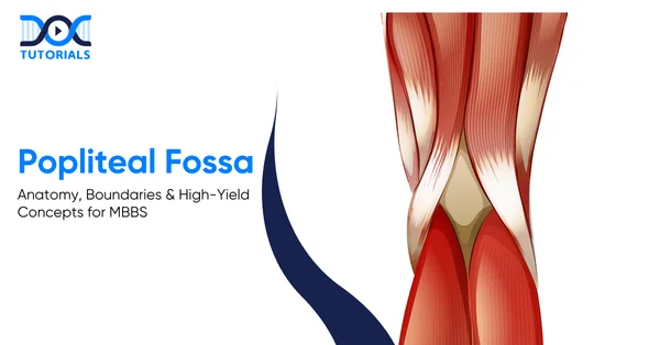
Popliteal Fossa: Anatomy, Boundaries & High-Yield Concepts for MBBS

Popliteal Fossa | DocTutorials

The popliteal fossa is a diamond-shaped depression at the back of the knee that serves as a significant point of transition for vessels and nerves passing between the leg and the thigh. Although it may appear as a simple depression, it contains several structures that are critical for lower-limb function.

For MBBS students, a clear explanation of its layout, relationships, and clinical significance is necessary. Let’s take an in-depth look at the popliteal fossa and its key anatomical and clinical features. Keep reading to learn more.

What is the Anatomy of the Popliteal Fossa?

The popliteal fossa contains the popliteal vessels, sciatic nerve branches, and the short saphenous vein. Its muscular boundaries provide stability, support knee movement, and protect these vital structures.

The popliteal fossa allows the passage of large nerves, arteries, veins, and lymphatics as they travel between the thigh and leg. Some of the structures passing through this region include the terminal branches of the sciatic nerve, the popliteal artery and vein, and the short saphenous vein.

It has a number of muscles that form its borders, and when combined, they form a stable yet flexible area that aids knee movement and protects the fine neurovascular elements. Knowledge of such muscular and fascial boundaries is the basis for clinical examination and surgery.​

What are the Boundaries of the Popliteal Fossa?

The popliteal fossa is bordered superiorly by the semimembranosus, semitendinosus, and biceps femoris, and inferiorly by the heads of the gastrocnemius. Its floor is formed by the distal femur, posterior capsule, oblique popliteal ligament, and popliteus. The popliteal fascia forms the roof and is pierced by the short saphenous vein and sural nerve.

Several muscles contribute to its borders, and together they create a stable yet flexible region that supports knee movement while safeguarding delicate neurovascular elements.

Understanding these muscular and fascial boundaries forms the foundation for clinical examination and surgical approaches:

  1. Superior Boundaries

The superior boundaries of the popliteal fossa consist of:

  • Superomedial: Formed by the semimembranosus and semitendinosus muscles
  • Superolateral: Formed by the long head of the biceps femoris
  1. Inferior Boundaries

The inferior boundaries of the popliteal fossa are:

  • Inferomedial: Medial head of the gastrocnemius
  • Inferolateral: Lateral head of the gastrocnemius (with plantaris occasionally contributing)
  1. Floor

The floor is composed of several structures arranged from superior to inferior:

  • The posterior aspect of the distal femur
  • The posterior capsule of the knee joint
  • The oblique popliteal ligament
  • The popliteus muscle
  1. Roof

The roof is formed by the strong popliteal fascia, which continues upward as the fascia lata and downward as the deep fascia of the leg. This tough fibrous layer acts as a protective cover while maintaining the structural integrity of the fossa.

The short saphenous vein and sural nerve pierce this fascia, making them easily identifiable during surgical procedures.

What are the Nerves in the Popliteal Fossa?

The popliteal fossa contains major superficial nerves that are prone to injury. The tibial nerve runs centrally, supplying posterior leg muscles and contributing to the sural nerve. The common fibular nerve follows the biceps femoris and curves around the fibular neck, where its exposed position makes it especially vulnerable before dividing into superficial and deep branches.

Although the fossa itself contains abundant fatty tissue, it is also home to several significant nerves. These are arranged superficially in the fossa, making them vulnerable to compression or trauma.

The primary nerves include:

  1. Tibial Nerve

The sciatic nerve divides into the tibial and common fibular nerves at the upper angle of the popliteal fossa. The tibial nerve, being the larger branch, travels vertically down the middle of the fossa before entering the posterior compartment of the leg.

It innervates several muscles, including the:

  • Soleus
  • Gastrocnemius
  • Plantaris
  • Popliteus

One of its branches, the medial sural cutaneous nerve, unites with the lateral sural cutaneous branch from the common fibular nerve to form the sural nerve, which supplies the posterolateral aspect of the leg and foot.

  1. Common Fibular Nerve

This nerve courses along the medial border of the biceps femoris before curving around the head and neck of the fibula. Due to its superficial position in this region, it is especially prone to injury. It eventually divides into 2 terminal branches: the superficial and deep fibular nerves.

What are the Blood Vessels of the Popliteal Fossa?

The popliteal artery runs deep in the fossa, giving off the genicular branches that form the knee’s vascular anastomosis. Superficial to it lies the popliteal vein, which becomes the femoral vein after receiving major tributaries.

The vascular structures lie deeper than the nerves, and they include the following:

  1. Popliteal Artery

The popliteal artery represents the continuation of the femoral artery after it exits the adductor hiatus. Within the popliteal fossa, it travels downward and laterally, giving off several branches collectively known as the genicular arteries.

These include:

  • Superior medial genicular artery
  • Superior lateral genicular artery
  • Middle genicular artery
  • Inferior medial genicular artery
  • Inferior lateral genicular artery

These arteries participate in the genicular anastomosis, an elaborate network that ensures continuous blood flow around the knee joint, particularly during flexion. The artery also gives muscular branches to the gastrocnemius, soleus, plantaris, and hamstring muscles.

  1. Popliteal Vein

Lying superficial to the popliteal artery, the popliteal vein forms a part of the deep venous system. As it exits the fossa superiorly, it transitions into the femoral vein. It receives blood from the posterior tibial and peroneal veins as well as the short saphenous vein.

  1. Short Saphenous Vein

This superficial vein ascends along the posterior leg, enters the fossa through the popliteal fascia, and ultimately drains into the popliteal vein. Its course makes it clinically important in venous access and varicose vein assessment.​

What is the Lymphatic Drainage for the Popliteal Fossa?

The popliteal fossa contains superficial and deep lymph nodes. The superficial nodes lie in the subcutaneous tissue and drain the lateral foot and leg via vessels accompanying the short saphenous vein. The deep nodes surround the popliteal vessels and receive lymph from deeper leg and foot structures before draining toward the deep inguinal nodes.

Two groups of lymph nodes are located within the popliteal fossa:

  1. Superficial Popliteal Nodes

Situated within the subcutaneous tissue, they drain lymph from the lateral foot and leg through vessels accompanying the short saphenous vein.

  1. Deep Popliteal Nodes

These nodes surround the popliteal vessels and receive lymph from the deeper structures of the leg and foot. From here, lymph drains toward the deep inguinal lymph nodes.

What is the ​Clinical Relevance of the Popliteal Fossa?

Swelling or pain in the popliteal fossa often results from a Baker’s cyst or a popliteal artery aneurysm. A popliteal aneurysm causes compression of nearby structures, especially the tibial nerve, leading to impaired plantar flexion and sensory changes, often accompanied by a pulsatile mass or bruit.

Swelling or pain in the popliteal fossa can represent a wide range of underlying conditions. Two of the most common causes include:

1. Baker’s Cyst

The popliteal cyst, also known as Baker’s cyst, develops as a result of the enlargement of the semimembranosus bursa, which is frequently caused by chronic inflammation or degenerative changes in the knee joint, such as osteoarthritis.

Even though most cysts spontaneously improve, ruptured Baker’s cysts may be imitations of deep vein thrombosis, and they manifest in the form of calf pain and swelling.

2. Popliteal Artery Aneurysm

A popliteal aneurysm represents a localised dilation of the artery. Due to the non-stretchable nature of the popliteal fascia, any enlargement compresses adjacent structures—particularly the tibial nerve.
Patients may experience:

  • Reduced or absent plantar flexion
  • Numbness or tingling along the foot and posterolateral leg

A pulsatile mass or an audible arterial bruit strongly suggests an aneurysm.

FAQs about the Popliteal Fossa

  1. Which popliteal fossa structure holds the greatest clinical significance?
    The popliteal artery is of paramount importance, as trauma or aneurysmal dilation of this vessel can rapidly compromise perfusion to the lower limb.
  2. Why is the common fibular nerve particularly susceptible to injury?
    Its superficial course around the neck of the fibula renders it highly exposed, predisposing it to injury from fractures, external compression, or even habitual leg-crossing.
  3. How is a popliteal aneurysm generally diagnosed?
    Diagnosis is typically based on the presence of a palpable pulsatile mass, the detection of a bruit on auscultation, and confirmation via Doppler ultrasonography.
  4. What conditions most commonly lead to the formation of Baker’s cysts?
    Baker’s cysts generally arise in the context of chronic intra-articular inflammation, most often associated with osteoarthritis, rheumatoid arthritis, or meniscal pathology.
  5. What is the optimal position for palpating the popliteal artery?
    Palpation is most effective when the knee is gently flexed, a position that brings the artery closer to the surface and facilitates examination.

Conclusion

The popliteal fossa, though often overlooked, is one of the most clinically significant regions of the lower limb. It contains vital nerves, blood vessels, and lymphatics that contribute to the stability, movement, and vascular integrity of the leg.

For MBBS students, mastering the anatomy and the clinical relevance of this region is indispensable. For added guidance, DocTutorials can be your study partner. We offer 3D & 2D animated videos, CBME-based video lectures, and expert mentorship.

Join DocTutorials today and explore our MBBS curriculum to excel in your medical career!

NEET PG Online Live Power Pack
DocTutorials App Download Now

Latest Blogs

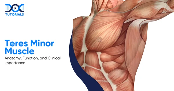
Teres Minor Muscle: Anatomy, Function, and Clinical Importance

Teres Minor Muscle | DocTutorials

The teres minor is the smallest posterior muscle out of the 4 muscles in the rotator cuff (a cluster of four muscles that permits wide mobility of the human shoulder) that connects the scapula to the humerus and primarily assists in external rotation of the arm.

This muscle, although less discussed than other muscles of the rotator cuff, plays a crucial role in stabilising the shoulder, particularly during overhead and rotational movements.

Read below to know more about the anatomy, functions, clinical relevance, and common pathologies of the teres minor muscle in detail.

What is the Teres Minor Muscle and its Anatomy?

The teres minor is a small but essential posterior shoulder muscle that forms part of the rotator cuff and connects the scapula to the humerus. It plays a key role in stabilising the shoulder joint and enabling external rotation and adduction of the arm.

Teres minor is an intrinsic muscle of the shoulder region. It is a crucial part of the rotator cuff muscle group. It is a posterior muscle of the shoulder, which connects the scapula to the humerus.

It, along with other muscles like the supraspinatus, infraspinatus, and subscapularis, helps in controlling shoulder movements. Although it is a small muscle, it cannot be overlooked, since it also plays a prominent role in providing shoulder joint stability.

Rotator muscles of the cuff work cooperatively to hold the head of the humerus fixed inside the glenoid cavity while moving in various directions. Every rotator cuff also has its own set of functions, and among them, the teres minor has its own role in external rotation and adduction of the arm.

What is the Origin of the Teres Minor Muscle?

The teres minor has its origins from the back of the scapula, more precisely from the upper two-thirds part of its lateral border. From there, the muscle fibres turn superolaterally to attach to the greater tubercle on the humerus. The teres minor can cause rotation on the humerus, thus facilitating the control of the movement of the arm.

  • Relations

The teres minor muscle lies superficial to the long head of the triceps brachii and between the large shoulder muscles. The medial aspect of the muscle is partly concealed by the trapezius muscle, and the deltoid muscle covers the superolateral part of the muscle. It relates to the infraspinatus muscle superiorly and to the teres major muscle inferiorly.

As it passes across the shoulder joint, the tendon of teres minor intermingles with the glenohumeral joint capsule on its inferoposterior aspect. This feature is valuable as it stabilises the joint. The muscle also defines the limits of two important axillary spaces.

It forms the superior boundary of the quadrangular space, which carries the axillary nerve and a posterior circumflex humoral artery, and also the superior boundary of the triangular region, through which passes the circumflex scapular artery.

Thus, any injury/compression injury related to this region should not be underestimated because of the critical nerves and blood vessels present.

  • Innervation

The teres minor muscle receives innervation from the axillary nerve, which also innervates the deltoid muscle and derives from nerve roots C5 and C6.

As the teres minor and deltoid muscles share a common nerve, injury to the former may compromise the stability or rotation of the shoulder joint, making weakness of the external rotation an important point not to miss during clinical examination.

  • Blood Supply

The supply of the teres minor muscle is from the circumflex scapular and posterior circumflex humoral arteries.

These arteries provide sufficient supply to this muscle so that it can carry out its roles in stabilising and rotating the shoulder joint during regular movements.

What is the Function of the Teres Minor Muscle?

The teres minor muscle has a major role in shoulder stability as well as shoulder movements. This muscle, together with all the rotator cuff muscles, is associated with shoulder joint stability, as well as maintaining the head of the humerus within a stable position in the glenoid cavity.

The functions of the teres minor muscle are explained in detail below:

1. Shoulder Stability

The muscle forms a functional musculotendinous unit that surrounds the shoulder joint like a sleeve. They work independently to produce specific movements. When they work together, they form a mechanism called concavity compression.

This compresses the humeral head into the glenoid fossa, thereby preventing dislocation. The teres minor supports the posterior aspect of the joint. Thus, the teres minor prevents the posterior displacement of the humeral head.

Without these supports, the shoulder cannot stabilise during the rapid movements that involve elevating the limbs.

2. External Rotation of the Arm

In the anatomical position, the teres minor is a lateral rotator of the shoulder joint. It assists in pulling the greater tubercle posteriorly, along with infraspinatus, in bringing about the rotation of the arm externally.

This is significant because it assists with shoulder functions and exerts force during the pre-load phase of throwing. The muscle contracts eccentrically at the end of this process so that the limb will not suddenly drop.

3. Arm Adduction

When the arm is lifted, the action of arm adduction is aided by the teres minor. Along with the latissimus dorsi and pectoralis major, the teres major assists in lowering the arm from an abducted position.

Clearly, this movement has particular significance in overhead activities and movements such as pulling or throwing, where controlled adduction of the arm helps in efficiently completing these acts.

What are the Pathologies Related to the Teres Minor Muscle?

Pathologies involving the teres minor muscle are relatively uncommon when compared with other muscles of the rotator cuff. However, in the context of an injury, shoulder stability and external rotation are greatly compromised.

The teres minor muscle hasn’t garnered much attention in the context of related pathologies. This is primarily because tears involving the teres minor are far less frequent than those affecting the supraspinatus, infraspinatus, or subscapularis muscles.

Additionally, the teres minor can be involved in specific cases of shoulder denervation injuries, a condition we’re still learning about.

  • Tears of the Teres Minor

Tears of the teres minor generally occur as part of large or massive rotator cuff tears involving multiple tendons. With failure of other rotator cuff tendons, the remaining muscles are exposed to increased mechanical stress, and thus, the teres minor might tear either insidiously or after acute trauma.

These tears can involve the muscle belly, tendon, or the broad muscular attachment to the proximal humerus, and they often present with teres minor pain during shoulder movement. Traumatic tears may also result from shoulder dislocation or subluxation, especially posterior shoulder instability.

In these situations, injury to the teres minor is often linked with posterior capsular disruption and posterior labral tears. Isolated teres minor tears do occur, but are less frequent; most commonly to posterior shoulder trauma.

Diagnosis of this problem is sometimes difficult, and cannot be based on the appearance of the tendon alone, as the tendon may be intact while the muscle belly is torn.

  • Injuries with Shoulder Dislocation

Although anterior shoulder dislocation is more common, the association between injury to the teres minor and posterior shoulder dislocation or subluxation has been well established. Posterior instability events have been documented in studies to involve both the teres minor and the posterior capsule frequently.

In many patients, injury to the teres minor can occur in the absence of injury to other rotator cuff tendons and thus necessitates clinically generated suspicion. These injuries typically have posterior shoulder pain and weakness in external rotation.

Given that posterior instability is not always clinically evident, imaging is highly important for delineating these injury patterns.

  • Denervation Injuries of the Teres Minor

Denervation injury to the teres minor is more common than structural tears. Denervation, often related to axillary nerve injury, is frequently seen on MRI (magnetic resonance imaging) and, depending on the stage of injury, may manifest as musoedema or fatty atrophy.

This has been attributed to quadrilateral space syndrome, in which the axillary nerve is compressed in the quadrilateral space. However, isolated denervation of the teres minor has now been increasingly recognised as a separate entity.

This occurs because the nerve branch to the teres minor follows a vulnerable course along the joint capsule and inferior glenoid rim. This nerve branch can be injured by trauma, repetitive microtrauma from overhead activity, or glenohumeral instability and still spare the deltoid muscle.

The absence of involvement of the deltoid does not exclude a nerve-related pathology.

What are the Clinical Tests for the Teres Minor Muscle?

The tear in the teres minor muscle needs to be evaluated carefully, as its function is important for shoulder joint function. Although imaging studies can help to some extent, they may not always give correct results; thus, clinical examinations remain important and cannot be ignored altogether.

The teres minor should be assessed in patients with massive rotator cuff tears because its integrity strongly influences shoulder function and surgical outcomes. Imaging is helpful, but not always reliable.

Clinical examination still matters in many regards, especially when considering management options. Various tests are used in practice; however, their accuracy varies. 

The following table provides a quick overview of the most appropriate clinical tests for assessing the function of the teres minor:

Clinical TestHow the Test is PerformedWhat a Positive Test MeansClinical Relevance
External Rotation Lag Sign (>40°)Patient sits with elbow flexed to 90° and shoulder slightly elevated in the scapular plane.
• The examiner places the arm in near-maximal external rotation and asks the patient to hold it.
Inability to maintain position with a lag >40°• Most accurate test for teres minor insufficiency.
• High specificity and sensitivity, hence strongly predictive of dysfunction.
Drop Sign• Arm abducted to 90° in the scapular plane, elbow flexed to 90°, shoulder externally rotated to 90°.
• The patient is asked to hold the position against gravity.
Arm drops into internal rotation.• Useful for assessing posterosuperior cuff function.
• Also helps identify the teres minor weakness when the infraspinatus is compromised.
Patte Test• The arm was abducted to 90° in the scapular plane with the elbow flexed.
• Patient performs external rotation against resistance.
External rotation strength < MRC Grade 4Only test that specifically helps isolate the teres minor strength, especially when the infraspinatus is deficient.
Hornblower’s Sign• Arm elevated to 90° in the scapular plane, elbow flexed.
• Patient attempts resisted external rotation (forming a “field-goal” position).
Inability to externally rotateSuggests teres minor tear or dysfunction. Often used as a quick screening tool.

FAQs about the Teres Minor Muscle

1. What conditions frequently affect the teres minor muscle?

Rotator cuff tears, tendinitis, and shoulder impingement syndrome may also affect the teres minor. It might also be involved in quadrangular space syndrome due to compression of the axillary nerve or posterior circumflex humoral artery.

These conditions more often than not present with shoulder pain and weakness, especially with overhead activities.

2. What is the primary function of the teres minor muscle?

It externally rotates the arm at the shoulder joint. The teres minor also assists in stabilising the humeral head within the glenoid cavity, especially during movements that involve medial rotation.

3. What nerve provides innervation to the teres minor muscle?

The teres minor is innervated by the axillary nerve. This arises from the posterior cord of the brachial plexus. It passes through the quadrangular space before supplying the muscle.

4. What blood vessels supply the teres minor muscle?

Teres minor is supplied by the circumflex scapular artery and by the posterior circumflex humeral artery. Both arteries arise from branches of the axillary artery. These vessels pass through defined anatomical spaces near the muscle.

5. How is an injury to the teres minor clinically evaluated?

Hornblower’s sign is utilised to examine the function of the teres minor. The test is considered positive when the patient is unable to externally rotate the shoulder against resistance, thus indicating possible pathology of the teres minor or its involvement in the rotator cuff.

Conclusion

The teres minor muscle is a small but functionally important part of the rotator cuff, contributing to shoulder stability, external rotation, and controlled arm movements. Having a detailed knowledge of the teres minor is essential for MBBS students, as it forms an important basis for evaluating rotator cuff pathology and shoulder dysfunction.

To understand such clinically important anatomy concepts with ease, DocTutorials can be your learning partner. We provide 3D video explanations and structured expert mentorship, and CBME-based video lectures to help MBBS students understand the concepts.

Join DocTutorials today, and explore our MBBS curriculum to excel in your medical career!

NEET PG Online Live Power Pack
DocTutorials App Download Now

Latest Blogs

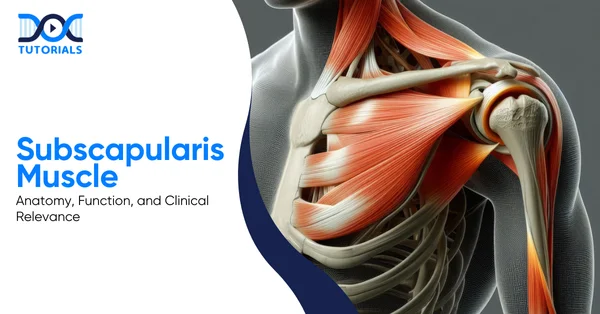
Subscapularis Muscle: Anatomy, Function, and Clinical Relevance

Subscapularis Muscle | DocTutorials

The subscapularis muscle is a very strong and important, though often neglected, portion of the rotator cuff; it is integral to the movement and stabilisation of the shoulder. One important fact is that it’s the only rotator cuff muscle responsible for internal rotation; thus, even a minor injury disrupts regular arm movement.

Understanding of the subscapularis is important for MBBS students, as it relates core anatomy to commonly recommended clinical tests, imaging, and shoulder pathologies in both exams and practice. Throughout this guide, we detail the anatomy, functions, pathologies, and clinical relevance of the subscapularis muscle. 

Keep reading!

What is the Subscapularis Muscle?

The subscapularis is a broad anterior shoulder muscle. It is also a prominent rotator cuff muscle. It is involved in stabilising the head of the humerus. The action of this muscle is unique and powerful, so it cannot be compared with the rest of the cuff muscles. Therefore, it must be tested separately because of the importance of its role in the shoulder joint stabilisation function.

The subscapularis is a broad, triangular muscle of the shoulder. The muscle is located on the anterior or costal surface of the scapula, where it attaches this bone to the humerus. As part of the rotator cuff muscles, which include supraspinatus, infraspinatus, and teres minor, this muscle is very important in maintaining shoulder stability.

The rotator cuff muscles coordinate to stabilise and assist in guiding the humeral head into the glenoid cavity during upper limb movement. However, the action of the subscapularis muscle is unique and powerful; therefore, it should be tested individually during a clinical assessment.

What is the Anatomy of the Subscapularis Muscle?

The subscapularis is the largest muscle in the rotator cuff and is triangular in shape. It has a close association with the axilla, nerves, blood vessels, bursae, and shoulder joint. Additionally, it has an extensive attachment, a strong tendon inserting into the lesser tubercle, a close association with the teres major, and innervation from C5-C6 and axillary roots.

The anatomy of the subscapularis muscle is explained in the sections below: 

Shape and Location

The subscapularis is a strong and triangular muscle in shape, which fills in completely the subscapular fossa of the scapula. It lies posterolateral to the thoracic cage and forms a major part of the posterior wall of the axilla. Since it is so large, it is considered the largest and most powerful muscle of the rotator cuff.

Origin

The subscapularis originates from:

  • The medial two-thirds of the subscapular fossa on the anterior surface of the scapula
  • Tendinous intramuscular septa along the ridges of the fossa
  • An aponeurosis covering the posterior surface of the lateral third of the muscle

Therefore, its origin is broad and able to generate a strong force.

Insertion

The muscle fibres converge laterally to form a thick, rounded tendon. This tendon:

  • Inserts on the lesser tubercle of the humerus
  • Blends with the anterior part of the glenohumeral joint capsule
  • The tendon, near its insertion, is continuous with the tendon of the teres major 

Since the two muscles also share somewhat similar actions and innervation, they are often considered as a functional unit.

A large subscapular bursa, which communicates with the shoulder joint, separates the tendon from the neck of the scapula. This reduces friction; hence, smooth movement is possible.

Relations

The subscapularis forms important anatomical relations; hence, it cannot be ignored in any examination:

  • Anteriorly: Faces the contents of the axilla
  • Superolaterally: Covered by serratus anterior
  • Inferomedially: The relation of coracobrachialis and biceps brachii
  • Centrally: Crossed by cords of the brachial plexus, axillary nerve, axillary artery, and vein

Its posteriorly blending tendon inserts into the fibrous capsule of the shoulder joint and continues with other tendons of the rotator cuff, forming the rotator cuff capsule.

The subscapularis also forms the boundaries of major axillary spaces:

  • Quadrangular space
  • Upper triangular space
  • Lower triangular space, also known as the triangular interval

Since these areas transmit major nerves and vessels, trauma or inflammation within this space has the potential to cause significant complications.

Innervation

The muscle is supplied by:

  • Upper subscapular nerve
  • Lower subscapular nerve

Originating from the posterior cord of the brachial plexus, both nerves carry fibres from C5–C6. The upper nerve supplies the upper part, and the lower nerve supplies the lower part of the muscle. In case of injury to these nerves, internal rotation strength won’t be normal.

Blood Supply

Blood supply is mainly derived from branches of the subclavian system:

  • Subscapular artery (branch of axillary artery)
  • Axillary artery
  • Suprascapular artery

Adequate blood supply is important; compromised vascularity will negatively impact tendon healing efficiency.

Lymphatic Drainage 

Clinically, lymph from the subscapularis muscle flows into the axillary lymph nodes, important during infections or malignancy spread.

What Are the Functions of the Subscapularis Muscle?

The subscapularis primarily produces internal rotation of the shoulder. The action of adduction of the joint also receives a certain amount of assistance from it, so actions such as reaching, walking, or reaching overhead would not be possible without the aid of this muscle. Also, it stabilises the humeral head by compressing the concavity, and if this ligament isn’t functioning properly, shoulder movement will not be normal, as the stability would be missing.

The subscapularis muscle functions are explained below: 

Primary Function: Internal (Medial) Rotation

The major role of the subscapularis muscle is to internally rotate the arm at the glenohumeral joint:

  • Medially rotates the humeral head in the glenoid fossa
  • Engages actions such as reaching across the body and arm swing in walking
  • Can work effectively in different arm positions, including overhead positions

As no other rotator cuff muscle assists in this action, a loss of function in this muscle leads to a weak internal rotation. Therefore, this action is strongly emphasised in both evaluation and rehabilitation.

Adduction of the Arm

To a lesser degree, the subscapularis muscle helps in the addition of the arm:

  • Draws the arm towards the body’s midline
  • Acts in conjunction with internal rotation
  • The sequence of rotation, followed by adduction, is a common sequence observed in normal arm movement during walking.

Otherwise, this is a subtle but important function, since everyday gestures do not exist in isolation.

Shoulder Stability and Concavity Compression

Apart from movement, another role of the subscapularis muscle is to provide stability to the shoulder joint:

  • This assists in concavity compression, which pushes the humeral head into the glenoid cavity
  • Stabilises and prevents anterior and superior migration of the humerus
  • Teaming with other rotator cuff muscles in opposition to the translating forces

But this function assumes a very important role when you do overhead work. When it comes to using heavy muscles such as the deltoid, pectoralis major, biceps, and triceps, this muscle prevents dislocation of the head of the humerus in an upward direction. But in these very functions, this particular muscle is most liable to impingement.

Shoulder and Scapular Movements Coordination

The subscapularis muscle is also responsible for the coordination of movement between the glenohumeral joints and scapulothoracic joints:

  • Facilitates scapular motion to parallel shoulder movements
  • Enables actions such as reaching for objects placed above
  • Acting in coordination with the back muscles, which mainly regulate scapular movement 

Owing to this function, shoulder motion will not have a fluid appearance if the subscapularis is not working properly.

The summary of the functions of the subscapularis muscle is given below in this table: 

FunctionRole of the Subscapularis Muscle
Internal RotationPrimary and exclusive rotator cuff action
Arm AdductionAssists movement toward the body
Joint StabilityPrevents dislocation via concavity compression
Movement CoordinationLinks glenohumeral and scapulothoracic actions

What Are the Pathologies Related to the Subscapularis Muscle?

Subscapularis pathologies include trigger points, tendonitis, and tendon tears, also causing referred pain, weakness, and limited internal rotation, so shoulder function cannot remain normal. Since the tendon endures high mechanical stress, overuse or trauma may lead to tears and instability; hence, early diagnosis and treatment are vital because symptoms won’t settle if ignored. 

Common pathologies associated with the muscular part of the subscapularis include overuse, degeneration, and traumatic injuries. Given the important role played by this muscle during rotation and stabilising functions of the shoulder joint, pathologies can have a marked impact on everyday functions.

Myofascial Trigger Points

The infraspinatus muscle can have myofascial pain sites, which most likely arise because of overuse. The subscapularis may have up to three trigger points. The common sites tend to be in the area towards the outer edge of the muscle. 

Referred pain is usually perceived in:

  • Posterior shoulder
  • Shoulder blade area
  • Back of the upper arm

In certain instances, a characteristic pain pattern may appear in a band form around the wrist. Wrist pain may be attributed by patients, but they do not connect it with a shoulder problem.

Subscapularis Tendonitis and Overuse Injuries

Subscapularis tendonitis can be common in people in jobs that involve constant overhead or throwing motions.

Common characteristics include:

  • Pain and tenderness at the tendon insertion in the inner upper arm.
  • Worsening pain with raising an arm above the level of the shoulder.
  • Discomfort during shoulder movement.

An overworked subscapularis can also present with a feeling of heavy arm or difficulty in lifting the arm. In other instances, it can present with a frozen shoulder because pain limits movement.

Subscapularis Muscle Tear

Subscapularis tendon tears can be considered a serious type of rotator cuff damage because they impact both internal rotation and anterior shoulder stability. The subscapularis tendon is considered to be the largest and most powerful among all rotator cuff muscles. 

As a consequence, heavy mechanical pressure is imposed on this tendon, especially in overhead and throwing motions. Tearing can be a consequence of acute trauma, overuse, and/or degeneration in elderly people. 

Most tears affect the part where this tendon attaches to the humerus. Additionally, they can relate to biceps tendon diseases.

AspectDescription
Common CausesTrauma or overextension (younger individuals); age-related degeneration (older individuals); chronic overuse; shoulder impingement
Site of TearUsually near the tendon insertion on the lesser tubercle of the humerus
General SymptomsAnterior shoulder pain; pain worsening at night; shoulder or arm weakness; pain during arm elevation
Specific Clinical SignsDifficulty reaching behind the back or into the back pocket; biceps weakness; pain beneath the clavicle; passive outward rotation of the arm without voluntary control
Associated FindingsClicking or catching sensation during shoulder rotation; possible biceps tendon involvement
Clinical TestsLift-off test; Bear hug test; Belly press test
ImagingMRI (confirms diagnosis and assesses size and severity of tear)
Conservative TreatmentRest; activity modification; ice or heat; NSAIDs; physiotherapy (preferred for small tears with minimal functional loss)
Surgical TreatmentArthroscopic repair for full-thickness tears or when symptoms haven’t improved after 3–6 months of conservative care
ComplicationsTear progression; muscle atrophy; increased stress on other rotator cuff muscles; secondary biceps pathology
PrognosisGenerally good; most patients regain near-normal shoulder function with appropriate treatment and rehabilitation

What Are the Tests for the Subscapularis Muscle? 

The integrity of the subscapularis is tested with the lift-off, bear hug, and belly press tests, as they emphasise internal rotation strength. Therefore, general rotator cuff testing is not adequate. The impairment in these tests is an indication of tendon dysfunction or tear, and early recognition helps in conservative management.

Clinical tests are essential for assessing the integrity and function of the subscapularis muscle, especially when a subscapularis muscle tear or dysfunction is suspected. These tests mainly evaluate internal rotation strength and help differentiate a torn subscapularis muscle from other rotator cuff pathologies.

Lift-Off Test (Gerber’s Test)

The “lift-off” test, first described by Gerber and Krushell in 1991, is one of the earliest tests and most specific for dysfunction of the subscapularis.

The patient is placed in a standing position, with a request to put the hand behind the back and place the dorsum of the hand over the middle part of the lumbar region. Then, the patient is asked to raise the hand away from the back by rotating the humerus maximally inwards and extending the shoulder.

The indication of a normal test is the capacity to actively raise the dorsum of your hand away from your back.

An abnormal test occurs when the patient can’t lift the hand away, which suggests subscapularis rupture or dysfunction. This usually points toward a significant or complete tear, hence it’s considered highly specific.

Bear Hug Test

The bear hug test is very helpful in assessing a partial tear of the subscapularis tendon.

To carry out this test, the patient holds the palm of the affected side on the shoulder opposite to it, with the elbow in front of the body. The patient is asked to hold this position while a rotational force is applied to the forearm by the examiner.

The test will be considered positive if the patient is unable to do this or if they have weak internal rotation when compared to the other side. This will reveal a tear in, or a weakening of, the subscapularis muscle. Additionally, this particular test will be simpler in patients with difficulty bringing their hands behind their backs.

Belly Press Test

Another assessment is the belly press test, which is especially relevant in circumstances where shoulder mobility is poor.

In this position, the patient’s affected limb is placed on the side with the arm bent at a 90-degree angle at the shoulder. The patient’s palm is placed on the stomach, and they are asked to press it against the stomach through internal rotation.

The test is positive if the patient shows weakness compared to the opposite side or compensates by extending the elbow or shoulder instead of internally rotating the arm. This compensation occurs because the subscapularis isn’t functioning properly, therefore indicating a tear or dysfunction.

After identification of a tear or tendinopathy in the subscapularis muscle, treatment will depend on the level of damage. Subscapularis tendonitis and tendinopathy of the subscapularis tendon can be successfully managed with non-operative methods, which include rest, modification of activity, pain medication, application of ice, and rehabilitation exercises. 

Applying ice can help to ease pain and reduce inflammation. Therefore, it is a common treatment in the early stages.

FAQs about the Subscapularis Muscle

  1. What is a subscapularis muscle tear?

A torn subscapularis muscle is an injury to the tendon of the subscapularis muscle, which attaches to the lesser tuberosity of the humerus. Such a tear can be partial or a full-thickness tear, which means a tear in part of the tendon or in the whole tendon. Such an injury can come about due to overuse, trauma, or degeneration.

  1. Why is the subscapularis important in shoulder stability?

As the subscapularis wraps around the front part of the shoulder joint and attaches to the humerus, this muscle assists in keeping the head of the humerus in the glenoid cavity of the scapula. In this particular function, it is termed concavity compression, where it presses the head into its socket to keep it from dislocation.

  1. What clinical problems can affect the subscapularis muscle?

The subscapularis can be affected in the following conditions:

  • Tendinopathies or overuse injuries, especially in throwers
  • Trigger points that produce referred pain in the shoulder region
  • Partial or complete tears of the tendon, especially in the region around the humeral attachment.

Such issues can lead to a weakness in internal rotation, pain in the shoulder joint, and functional impairment. Tearing and tendinosis are usually assessed via tests and imaging studies.

  1. How is the subscapularis muscle supplied with nerves and blood?

The subscapularis receives its nerve supply from the upper and lower subscapular nerves, which are derived from the posterior cord of the brachial plexus. The blood supply can be traced to various branches of the axillary artery, which include the subscapular and suprascapular arteries.

  1. What are the main functions of the subscapularis muscle?

Its main function is the medial rotation of the arm, which means rotating the arm towards the mid-plane of the body. Medial rotation of the arm is very important in a lot of functions, such as reaching across the chest or holding up a load above the head. Secondly, this muscle assists in maintaining the head of the humerus in the scapular socket to prevent it from coming out of place during a movement. To a smaller degree, it assists in the adduction of the arm to bring it towards the side of the body.

Conclusion

Given the significance of the subscapularis muscle in supporting rotation and stability in the shoulders, any damage to this part can have a great impact on upper limb functions. For MBBS students, the relevance of the subscapularis lies in its connection with different aspects of medicine. 

An understanding of this topic can help in comprehending rotator cuff tears, shoulder pain, and correlations during examinations. Hence, it can in no way be overlooked when learning or during testing.

DocTutorials assists medical students with high-yield video lectures, Qbanks, and revision programmes, which help students master difficult topics quickly. Additionally, it assists in filling the gap between learning and application in medical studies.

Check out our MBBS courses today!

NEET PG Online Live Power Pack
DocTutorials App Download Now

Latest Blogs

Infraspinatus Muscle: Structure, Function, and Clinical Relevance

Infraspinatus Muscle | DocTutorials

The infraspinatus muscle is a vital stabiliser and functional powerhouse of the shoulder joint, forming an integral part of the rotator cuff complex. The anatomy and action of the infraspinatus, along with its clinical implications, form a solid foundation for medical students, both for exams and future clinical practice.

Although small in size compared to larger back and shoulder muscles, its contribution to upper-limb mobility and shoulder joint stability is immense. Keep reading to learn about the infraspinatus muscle, its structure, functions, clinical significance, and more.

What is the Infraspinatus Muscle?

The infraspinatus is one of the most important rotator cuff muscles, which primarily manage external rotation and stabilise the shoulder joint. It originates from the infraspinous fossa and inserts on the greater tubercle of the humerus, playing a vital role in the upper-limb biomechanics.

The infraspinatus is a thick, triangular muscle occupying most of the infraspinous fossa of the scapula and forming an integral part of the rotator cuff. With the supraspinatus, teres minor, and subscapularis, it acts to stabilise the glenohumeral joint.

It is inserted onto the middle portion of the greater tubercle of the humerus, and its innervation is via the suprascapular nerve (C5–6). The suprascapular and circumflex scapular arteries provide blood to it, and its primary function is external rotation of the humerus and assisting in shoulder stability.

What is the Anatomy of the Infraspinatus Muscle?

The infraspinatus is one of the most important rotator cuff muscles, which primarily manage external rotation and stabilise the shoulder joint. It originates from the infraspinous fossa and inserts on the greater tubercle of the humerus, playing a vital role in the upper-limb biomechanics.

The following points break down the anatomy of the infraspinatus muscle:

  • Location and General Structure

The infraspinatus muscle is a thick, triangular muscle that covers most of the infraspinous fossa on the posterior surface of the scapula.

It is one of the four rotator cuff muscles—along with supraspinatus, teres minor, and subscapularis—and plays a major role in stabilising the glenohumeral joint.

  • Origin

It originates from the medial two-thirds of the infraspinous fossa, including fleshy fibres from the fossa itself and tendinous fibres from the ridges running across it.

Additional fibres arise from the infraspinatus fascia, which also separates it from the teres major and teres minor muscles.

  • Insertion and Tendon Pathway

The muscle fibres converge into a narrow tendon that crosses the posterior part of the shoulder joint capsule. This tendon inserts onto the middle facet of the greater tubercle of the humerus and partially blends with the posterior shoulder capsule.

  • Anatomical Relations

The infraspinatus is located deeper into the trapezius and portions of the deltoid and latissimus dorsi. It sits inferior to the supraspinatus and superior to the teres major and minor. As they are separated only by the scapular spine, the supraspinatus and infraspinatus often behave as a functional unit.

  • Bursa and Surrounding Structures

In some individuals, a small bursa lies between the infraspinatus tendon and the shoulder capsule. This bursa may communicate with the glenohumeral joint cavity, facilitating smooth tendon movement during shoulder rotation.

  • Innervation

The muscle is innervated by the suprascapular nerve (C5–C6), originating from the superior trunk of the brachial plexus. The nerve passes through the scapular notch, supplying both supraspinatus and infraspinatus before giving articular branches to the shoulder capsule.

  • Blood and Lymphatic Supply

Arterial supply comes from the suprascapular artery and the circumflex scapular arteries. Lymphatic drainage primarily occurs through the subscapular (posterior) axillary nodes, which are a crucial group in the lymphatic pathways of the shoulder area.

  • Infraspinatus Muscle Action

The infraspinatus is the primary external rotator of the humerus and is crucial for maintaining glenohumeral joint alignment during movement.

Working with the teres minor and the rest of the rotator cuff, it prevents upward displacement of the humeral head. It also contributes to posterior shoulder stability, assists in extension, and helps abduct the inferior angle of the scapula when the arm is fixed.

What are the Functions of the Infraspinatus Muscle?

As a key rotator cuff muscle, the infraspinatus muscle maintains humeral head alignment while enabling efficient rotational and overhead actions. It’s coordinated activity with surrounding muscles ensures smooth, powerful, and safe shoulder mechanics.

Following is a breakdown of the functions of the infraspinatus muscle into clear, captioned explanations for better understanding and MBBS-level retention:

  • Glenohumeral Joint Stabilisation

The infraspinatus acts as part of the rotator cuff musculotendinous unit to maintain the humeral head centralised within the glenoid cavity. It inserts its tendon into the fibrous capsule of the shoulder, contributing to the formation of the rotator capsule.

This provides a concavity compression mechanism that compresses the humeral head into the glenoid fossa and prevents dislocation. This stabilising effect ensures seamless arm movement.

  • External (Lateral) Rotation of the Humerus

The infraspinatus muscle creates strong lateral rotation at the shoulder joint and is considered to be the primary external rotator. This movement is important in activities that require backward extension and preloading of the upper limb, such as throwing or overhead hitting.

Its mechanical axis, in conjunction with its large humeral footprint, makes it one of the most powerful contributors to external rotation, working synergistically with the teres minor.

  • Control of Humeral Head Position During Abduction

During abduction of the shoulder, an upward pull on the humeral head is provided by the deltoid. The infraspinatus provides a downward and stabilising force in cooperation with the teres minor, thus preventing upward migration and impingement against the coracoid process.

While the supraspinatus acts to some degree, it is not known precisely to what extent, so the infraspinatus-teres minor pair constitutes the principal stabilisers for this action.

  • Anterior–Posterior Force Balance

The infraspinatus provides the posterior stabilising force in the glenohumeral joint, balancing the anterior pull of the subscapularis. These muscles, together, prevent anterior or posterior translation of the humeral head and allow coordinated, effective shoulder movements.

A deficiency or tear in the infraspinatus would disrupt the balance and often result in the elevation of the humeral head, coupled with the reduced effectiveness of deltoid-driven abduction.

  • Eccentric Control During Overhead Movements

During overhead actions at high velocity, such as throwing, the infraspinatus contracts eccentrically to decelerate the arm and prevent excessive internal rotation. This fine-tuned braking function protects the shoulder against injury and ensures smooth termination of powerful movements.

  • Contribution to Scapular Motion (Scaption)

When the shoulder is in anatomical position, the infraspinatus assists in the abduction of the scapula’s inferior angle, providing a motion described as scaption. This action increases scapular mobility, allowing full shoulder abduction by optimising the position and movement of the scapulothoracic joint.

  • Role in the Rotator Cuff Force-Couple System

The muscles of the rotator cuff are nonlinear, viscoelastic tendons capable of producing balanced opposing forces around the shoulder. The infraspinatus contributes to this dynamic force-couple system through its collagen-rich tendon and its alignment with the other cuff muscles. This ensures optimal biomechanics during complex, multi-plane shoulder actions.

  • Support in the Movements of Shoulder Extension

Although this is not a ‘prime’ extensor, the infraspinatus assists in shoulder extension by providing a supportive backward force that helps coordinate posterior chain actions, involving the latissimus dorsi and posterior deltoid, when external rotation is combined with retraction movements.

FAQs about the Infraspinatus Muscle

  1. What is the primary function of the infraspinatus muscle?

The infraspinatus, as a rotator cuff muscle, plays a crucial role in shoulder movement and contributes to glenohumeral stability. It exerts compression on the humeral head, which facilitates the stabilisation of the humeral head during shoulder abduction.

  1. Which nerve is responsible for controlling the infraspinatus muscle?

The suprascapular nerve provides both motor and sensory innervation. Its motor function targets the supraspinatus and infraspinatus muscles.

  1. Is it necessary to stretch the infraspinatus muscles?

Yes, individuals need to stretch their infraspinatus muscles to keep shoulder injuries at bay.

  1. What are the injuries that can affect the infraspinatus?

The infraspinatus muscle can be injured through activities that involve repetitive overhead, overloading of the shoulder, or even episodes of shoulder instability.

Most cases of overuse in tennis, swimming, baseball, and volleyball result in strains or partial tears of the tendon. Chronic mechanical irritation or degeneration of the infraspinatus might also arise in individuals with heavy manual work.

  1. What is infraspinatus muscle pain?

Infraspinatus muscle pain is a type of discomfort that occurs at the back or top of the shoulder and may radiate towards the front and side of the upper arm or shoulder blade. This discomfort can worsen if left untreated.

Conclusion

The infraspinatus muscle is a structure of immense functional and clinical importance, especially for medical students studying musculoskeletal anatomy and shoulder biomechanics.

Understanding its anatomy, action, nerve supply, and clinical relevance not only improves academic scores but also lays a foundation for sound clinical decision-making in the future. For added guidance, DocTutorials can be your study partner. We offer 3D animated video lectures, structured mentorship by experts, and much more.

Join DocTutorials today and explore our MBBS curriculum to excel in your medical career!

NEET PG Online Live Power Pack
DocTutorials App Download Now

Latest Blogs

Urea Cycle Steps: Mechanism, Regulation, and Clinical Significance

Urea Cycle Steps | DocTutorials

The urea cycle is a fundamental metabolic pathway that detoxifies ammonia, a byproduct of protein metabolism, by converting it into urea for safe excretion. Having comprehensive knowledge of this cycle is crucial for a medical student as it bridges biochemistry, physiology, and clinical medicine.

From the liver’s organ-level role to the molecular interplay among enzymes, the urea cycle is essential to mastering nitrogen metabolism and related disorders. Let’s explore the overview of this cycle, its step-by-step mechanism, and regulation.

Keep reading to learn more.

Urea Cycle: An Overview

The urea cycle is a biochemical pathway in the liver that converts toxic ammonia into urea for safe excretion. The cycle involves cooperation between mitochondrial and cytosolic enzymes for the detoxification of waste nitrogen.

The urea cycle, also known as the ornithine cycle, is a sequence of biochemical reactions in the liver in which highly toxic ammonia, produced by amino acid breakdown, is converted to urea, a safe, water-soluble compound excreted via the urine.

First described by Hans Krebs and Kurt Henseleit in 1932, it operates principally in periportal hepatocytes, using both mitochondrial and cytosolic enzymes. Since mammals and amphibians cannot excrete ammonia directly, this pathway is paramount in preventing nitrogen toxicity.

The cycle is irreversible and requires ATP (adenosine triphosphate). The key enzymes involved include CPS-I, ornithine transcarbamylase, argininosuccinate synthetase, argininosuccinate lyase, and arginase.

What are the Steps of the Urea Cycle?

The urea cycle consists of five enzymatic steps that convert toxic ammonia into urea for safe excretion. It starts in the mitochondria and concludes in the cytosol of liver cells. Knowledge of these steps is important for MBBS students to master the biochemical basis of nitrogen detoxification.

The urea cycle is a five-step biochemical pathway in hepatocytes that detoxifies ammonia by converting it into urea. The steps are discussed below:

  • Step 1: Formation of Carbamoyl Phosphate (Mitochondria)

Ammonia, CO₂, and 2 ATP combine to form carbamoyl phosphate, catalysed by Carbamoyl Phosphate Synthetase I (CPS-I). It requires N-acetylglutamate (NAG) as an essential activator.

Reaction: NH₃ + CO₂ + 2ATP → Carbamoyl phosphate + 2ADP + Pi

  • Step 2: Formation of Citrulline (Mitochondria → Cytosol)

Carbamoyl phosphate transfers its carbamoyl group to ornithine, forming citrulline, catalysed by Ornithine Transcarbamylase (OTC). Citrulline is then transported into the cytosol by ornithine translocase.

Reaction: Carbamoyl phosphate + Ornithine → Citrulline + Pi

  • Step 3: Formation of Argininosuccinate (Cytosol)

Citrulline condenses with aspartate in an ATP-dependent reaction catalysed by Argininosuccinate Synthetase. This adds the second amino group to urea.

Reaction: Citrulline + ATP + Aspartate → Argininosuccinate + AMP + PPi

  • Step 4: Cleavage to Arginine and Fumarate (Cytosol)

Argininosuccinate Lyase splits argininosuccinate into arginine and fumarate. Fumarate enters the TCA cycle, linking the two pathways.

Reaction: Argininosuccinate → Arginine + Fumarate

  • Step 5: Formation of Urea and Regeneration of Ornithine (Cytosol)

Arginase hydrolyses arginine to produce urea and ornithine. Ornithine re-enters the mitochondria to restart the cycle.

Reaction: Arginine + H₂O → Urea + Ornithine

Overall Reaction of the Urea Cycle

NH₃ + CO₂ + Aspartate + 3 ATP + H₂O →

Urea + Fumarate + 2 ADP + Pi + AMP + PPi

What is the Significance of the Urea Cycle?

The urea cycle is essential for detoxifying ammonia, a highly toxic by-product of protein metabolism. Its proper functioning prevents hyperammonemia and maintains metabolic balance. Understanding the importance of the urea cycle helps explain its clinical consequences and the associated disorders resulting from enzyme deficiencies.

The urea cycle is critical for maintaining nitrogen balance and preventing toxic accumulation of ammonia in the body. Mentioned below are the key reasons why it is so important:

  • Ammonia Detoxification: Highly toxic ammonia is converted into urea, thus preventing accumulation and protecting the brain and other organs from irreversible damage.
  • Major Pathway for Nitrogen Excretion: Accounts for almost 80% of nitrogenous waste excretion, ensuring nitrogen is eliminated safely through urine.
  • Prevention of Hyperammonemia: A functional cycle prevents dangerous conditions such as hyperammonemia. This condition can lead to vomiting, lethargy, coma, convulsions, and even death in newborns.
  • Clinical Relevance of Enzyme Deficiencies: Deficiencies in key enzymes (CPS-I, OTC, ASS, ASL, or arginase) result in urea cycle disorders, which are often misdiagnosed as septicemia and treated with unnecessarily high doses of medication.
  • Support for Biosynthesis: Provides intermediates necessary for the recycling of TCA cycle components, amino acids, and keto acids, thereby linking energy metabolism with nitrogen metabolism.
  • Daily Ammonia Clearance: Ensures the safe removal of 10–20 g of ammonia that is produced daily in a healthy adult through normal protein turnover.

FAQs about Urea Cycle Steps

  1. What activates the urea cycle?

N-acetylglutamate (NAG) activates the urea cycle by activating carbamoyl phosphate synthetase I, the rate-limiting enzyme of the pathway. NAG levels rise after a protein-rich meal, helping the liver detoxify increased ammonia more efficiently.

  1. What is Ornithine Transcarbamylase (OTC) deficiency?

OTC deficiency represents the only X-linked recessive disorder among the urea cycle disorders.

As a result of this disorder, carbamoyl phosphate accumulates and is diverted into pyrimidine synthesis, forming orotic acid, which appears in blood and urine and is often observed as orange crystals in infant diapers.

  1. What is Argininosuccinate Synthetase Deficiency?

Also known as Citrullinemia Type I, this autosomal recessive disorder presents with elevated citrulline and ammonia. This disorder presents with seizures, lethargy, and respiratory distress in infants, while adults experience much milder symptoms like vomiting, ataxia, and lethargy.

  1. How are urea cycle defects diagnosed?

Diagnosis is often based on a plasma amino acid profile, which, in the case of a defect, may show elevated glutamine, alanine, citrulline, or argininosuccinate. OTC deficiency is suggested by elevated urinary orotic acid. Monitoring for ammonia is necessary if unexplained neurological symptoms occur.

  1. In which areas is the concept of the urea cycle steps applied?

The concept of the urea cycle steps is used in fields such as medicine (diagnosing metabolic disorders and liver function), genetic counselling (inherited urea cycle disorders), and environmental science (understanding nitrogen metabolism).

Conclusion

Mastering the urea cycle helps medical students correlate metabolic pathways with clinical practice, particularly in understanding ammonia detoxification and its disorders. The mechanisms, regulation, and clinical implications constitute the necessary knowledge base for MBBS students.

DocTutorials makes complex biochemical pathways easier to understand and remember through 3D-animated videos and structured mentorship and guidance from experts.

Reinforce your concepts, improve exam scores, and boost your medical journey—opt for DocTutorials’ MBBS curriculum and build the confidence to excel as a future doctor.

NEET PG Online Live Power Pack
DocTutorials App Download Now

Latest Blogs

Complete MBBS Subject List: Syllabus, Year-Wise Subjects and Best Books

Complete mbbs subject List | DocTutorials

The MBBS course was designed to transform learners into efficient medical professionals. Since medicine can never be learned in isolation, the course begins with the basic sciences and proceeds to clinical subjects.

Therefore, having a clear understanding of the MBBS syllabus and subject distribution is essential from the start. It helps students gain some knowledge of what they are facing and reduces confusion and stress associated with academics.

Keep reading to learn more about the comprehensive list of MBBS subjects and the syllabus and textbooks associated with them.

What is the MBBS Syllabus and Subjects Structure?

The MBBS course is of 5.5 years, including semesters and three phases, and hence, students cannot directly enter into practicals and must develop robust basics first. It comprises 19 subjects, categorised into core and elective streams, which ensures a well-rounded development of a medical professional.

The course structure for the MBBS programs includes the establishment of a sound foundation in medical science and the subsequent exposure to clinical practice. The course consists of a total of 5.5 years; this refers to the study period of 4.5 years and also includes a mandatory single-year internship at a hospital.

The MBBS course structure follows a semester pattern with each semester lasting 6 months. The MBBS curriculum is divided into 3 phases: the Pre-Clinical phase, the Para-Clinical phase, and the Clinical phase, which together deliver education in 19 disciplines. The curriculum also divides study topics into core and elective courses.

What are the 19 Subjects of MBBS?

The MBBS course structure also contains a list of 19 subjects that need to be covered during the entire duration of the course. The subjects are divided into several phases and semesters.

The 19 subjects for MBBS are:

  1. Anatomy
  2. Biochemistry
  3. Physiology
  4. Pathology
  5. Pharmacology
  6. Microbiology
  7. Community Medicine
  8. Forensic Medicine and Toxicology
  9. ENT (Otorhinolaryngology)
  10. Ophthalmology
  11. Psychiatry
  12. Dermatology and Venereology
  13. Pediatrics
  14. Orthopaedics
  15. Obstetrics and Gynaecology
  16. Anesthesiology
  17. Medicine
  18. Surgery
  19. Clinical postings in Wards and OPDs

What are the Elective Subjects in MBBS?

Elective subjects taught in MBBS programs orient a student to a particular area of medicine, in addition to his/her core training. The elective subjects that are considered in the MBBS syllabus are:

  • Psychiatry
  • Anaesthesia
  • Radio-diagnosis
  • Blood Bank & Transfusion
  • Forensic Medicine & Toxicology

What is the Phase-Wise MBBS Subject List?

The MBBS curriculum is structured in 3 phases: Pre-Clinical, Para-Clinical, and Clinical.  This allows students to move seamlessly from learning the basics to studying diseases and disorders, helping them acquire practical clinical knowledge rather than mere classroom learning.

The list of MBBS subjects is segregated into three distinct phases. This ensures that the students are able to lay a strong foundation first and only then move on to disease-oriented subjects, and finally develop hands-on clinical skills.

As all phases are connected, students cannot skip or rush any phase. The phases in the MBBS syllabus include:

  • Pre-Clinical Phase: 1 year
  • Para-Clinical Phase: 1 year
  • Clinical Phase: 2.5 years

The phase-wise subject list for MBBS is given in the table below:

PhaseCore Subjects
Pre-Clinical PhaseAnatomy, Physiology, Biochemistry
Para-Clinical PhasePathology, Microbiology, Pharmacology, Forensic Medicine, Community Medicine
Clinical PhaseMedicine, Surgery, Paediatrics, Obstetrics, Gynaecology, Ophthalmology, Otorhinolaryngology (ENT), Orthopaedics, Dermatology, Venereology and Leprosy, Psychiatry, Anaesthesiology, Radiology

These are the 19 MBBS subjects studied by students during this course. Every stage is based on a certain aspect of MBBS education; thus, students are not burdened with everything right from the initial stages.

What is the Phase-Wise Exam Pattern for MBBS?

The MBBS course has a structured system of exams that test not only theoretical concepts but also practical skills.

  • Each semester typically takes about 6 months, with 120 teaching days
  • Mid-semester, end-semester, and clinical posting assessments
  • Internal evaluations are allotted 50% weightage in both theory and practical/clinical exams
  • After successful completion of all academic stages, there is a 1-year compulsory rotatory internship that students must participate in

This phase-wise system ensures a smooth transition from textbooks to patients. It also enables students to apply the knowledge they have gained in practical settings, and therefore, they can face the realities of medical practice.

Since the process is gradual, students don’t base their knowledge solely on memorisation, as they also need to develop clinical acumen.

What is the Semester or Year-Wise MBBS Subject List?

The MBBS program follows a clear year- and semester-wise structure, so students move gradually from basic sciences to advanced clinical subjects without being overwhelmed. Since clinical postings run alongside academics from early years, students can’t skip any stage and can strengthen their understanding of patient care.

The MBBS program is designed on a yearly and semester basis to provide a seamless transition from basic sciences to clinical work. Concepts are introduced incrementally to allow students to gather knowledge layer by layer without burdening them at a very early stage of their academic journey.

 In this way, since a particular academic year defines a particular phase of learning, things cannot be skipped in a particular stage by students. During the MBBS course, clinical postings also take place simultaneously, thus ensuring early practical exposure to hospitals.

This enables them to apply what they have learnt and thus enhances their understanding of clinical subjects.

What are the MBBS Subjects in the 1st Year?

The first year of MBBS is based on pre-clinical courses. These courses are considered basics in MBBS. Pre-clinical subjects provide adequate information on how the human body is structured and functions.

MBBS SubjectDescription
AnatomyMicroanatomy, Embryology, and Genetics
BiochemistryMolecular Biology, Biological Cells, Hormones, Metabolic Pathways, Enzymes
PhysiologyGeneral Physiology, Gastrointestinal System, Cardiovascular System, Respiratory System

What are the MBBS Subjects in the 2nd Year?

The second year encompasses para-clinical subjects, marking the onset of exposure to clinics. The students are also permitted to visit the posting and OPDs, making the learning process more practical.

MBBS SubjectDescription
PharmacologyGeneral pharmacology, drug dosages, drug formulations, and drug calculations
PathologyGeneral pathology, systemic pathology, practical pathology
MicrobiologyBacteriology, virology, sterilisation, vaccines, and host-parasite relationships
Clinical Postings (Inwards)12 weeks of clinical postings focused on community medicine
OPDExposure to outpatient care for patients not admitted as inpatients

What are the MBBS Subjects in the 3rd Year?

The third year involves key clinical topics and community-based learning in healthcare. At this point, students are confident with patient interaction.

MBBS SubjectDescription
Community MedicinePopulation health and services provided at primary healthcare centres
Forensic Medicine and ToxicologyForensic medicine, toxicology
ENT (Otorhinolaryngology)Disorders of the ear, nose, throat, head, and neck
OphthalmologyDiagnosis and treatment of eye disorders

What are the MBBS Subjects in the Final Year?

The last year has been more clinically focused and has concentrated on the important specialities. The learning now incorporates the knowledge from the previous years because it is now centred on patient care.

MBBS SubjectsDescription
PsychiatryPsychiatric disorders, behavioural sciences, cognitive processes, OCD
MedicineStudy and treatment of disease.
Dermatology and VenereologySkin diseases and venereal diseases
PaediatricsDiseases of infants and children
AnesthesiologyCannulation, airway insertion techniques
OrthopaedicsOrthopaedic neurology, paediatric orthopaedics, spine disorders, fractures
Obstetrics and GynaecologyObstetrics, gynaecology, neonatology, contraception

What are the Popular MBBS Books and Authors?

MBBS students have access to a wide range of textbooks that cover both theoretical concepts and clinical applications. These books are widely recommended because they explain concepts clearly and align well with the syllabus.

The selection of textbooks is important because students cannot read all textbooks simultaneously; rather, they have to rely on credible sources.

Thus, the books and authors mentioned below have been regarded as common textbooks of most Indian medical universities.

Popular MBBS Books (General Recommendations)

The general recommendations for MBBS books are given below:

Book NameAuthor
Grey’s AnatomyRichard L. Drake, Wayne Vogl, Adam W.M. Mitchell
Guyton and Hall Textbook of Medical PhysiologyArthur C. Guyton, John E. Hall
Harper’s Illustrated BiochemistryRobert K. Murray et al.

Subject-Wise Popular MBBS Books and Authors

The subject-wise popular MBBS Books that most students read are given below:

SubjectBook NameAuthor
AnatomyCunningham’s Manual of Practical Anatomy (Vol I–III)G.L. Romanes
Textbook of Human HistologyInderbir Singh
Medical EmbryologyJan Langman
The Human Nervous System: An Anatomical View PointMurray L. Barr & Kiernan
Surface and Radiological AnatomyA. Halim
Thomson and Thomson Genetics in MedicineRobert L. Nussbaum et al.
BiochemistryPrinciples of BiochemistryLehninger, Nelson & Cox
Tietz Textbook of Clinical ChemistryBurtis & Ashwood
PhysiologyUnderstanding Medical PhysiologyR.L. Bijlani
Review of Medical PhysiologyW.F. Ganong
Forensic MedicineEssentials of Forensic Medicine & ToxicologyK.S.N. Reddy
Principles of Forensic MedicineApurba Nandy
MicrobiologyMedical MicrobiologyGreenwood, Slack & Peutherer
Jawetz, Melnick & Adelberg’s Medical MicrobiologyGeo F. Brooks et al.
PathologyTextbook of PathologyHarsh Mohan
Pathological Basis of DiseaseRobbins
PharmacologyGoodman & Gilman’s Pharmacological Basis of TherapeuticsGoodman & Gilman
Basic & Clinical PharmacologyBertram G. Katzung
Community MedicineTextbook of Preventive MedicinePark
Methods in BiostatisticsB.K. Mahajan
MedicinePrinciples of Internal MedicineHarrison
Principles and Practice of MedicineDavidson
SurgeryShort Practice of SurgeryBailey & Love
PediatricsEssentials of PediatricsO.P. Ghai et al.
Obstetrics & GynaecologyTextbook of Obstetrics for UndergraduatesV.L. Bhargava
OrthopaedicsOutlines of OrthopaedicsCrawford Adams
ENTTextbook of ENTLogan Turner
OphthalmologyClinical OphthalmologyKanski
PsychiatryTextbook on PsychiatryNeeraj Ahuja

Popular MBBS Books in AIIMS

At AIIMS (All India Institute of Medical Sciences), the learning atmosphere is quite tough and competitive. The students are expected to use text guides with very comprehensive and clinical conceptual knowledge.

The following are the recommended text guides used by AIIMS students for preparation of MBBS entrance exams:

Book NameAuthor
Understanding Medical PhysiologyR.L. Bijlani
Essentials of Forensic Medicine & Toxicology (21st Edition)K.S.N. Reddy
Textbook of Forensic Medicine & ToxicologyKrishan Vij
Medical MicrobiologyGreenwood, Slack & Peutherer

FAQs about the MBBS Subject List

1. What is the pattern of the 5.5-year MBBS course?

The duration of the MBBS program is 5.5 years, with the last year as an internship, which is compulsory. The next 4.5 years involve classroom sessions and practical training. During this time, the student undergoes several sets of written tests, practical evaluations, and clinical evaluations.

2. Which books should be followed in the 2nd year of MBBS?

The second year of MBBS deals with para-clinical and early clinical subjects. It is relevant to pay attention to proper textbook selection for these subjects. Students cannot merely depend on class notes and have to stick to standard reference books.

Below are some of the top MBBS books for 2nd year students:

  • Robbins Basic Pathology by Vinay Kumar
  • Guyton & Hall Textbook of Medical Physiology by John E. Hall
  • Harrison’s Principles of Internal Medicine by Dennis L. Kasper
  • Clinical Medicine Parveen Kumar & Michael L. Clark
  • Pharmacology by K.D. Tripath

3. What is the importance of research in the MBBS syllabus?

Research is an integral part of the MBBS syllabus, as it enhances critical thinking skills in students. It also increases clinical knowledge and helps in practising evidence-based medicine.

As new advancements are taking place in modern medicine on a constant basis, students cannot be dependent on conventional learning patterns alone.

4. What is the internship period for MBBS students?

The MBBS course is spread over 5.5 years, out of which the final year is dedicated to internship, which is compulsory. Qualified doctors supervise students during their internship.

They are involved in diagnosing, treating, and taking care of the patients, hence acquiring direct practical knowledge. The internship acts as a link between academics and independent practice.

5. How many subjects are there in the entire MBBS course?

The student is required to study a total of 19 basic subjects. The subjects encompass a broad curriculum of different areas in the medical field, from basic sciences to specialities.

Attending classes for each subject is essential, as each one of them contributes to a different area of expertise in providing medical care.

Conclusion

Understanding the entire MBBS syllabus and subject pattern is crucial for every medical student, as it is the blueprint of their entire medical journey. Therefore, knowing the subjects and books for each year helps plan accordingly and avoid unnecessary academics.
DocTutorials help simplify complex subjects through structured video lectures, expert guidance, and high-yield study resources. Join DocTutorials today and explore our MBBS curriculum to excel in your medical career!

DocTutorials App Download Now

Latest Blogs

NEET PG Online Live Power Pack
DocTutorials App Download Now