The Ultimate PG Resident’s Guide to Dysmorphic Syndromes and CAR T Cell Therapy
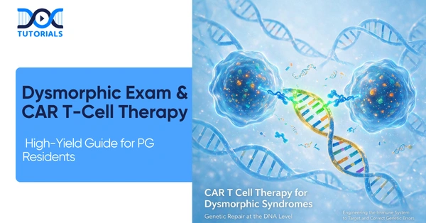
Mastering clinical genetics, dysmorphic assessments, and advanced molecular treatments like CAR T Cell Therapy is no longer optional for today’s post-graduate (PG) medical residents. This comprehensive guide connects foundational pediatric bedside examinations with the cutting-edge frontiers of gene modification and CAR T Cell Therapy.
Get High Quality, High Yield PG Residency Notes for Free!
The Foundation of Dysmorphic Examination
Evaluating a child for genetic syndromes begins with a meticulous general assessment and anthropometry. Understanding foundational metrics helps clinicians identify underlying genetic abnormalities before moving to advanced treatments like CAR T Cell Therapy.
Anthropometry and Crucial Metrics
A non-negotiable metric for any syndromic assessment is the Upper Segment to Lower Segment (US:LS) ratio. This ratio must always be correlated with age-specific norms. As a rule for all dysmorphic children, clinicians must document the US:LS ratio even if it falls within normal limits. Alongside this, the clinical checklist should include assessing consciousness, vital signs, and malnutrition staging (acute, chronic, or acute-on-chronic). A rapid screen for general signs like pallor, icterus, cyanosis, and clubbing is also essential.
Head, Skull, and Facial Features
Plotting the occipitofrontal circumference (OFC) on a growth chart determines whether a patient has microcephaly, macrocephaly, or normal head growth. Furthermore, analyzing the anterior fontanel for size, shape, and bulging (if open) is critical. Skull shapes vary significantly in craniosynostosis: scaphocephaly presents with increased AP diameter, while brachycephaly shows increased biparietal diameter.
Facial features provide immense diagnostic clues. For instance, an upward lateral canthus (mongoloid slant) is associated with Down Syndrome, whereas a downward lateral canthus (antimongoloid slant) points to Noonan Syndrome.
Below is a table summarizing key facies types and their associated conditions.
Common Facies and Associated Syndromes
| Facies Type | Key Clinical Features | Associated Conditions |
| Coarse Facies | Thick lips, flat bridge, large tongue | MPS, Cretinism |
| Elfin Facies | Wide mouth, smooth philtrum, pointed teeth | Williams Syndrome |
| Hemolytic Facies | Frontal bossing, crowded teeth | Thalassemia |
| Other Facies | Moon, Hepatic, Mask-like | Cushing’s, CLD, Moebius |
The Evolution of Gene Therapy Strategies
While physical examinations identify the phenotypic expressions of genetic disorders, the treatment landscape is rapidly shifting toward molecular interventions, laying the groundwork for complex modalities like CAR T Cell Therapy. Gene therapy strategies are broadly categorized into In Vivo and Ex Vivo mechanisms.
In Vivo Therapies
In Vivo gene therapy involves direct administration inside the body, where gene modification occurs internally. The primary methods for delivery include viral vectors (like AAV), nanoparticles, and RNA/Ribosomes.
How do Antisense Oligonucleotides (ASO) function in the treatment of Duchenne Muscular Dystrophy (DMD)?
In DMD, which is an X-linked recessive disorder characterized by deficient dystrophin, ASO therapy works via exon skipping. The ASO binds to the pre-mRNA and masks specific exons—such as skipping Exon 51 with Eteplirsen—to bypass the mutation stop codon, ultimately allowing translation into a shorter but functional dystrophin protein.
In Vivo delivery systems heavily rely on advanced transport mechanisms, such as GalNAc conjugates which are receptor-dependent and liver-specific (administered subcutaneously), and Lipid Nanoparticles (LNP) which utilize non-receptor dependent entry via membrane fusion or endocytosis.
In Vivo ASO Therapeutics Overview
| Disease | Drug Name | Gene Target | Route of Administration |
| Spinal Muscular Atrophy | Nusinersen (Spinraza) | SMN2 Gene | Intrathecal |
| TTR-Amyloidosis | Inotersen | Transthyretin (TTR) | Subcutaneous |
| Hyperlipidaemia | Mipomersen | Apolipoprotein B100 | Subcutaneous |
| Familial Chylomicronaemia | Volanesorsen | Apolipoprotein C3 | Subcutaneous |
The Breakthrough of CAR T Cell Therapy (Ex Vivo)
Ex Vivo gene therapy takes cells harvested from the patient, modifies them in a laboratory setting, and returns them to the patient. This framework is exactly how CAR T Cell Therapy operates.
CAR T Cell Therapy is a revolutionary immunotherapy primarily utilized in pediatrics for the treatment of Acute Lymphoblastic Leukaemia (ALL). The process of CAR T Cell Therapy begins with the isolation of the patient’s own T-cells. Once isolated, these cells undergo modification ex vivo to express the Chimeric Antigen Receptor (CAR).
After being successfully modified to become CAR T-cells, they are expanded in the laboratory and subsequently re-infused into the patient’s bloodstream. The core action of CAR T Cell Therapy is to grant specific cytotoxic activity against target cancer cells. By leveraging the body’s own immune system, CAR T Cell Therapy ensures the direct and targeted destruction of leukemia cells.
The integration of lentiviral vectors is frequently utilized in these Ex Vivo additions to ensure functional gene expression. Thus, CAR T Cell Therapy represents a paradigm shift in how hematological malignancies are treated in pediatric patients.
Advanced Ex Vivo Gene Editing: CRISPR/Cas9
Beyond CAR T Cell Therapy, Ex Vivo strategies also utilize CRISPR/Cas9 technology, functioning as “molecular scissors.” A prominent example is the treatment for Sickle Cell Anaemia using Exagamglogene Autotemcel (Exa-cel/Casgevy). This mechanism involves the inactivation of the BCL11A gene, which serves as the switch that normally stops fetal hemoglobin (HbF) production. The resulting increase in HbF successfully prevents red blood cell sickling and subsequent crises.
However, much like the stringent monitoring required after CAR T Cell Therapy, CRISPR therapies demand careful safety oversight. Clinicians must monitor for off-target effects and potential malignant transformations, as these represent permanent genetic changes compared to treatments like Hydroxyurea.
Frequently Asked Questions (FAQs)
1. What is the primary pediatric indication for CAR T Cell Therapy?
The primary pediatric use for CAR T Cell Therapy is the treatment of Acute Lymphoblastic Leukaemia (ALL).
2. How are cells modified in CAR T Cell Therapy?
In CAR T Cell Therapy, a patient’s T-cells are isolated, modified ex vivo to express Chimeric Antigen Receptors (CAR), expanded, and then re-infused.
3. What is the mechanism of action for CAR T Cell Therapy?
CAR T Cell Therapy grants the modified T-cells specific cytotoxic activity directed against target cancer cells, leading to cancer cell destruction.
4. How does CAR T Cell Therapy differ from In Vivo gene therapy?
CAR T Cell Therapy is an Ex Vivo method, meaning cells are harvested, modified outside the body in a lab, and then returned. In Vivo therapy involves direct administration and internal modification.
5. What is the US:LS ratio in dysmorphic examinations?
The Upper Segment to Lower Segment (US:LS) ratio is a non-negotiable metric essential for all dysmorphic children and must correlate with age-specific norms.
6. Which condition is indicated by an upward lateral canthus?
An upward lateral canthus, also known as a mongoloid slant, is a feature associated with Down Syndrome.
7. How is Spinal Muscular Atrophy (SMA) treated using gene therapy?
SMA is treated using Nusinersen (Spinraza), an In Vivo therapy administered intrathecally that activates the inactive SMN2 gene to compensate for the missing SMN1.
8. What vectors are used in Ex Vivo therapies like CAR T Cell Therapy?
Ex Vivo therapies often utilize Lentiviral vectors for the laboratory integration of functional genes.
9. How does CRISPR/Cas9 help Sickle Cell Anaemia patients?
It inactivates the BCL11A gene, increasing Fetal Hemoglobin (HbF) production, which prevents sickling and crises.
10. What dermatological marker indicates Tuberous Sclerosis?
The presence of an Ash Leaf Macule (a hypopigmented spot) is a hidden dermatological marker for Tuberous Sclerosis.

Latest Blogs
-

NEET SS Exam 2024: Analysis, Key Dates, Counselling
The NEET SS 2024 exam kicked off on March 29, 2025. Over two days and two slots, candidates across 13…
-
INI SS Topper: 1 Dream, 1 Study Buddy, 1 Rank – The Journey of Dr. Prithivi Raaj, CML 1 & (AIIMS) AML 1 in DM Cardiology
Achieving the top rank in a competitive exam like the INI SS DM Cardiology requires tireless dedication, a structured approach,…
-

INI SS Results: Merit List, Exam Insights, and What Comes Next
The INI SS results for October 2024 are just around the corner. This is a life-changing moment for thousands of…