guide Archives - Page 4 of 4 -

Glycogen Storage Diseases: Types, Causes, Symptoms, Diagnosis, and Treatment
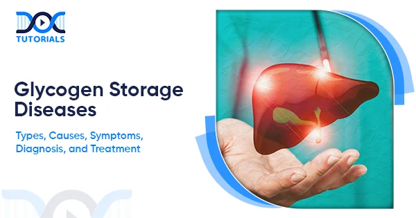
Glycogen Storage Diseases

Glycogen storage diseases, commonly referred to as GSDs, are sporadic genetic disorders that impact the storage and release of glucose in the body. Although these disorders are not common, they can affect growth, metabolism, and different body functions, especially in infants and pediatric patients.

This guide provides a comprehensive overview of glycogen storage diseases, covering their types, causes, and symptoms, as well as diagnosis and treatment, enabling NEET PG aspirants to develop a clear understanding of these complex conditions. Keep reading to learn more.

What are Glycogen Storage Diseases?

GSDs are numbered systemic disorders, naming the order in which the enzyme defects were described. The age of presentation can range from the neonatal period to adulthood, so patients may not develop symptoms in infancy.

Glycogen storage diseases are a set of rare genetic metabolic disorders that affect the body’s storage or degradation of glycogen, the storage form of glucose. Consumption of carbohydrates produces glucose, which is either used as fuel or stored as glycogen through the process of glycogenesis.

Glycogen is stored primarily in the liver and muscles, and when the body is fasting or exercising, it is converted into glucose through a process called glycogenolysis. Lack of one or more enzymes required for glycogen production or breakdown results in GSDs. Since the body cannot maintain an appropriate blood glucose level, it stores excess glycogen. 

Depending on the organs they affect, GSDs might result in hypoglycemia, hepatomegaly, weakness, and metabolic issues. Although the liver has a higher glycogen concentration, muscles have more glycogen due to their size; hence, changes in either the liver or the muscles cause variances in the presentations of individuals with GSDs.

What are the Types of Glycogen Storage Disease?

The glycogen storage diseases are inherited metabolic disorders, each caused by a particular enzyme deficiency. They can be classified based on the organs involved: liver, skeletal muscle, or both. For each type of disease, a specific set of signs, severity, and clinical outcomes exists.

GSDs primarily affecting the liver include disorders that impair hepatic glucose release, leading to fasting hypoglycemia and hepatomegaly. They are listed in the table below:

DisorderGSD Type / Enzyme Defect
Glycogen synthase-2 deficiencyGSD type 0a
Glucose-6-phosphatase deficiencyGSD type Ia
Glucose-6-phosphate transporter deficiencyGSD type Ib
Glycogen debrancher enzyme deficiencyGSD type III
Glycogen branching enzyme deficiencyGSD type IV
Liver phosphorylase deficiencyGSD type VI
Phosphorylase kinase deficiencyGSD type IXa
GLUT2 deficiency (Fanconi–Bickel disease)GSD type XI

These disorders cannot maintain normal glucose homeostasis during fasting. Therefore, dietary therapy is often the cornerstone of treatment.

GSDs primarily affecting skeletal muscle interfere with energy generation during exercise and usually present later in life. They are listed below:

DisorderGSD Type / Enzyme Defect
Muscle phosphorylase deficiency (McArdle disease)GSD type V
Phosphofructokinase deficiencyGSD type VII
Phosphoglycerate mutase deficiencyGSD type X
Lactate dehydrogenase A deficiencyGSD type XI
Aldolase A deficiencyGSD type XII
β-enolase deficiencyGSD type XIII
Phosphoglucomutase-1 deficiencyGSD type XIV

These individuals won’t tolerate strenuous exercise because their muscles can’t efficiently access stored glycogen. GSDs affecting both skeletal and cardiac muscle are often more severe and include the following:

DisorderGSD Type / Enzyme Defect
Lysosomal acid maltase deficiency (Pompe disease)GSD type IIa
Lysosome-associated membrane protein-2 deficiencyGSD type IIb
Glycogenin-1 deficiencyGSD type XV
Muscle glycogen synthase deficiencyGSD type 0b

1. Glycogen Storage Disease Type I (Von Gierke Disease)

Type I glycogen storage disease (von Gierke disease) is an autosomal recessive disorder caused by defects in the glucose-6-phosphatase system, essential for glucose homeostasis.

Type Ia results from glucose-6-phosphatase deficiency, while type Ib is due to glucose-6-phosphate translocase deficiency and is associated with recurrent infections due to neutrophil dysfunction. Types Ia and Ib are the primary clinically relevant forms.

2. Glycogen Storage Disease Type II-Pompe Disease

GSD type II, also known as Pompe disease or acid maltase deficiency, is a lysosomal storage disorder. It was first described by Pompe in 1932 and differs significantly from other GSDs because glycogen accumulates inside lysosomes rather than the cytoplasm.

The disease is caused by a deficiency of the lysosomal enzyme alpha-1,4-glucosidase, resulting in widespread glycogen accumulation in many tissues. There are 3 clinical forms recognised:

  • an infantile-onset form
  • a juvenile-onset form
  • an adult-onset form

In the classic infantile form, signs such as cardiomyopathy and severe muscle hypotonia dominate the presentation. The juvenile and adult forms mainly cause skeletal muscle impairment; thus, the primary concern becomes progressive muscle weakness.

3. Glycogen Storage Disease Type III (Forbes–Cori Disease)

GSD type III, also known as Forbes–Cori disease or limited dextrinosis, is an autosomal recessive disorder caused by mutations in the AGL gene. This leads to a deficiency of the glycogen debranching enzyme and accumulation of abnormal glycogen, also called limit dextrin.

Here, both the liver and the skeleton are affected. GSD type IIIa, being the most common form, accounts for approximately 85% of the cases, while type IIIb is less common and generally milder.

Clinical features overlap, so it is not always possible to distinguish GSD type III from GSD type I based on physical features.

4. Glycogen Storage Disease Type IV (Andersen Disease)

GSD type IV or Andersen disease (glycogen branching enzyme deficiency) is a rare and serious disorder. Andersen first reported the disease in 1956 in patients with progressive hepatosplenomegaly and with an accumulation of abnormal polysaccharides.

Abnormal glycogen accumulates in tissues due to defective glycogen branching, resulting in liver failure and involvement of the heart and nervous system. The course of the disease is often fulminating, with death occurring early in life; hence, early recognition is essential.

5. Glycogen Storage Disease Type V-McArdle Disease

McArdle disease, or GSD type V, mainly involves skeletal muscle. Muscle glycogen phosphorylase deficiency is the cause of this disease, which was first reported by McArdle in 1951.

Symptoms typically first appear during adolescence or adulthood. Common symptoms include exercise intolerance and muscle fatigue. Due to impaired breakdown of muscle glycogen, the glycolytic pathway is impaired during exercise.

Like other GSDs, McArdle disease is heterogeneous, with symptom severity varying from one individual to another.

6. Glycogen Storage Disease Type VI Hers Disease

GSD type VI, also called Hers disease, is a hepatic glycogenosis and represents a heterogeneous group of disorders. It results from hepatic phosphorylase deficiency or defects in enzymes involved in phosphorylase activation.

Hers first described the disease in 1959. Since the liver is primarily affected, patients usually present with hepatomegaly and mild hypoglycemia. Since the condition has milder symptoms than GSD type I, long-term outcomes are often positive.

7. Glycogen Storage Disease Type VII (Tarui Disease)

GSD type VII, also known as Tarui disease, results from a deficiency of phosphofructokinase (PFK). The enzyme is expressed in skeletal muscle and erythrocytes, and Tarui first reported affected individuals in 1965.

Clinically, GSD type VII is similar to GSD type V, with symptoms mainly centred on exercise intolerance and muscle weakness. Due to impairment at a different step of the glycolytic pathway, there is an inability to generate adequate energy during exercise; thus, the similarity in presentation.

What are the Causes of Glycogen Storage Diseases?

The glycogen storage diseases are inherited due to genetic mutations that affect enzymes responsible for glycogen synthesis, degradation, or transport. This genetic problem makes it impossible for the body to release energy from glycogen, which is inherited in an autosomal recessive trait, but sometimes X-linked.

The causes of GSD are given below:

  • Defects in Glycogen Synthesis

Alterations in glycogen synthesis cause certain types of glycogen storage disease. The gene responsible for the primary enzyme for synthesising glycogen from glucose is GYS2, which is expressed in the liver, while that of the enzyme expressed in muscles is GYS1.

The resulting diseases are known as GSD type 0a (liver cells) and type 0b (muscle cells), which are characterised by low glycogen deposits, thereby affecting energy production.

Another vital enzyme is the branching enzyme, which is encoded by the GBE1 gene and adds glycogen branches. The resulting disease is GSD type IV, brought on by deposits of abnormal glycogen (polyglucosan bodies).

  • Deficiencies in  Glycogen Breakdown

Most types of GSD are caused by glycogenolysis, the mechanism by which glycogen is metabolised into glucose. Mutations in the gene for muscle glycogen phosphorylase (PYGM), encoded by PYGM, underlie GSD type V, while mutations in liver phosphorylase (PYGL), encoded by PYGL, result in GSD type VI.

The metabolism of glucose from glucose-6-phosphate in the liver is dependent on glucose-6-phosphatase (G6PC); a deficiency in this gene results in GSD type Ia, known as von Gierke disease.

In GSD type Ib, a deficiency of glucose-6-phosphate transport into the endoplasmic reticulum, encoded by SLC37A4, leads to the accumulation of glycogen because of the inability to release glucose.

  • Other Enzyme Defects

In some cases, GSDs develop because of problems with the removal of glycogen branch points. This is because the glycogen-debranching enzyme, encoded by the AGL gene, is involved in the process. Deletions in the AGL gene cause GSD type III, which depends on whether the gene is inactive or partially active.

The uniqueness of GSD type II is that it is a lysosomal storage disorder, in addition to being a glycogen storage disorder. The genetic cause is mutations in the GAA gene, which encodes lysosomal acid α-glucosidase.

  • Inheritance Patterns

Most glycogen storage conditions are inherited in an autosomal recessive manner. This is because a child has to receive the gene from both parents to develop the condition. The parents may not have been ill; therefore, they might not know that they are carriers.

On the other hand, some types, like GSD type IX, are X-linked. In X-linked disorders, male carriers of the genetic mutation develop the disorder because they have only one X chromosome.

Taken together, these genetic abnormalities contribute to the variability in disease onset, severity, and presentation in GSDs because different enzymes are targeted.

What are the Symptoms of Glycogen Storage Diseases? 

Symptoms of glycogen storage disease typically occur during infancy or childhood, although these vary depending on the type, with hypoglycemia and exercise intolerance being the most common presentations.

The two most frequent symptoms common to most GSDs are hypoglycemia, which means low blood sugar, and exercise intolerance. Hypoglycemia is caused by the body’s inability to release glucose from storage.

This means that when the body experiences hypoglycemia, the brain and muscles do not receive sufficient energy. Possible symptoms of low blood sugar may include:

  • Shaking or Trembling
  • Sweating and chills
  • “Dizziness” is most
  • Weakness & Fatigue
  • An increased heart rate
  • Severe hunger (Hyperphagia)
  • Difficulty thinking or concentrating
  • Irritability or Anxiousness
  • Easy bruising
  • Seizures in Severe Cases

Exercise intolerance is also common, particularly in cases involving muscles. The reason is that the muscles cannot readily derive energy from storage. Thus, exercise may result in weakness, soreness, or spasms even when the exercise is mild.

  • Other manifestations of glycogen storage disease may include:
  • Muscle weakness/muscle tone that is low
  • Muscle spasms and aches
  • Delayed growth and failure to gain weight in children
  • Enlargement of the liver (hepatomegaly), sometimes resulting in a distended abdomen
  • Easy bruising
  • Chronic hunger
  • High cholesterol (hyperlipidemia)

In a child, persistent low blood sugar and limited energy availability might affect growth. This is because growth might be slowed because the body has been unable to manage glucose effectively, hence, weight gain might be insufficient.

How are Glycogen Storage Diseases Diagnosed?

Glycogen storage disease diagnoses are made using a variety of tests, including blood and urine tests, imaging studies, and genetic tests for abnormalities in glucose metabolism, the liver, muscles, specific enzymes, and genes.

This table gives a detailed overview of how GSDs are diagnosed:

TestWhat it shows
Fasting Blood Sugar TestLow fasting blood glucose suggests GSD because the body can’t properly release stored glucose.
Ketone Blood TestElevated ketones indicate the body is using fat for energy instead of glucose, which is common in GSD.
Basic Metabolic PanelProvides an overview of metabolic health and helps rule out other conditions.
Lipid PanelDetects high cholesterol levels (hyperlipidemia), which are common in several types of GSD.
Liver Function TestsAbnormal results may indicate liver involvement, a key feature of many GSDs.
UrinalysisMeasures kidney function and uric acid levels; GSD often causes hyperuricemia.
Abdominal UltrasoundChecks for an enlarged liver (hepatomegaly).
Genetic TestingIdentifies gene mutations affecting glycogen-related enzymes and helps confirm the GSD type.
Muscle or Liver BiopsyPerformed when genetic testing isn’t conclusive to confirm glycogen accumulation and enzyme defects.

What are the Treatment and Management Options for Glycogen Storage Diseases?

Glycogen storage diseases are managed through a lifelong diet, medications, and treatment plans to keep blood sugar levels stable, with conditions such as enzyme replacement therapy or a liver transplant used in particular cases.

There is no treatment available for glycogen storage disease (GSD), so management is symptomatic. It depends on the type of GSD and which organs are affected. Management aims to maintain stable blood sugar levels, minimise the accumulation of abnormal metabolites, and preserve the involved organs.

  • Prevention and Management of Hypoglycemia

The control of hypoglycemia is a fundamental part of the treatment of GSD. Most patients require a daily diet of unsweetened cornstarch. Cornstarch is a complex carbohydrate that is slowly digested, ensuring that glucose is released gradually into the bloodstream. 

Newer preparations of longer-acting agents are also available, so patients may not need to be fed at night. In most cases, when hypoglycemia manifests, it has to be treated immediately by consuming high-carb foods.

In cases where this condition is left untreated, it has a high tendency to result in severe conditions such as the onset of a seizure or coma.

  • Handling Metabolic Complications

Some instances of GSD are linked with metabolic abnormalities, which require attention as well. High cholesterol is typically treated with statin medications. High uric acid levels can be managed with allopurinol, which decreases uric acid production.

  • Enzyme Replacement Therapy

In some types of GSD, treatment is more direct. For instance, type II GSD can be treated with enzyme replacement therapy (ERT).

The treatment consists of frequent intravenous injections to compensate for the deficient enzyme, resulting in a buildup of glycogen in the body. Researchers are working on a way to extend the treatment to other types of GSD.

  • Advanced and Surgical Options

In more serious cases, particularly when there is progressive liver damage, liver transplantation has been suggested. Although it doesn’t affect the genetic problem, it has been shown to result in marked improvement in metabolism in certain patients.

In summary, the management of GSD is a long-term personalised approach. The reason is that, in the case of GSD, the condition cannot be managed on its own, unlike in healthy patients.

FAQs about Glycogen Storage Diseases

  1. What is the prognosis of glycogen storage disease?

Early diagnosis and proper care promise a bright future for most people with GSD. But in some cases, GSD is hard to cope with.

  1. How is glycogen storage disease diagnosed in a child?

The diagnosis involves reviewing symptoms, conducting a physical exam, and obtaining blood work. In some cases, genetic screening, as well as a liver or muscle biopsy, might be necessary.

  1. What are the complications of glycogen storage disorder? 

Complications that can arise are serious cases of hypoglycemia, growth delays, liver problems, heart problems, muscle damage, and/or renal difficulties, depending on the type of GSD.

  1. How can glycogen storage disease be prevented?

GSD is not preventable because it is inherited. Early treatment can, though, keep the symptoms at bay.

  1. What is the life span of a person with glycogen storage disease?

The lifespan depends on the type, age of onset, and the quality of treatment management. In some types, the conditions are serious, but in others, a close-to-normal lifespan is possible.

Conclusion

Glycogen storage disease is a broad category of genetic disorders that vary significantly from one type to another. Although a cure is not available, a lot can still be done to make life better with proper management. In fact, diet, treatment, enzyme replacement, and support for patients with GSD have improved significantly, necessitating care.

For pursuing NEET PG, DocTutorials can be a valuable companion for studying metabolic disorders, among other high-yield topics. We offer crisp videos, Qbank, exam-focused notes, flashcards, mindmaps, and much more to ensure aspirants gain complete clarity over complex NEET PG concepts.

Join DocTutorials today and explore our NEET PG course to excel in your medical career!

NEET PG Online Live Power Pack
DocTutorials App Download Now

Latest Blogs

Twin Pregnancy: Risk Factors, Symptoms, and Complications

Twin Pregnancy

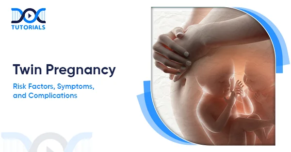
Twin pregnancy is an important concept in Obstetrics and Gynaecology, as it is a special type of multiple gestation in which two fetuses grow simultaneously in the womb.

This knowledge is essential because twin gestation is associated with greater maternal and fetal risks than singleton gestation. Thus, early detection, adequate monitoring, and timely intervention are necessary for a safe result.

This guide describes the types, causes, symptoms, and complications of twin pregnancy and helps students and practitioners establish a reliable background of knowledge. Keep reading to learn more.

What is a Twin Pregnancy?

A twin pregnancy implies the transfer of two embryos. These twins may produce either of two fertilised eggs (identical twins) or one (fraternal twins), and they may have a common or dissimilar placenta and sacs.

The most prevalent type of multiple gestation is a twin pregnancy, where the woman is carrying two fetuses concurrently. Identical (monozygotic) twins occur when a fertilised egg divides into two embryos, resulting in babies of the same sex whose genes never mix. 

This division makes the difference between separate and common placentas as well as common and separate amniotic sacs, depending on the time of this split. Non-identical (dizygotic or fraternal) twins are where two distinct eggs are fertilised by two different sperm in the same cycle.

Other terms used to describe twin pregnancies include chorionicity (number of placentas) and amnionicity (number of amniotic sacs).

The NICE (National Institute for Health and Care Excellence) guidelines include the following primary forms:

  1. Dichorionic–Diamniotic (DCDA): Indicates two placentas and two sacs. Every fetus contains its placenta and amniotic sac. (The majority of fraternal twins, a few early-separated identical twins).
  2. Monochorionic–Diamniotic (MCDA): Depicts one shared placenta and two sacs. The twins’ condition is characterised by a common placenta and distinct amniotic sacs. This is the case with the twins. (Occurs only in identical twins).
  3. Monochorionic–Monoamniotic (MCMA): Means one shared placenta and one sac. The two twins share the same placenta and amniotic sac. (Hardly, only identical twins separating late).
  4. Conjoined Twins: Indicates a scarce variant, in which the twins are physically attached; they are invariably monochorionic-monoamniotic.

The twin pregnancies in most cases are diagnosed early in the form of a first-trimester ultrasound, which may reveal two gestational sacs or embryos.

In some cases, a second sac develops later (with the onset of the twin’s appearance), or there are clinical indications that raise suspicion. Twin gestations are high-yield topics in Obstetrics and Gynaecology due to their distinct anatomy and management.

What are the Risk Factors of Twin Pregnancy?

Twin pregnancy has a range of factors that tend to increase the chances of an occurrence: genetic predisposition (preferentially fraternal twins), maternal age, high parity, and fertility treatment.

The reasons for twin pregnancy are different between identical and fraternal twins. Identical (monozygotic) twinning is said to be random during the development of the embryo and is normally not influenced by the family background or maternal characteristics. In contrast, fraternal (dizygotic) twinning is influenced by genetics and maternal factors.

The risk factors that significantly increase the risk of twin pregnancy are:

  • Genetic Predisposition (Maternal Lineage): The presence of twins in the family raises the chances of fraternal twins. (Fathers’ twin history has much less effect.)
  • Advanced Maternal Age: Females above 35 are much more exposed to a higher chance of getting twins as compared to younger females. Optimal ovulation at the age of approximately 37 raises the probability of releasing multiple eggs.
  • High parity (Many Prior Pregnancies): With each additional pregnancy, the probability of twins increases. Women with multiple previous pregnancies have 2–3 times the twin rate of nulliparous women.
  • Use of Fertility Drugs or Assisted Reproductive Technology (ART): Ovulation-inducing drugs such as clomiphene and gonadotropins may result in numerous ovulations. In one out of every five to twelve pregnancies, clomiphene may lead to twins.
  • Maternal Nutrition and Body Habitus: Females who are well-nourished, taller, or heavier are more likely to have fraternal twins due to better, healthier physiology and hormonal levels. In the past, the rate of twins was lower in regions affected by famine.

Localised research indicates that twinning occurs at 10-12 per 1000 births, but the prevalence is higher in fraternal twins. This increase is attributed to an increased maternal age, fertility treatment, yet nonetheless, a disproportional number of perinatal deaths is presented through twin pregnancies.

What are the Symptoms and Diagnosis of Twin Pregnancy?

Twin pregnancy has exaggerated initial symptoms (e.g., severe nausea, rapid growth of the uterus), and diagnosis depends upon ultrasound and prenatal screening.

Clinically, the early signs of a pregnancy in twins can be very insidious or confused with a single pregnancy. A woman with pregnant twins may experience the following symptoms:

  • Severe Fatigue: Most women experience more severe fatigue or nausea (elevated hCG hormone levels) with having twins, although this is not universal.
  • Rapid Uterine Growth: Fundal height (compared to the distance between the pubic bone and that of the uterus) is usually large when compared to the gestational age.
  • Elevated Pregnancy Blood Tests: Maternal serum screening markers may be abnormal. As an illustration, a markedly elevated AFP (alpha-fetoprotein) or β-hCG during second-trimester screening may indicate twins (though it may also indicate other conditions).
  • Physical Exam Clues: An experienced clinician or Doppler device may hear more than one fetal heartbeat later in the first or early second trimester. But the separation of two heartbeats can hardly be detected, and therefore, this is only a suggestion, not an absolute.

Diagnosis of Twin Pregnancy

Diagnostic ClueDescription
Stronger Pregnancy SymptomsMore severe nausea or fatigue, which is usually caused by high levels of hCG.
Rapid Uterine GrowthThe fundal height is more than expected for gestational age.
Abnormal Maternal Serum MarkersHigh levels of AFP or β-hCG in second-trimester screening.
Physical Examination CluesThere is a possibility of more than one fetal heartbeat detected using Doppler (not definitive).
Ultrasound (Gold Standard)Confirms there are said fetal number, chorionicity, and gestational age; recognises two sacs, yolk sacs or fetal poles by week 6-8.
Early Ultrasound FindingsOne placenta with a membrane → monochorionic-diamniotic; two placentas → dichorionic twins.
Special CasesVanishing twin syndrome or appearing twin syndrome is identified by serial scans.

Practically, when a twin pregnancy is suspected clinically, the doctors will request a serial scan and prenatal tests.

What are the Complications of a Twin Pregnancy?

Twin gestation is associated with high maternal and fetal risks compared to singletons. Preterm birth, fetal growth issues, and placenta syndromes are the most significant complications.

A twin pregnancy is said to be high-risk because of a range of potential complications. Crowding in the uterus leads to fetal risks. Common complications include:

  • Preterm Birth: The greatest risk during pregnancy of twins is preterm birth. Premature infants can be both spontaneous and planned, and they tend to cause neonatal problems of respiratory distress, feeding disorders, and prolonged NICU (neonatal intensive care unit) stays.
  • Preeclampsia and Hypertension: The risk of developing hypertensive disorders of pregnancy (gestational hypertension or preeclampsia) is high in twins due to the size of the placenta. It is also recommended to carefully monitor blood pressure; low-dose aspirin can be considered in case of other risk factors.
  • Gestational Diabetes Mellitus (GDM): Being pregnant with more than one fetus increases insulin resistance as the levels of pregnancy hormones increase. Twin pregnancy has been linked with increased cases of GDM.
  • Maternal Anaemia: Twin pregnancies often demonstrate decreased maternal hemoglobin because of increased volume of blood and iron requirement of two fetuses. Iron supplementation is frequently required to prevent or cure anemia.
  • Placental Complications: Having two placentas (or an abnormally large single placenta), the problems of placenta previa or abruption may become more prevalent.
  • Fetal Growth Restriction: Even in uncomplicated twins, each fetus is likely to develop slightly poorer than singletons because of the limits of uterine space. The IUGR (Intrauterine Growth Restriction) or slow growth (10th percentile or below) is usual; approximately (40-50)% twins have some growth retardation.
  • Congenital Anomalies: The rate of anomalies with twin pregnancies is a bit greater, and that could be due to intrinsic factors that tend to cause a twin pregnancy. In addition, identical twins have all the genes in common, and therefore the genetic disorder will be observed in both.
  • Delivery Complications: A mother is at risk of postpartum bleeding since it occurs due to overdistension of the uterus. In case of a first twin being vertex, vaginal delivery of twins can be tried; C-section is more commonly used, particularly when the first twin is breech, or when there are complications.

The challenges that require close monitoring among the carriers of twin gestations include hypertension, diabetes, and premature labor. Early identification of such complications is one of the key elements in obstetric management.

What are the Treatment Options for Twin Pregnancy?

The treatment of twin pregnancy is aimed at close attention and care. Carrying twins has no cure; however, they can be alleviated through measures taken to reduce risks.

Although no particular form of treatment is needed to change the state of preventing a twin pregnancy, the current obstetric management of the issue strives to maximise the results of both the mother and the babies. Key approaches include:

  • Frequent Prenatal Care and Monitoring: Women carrying twins require more frequent antenatal check-ups and ultrasounds. Getting scans every 3-4 weeks- or sooner in the case of monochorionic twins- measures growth, amniotic fluid, and placental activity. Visits involve blood pressure, weight, and urine protein checks, and later a non-stress test or biophysical profile to gauge the health of the fetus.
  • Nutritional Support: Twin pregnancies have higher caloric requirements and weight-gain needs. Higher protein diets and sufficient folate and iron (including in the form of supplements) are crucial, and positive birth weights of twins correlate with better health.
  • Lifestyle Adjustments: Moderate exercise (such as walking or swimming) has generally been advised except in cases of contraindication, and full bed rest is discouraged. Cleanse up tobacco, alcohol, and other threats to pregnancy, and focus on management and rest since the physical demands are increased in the case of twin pregnancies.
  • Specialist Interventions: Fetal therapies may be used in complex pregnancies that are monochorionic. In case of severe polyhydramnios, Amnioreduction (removal of excess fluid) may be undertaken. These processes involve making a referral to a fetal medicine centre.
  • Planning Delivery: Planned cesarean or induction is usual, especially when the first of the twins is not head-down. Pregnancy expansion to more than the suggested weeks is associated with higher chances of stillbirth; hence, early birth is safer.
  • Birth Preparedness and Support: Planning the postpartum assistance at home, since taking care of two newborns is quite challenging. Often, nutritional counseling and even lactation consultation are provided that way because by means of it, some women are able to breastfeed twins.

Altogether, managing twin pregnancies can be supportive through healthy nutrition, diabetes or hypertension management, and early warning sign check-ups.

It is best followed up by a special obstetric team frequently. Risks are more, but most twins can be fine with adequate care, and realistic counselling lets families realise that in-care antenatal management has a significant enhancement effect on outcome.

FAQs about Twin Pregnancy

  1. Are identical twins hereditary?

No. There is no correlation between family history and identical (monozygotic) twins. The primary effect of heredity is that it elevates the probability of fraternal twins as a result of a predilection to multiple ovulation.

  1. Can I safely prolong a twin pregnancy to full term?

Usually not. Exceeding the recommended boundaries of 37+6 weeks in dichorionic and 36 +6 weeks in monochorionic twins increases stillbirth chances. Doctors strike a balance between prematurity risks and the risks of remaining in the womb, and usually examine near-term weekly.

  1. Are twin pregnancies high-risk?

Yes. Twin pregnancies are associated with increased risks of complications such as preterm delivery, preeclampsia, gestational diabetes, and birth problems. It is the reason why high-risk obstetric care of specialists is recommended.

  1. Will I definitely need a C-section?

Not necessarily. Delivery can occur at the vagina when the first twins are in a head-down position, and there are no complications. But C-section is standard – of course, not prevalent in case Twin A is not in a cephalic position. There could be cases when the second twin might require an emergency C-section.

  1. How can I prepare for having twins?

Find out about twin-specific care, write prenatal counseling, and make additional home arrangements. You can be prepared in case you need to receive NICU care and plan out exercises, traveling, and feeding with your doctor to be on track.

Conclusion

Twin pregnancies are high risk, both identical and fraternal. It is important to learn their causes, early symptoms, and complications, such as preterm birth and growth complications, and to be able to effectively provide antenatal care. Timely interventions, close monitoring, and early diagnosis can be used to guarantee better outcomes for mothers and babies.

To understand more such complex NEET PG concepts, DocTutorials can be your study partner. We offer crisp video lectures, clinical Qbank, exam-focused notes, flashcards, and mind maps to help aspirants gain complete clarity.

Join DocTutorials today and explore our NEET PG course to excel in your medical career!

NEET PG Online Live Power Pack
DocTutorials App Download Now

Latest Blogs

Lip Cancer: Causes, Symptoms, Diagnosis, and Treatment

Lip Cancer

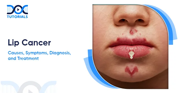
Lip​‍​‌‍​‍‌​‍​‌‍​‍‌ cancer is one of the most common types of cancer found in the mouth. It develops when these cells, called squamous cells, cover the outer part of the lips and begin to multiply abnormally.

Generally, lip cancer appears just like other cancers of the oral cavity, i.e., arising from the cells of the lining of the lips, tongue, cheeks, and throat. It may call for interventions like surgery, radiation therapy, and chemotherapy. With a frequency of 0.5 cases per 100,000 individuals per year, preventive measures and early diagnosis play a significant role.

In this article, we will explore the causes, early symptoms, diagnostic methods, and the most effective treatment options available for lip cancer. Read on for a comprehensive analysis.

What is Lip Cancer?

Lip cancer is a kind of oral cancer that comes from the squamous cells of the lips and is most commonly related to smoking, heavy alcohol use, and sun exposure. The cancer is highly curable if it is caught at an early stage, usually by a dentist.

Lip cancer develops when the cells in the lips become abnormal and start to multiply rapidly, eventually forming sores or tumours. Basically, it is a cancer of the oral cavity. The cancer develops in the cells that make up a single layer of the cells that form the:

  • Lips
  • Mouth
  • Tongue
  • Cheeks
  • Sinuses
  • Throat
  • Hard and soft palates

Lip cancer, like many other oral cancers, falls under the broader group of head and neck cancers.

Dentists often identify the earliest signs of lip cancer during routine checkups, spotting subtle changes that may otherwise go unnoticed. The positive side is that lip cancer has an excellent prognosis when detected at an early stage, making timely evaluation incredibly important.

What are the Causes of Lip Cancer?

The primary causes include UV radiation from the sun or tanning beds and tobacco use. The risk is significantly heightened by heavy alcohol use and a history of infections like HIV/HPV or use of immunosuppressive drugs.

As with most cancers, lip cancer develops due to multiple factors. However, several causes are pointed out that increase the chance of lip cancer:

  • UV (ultraviolet) radiation from the sun or tanning beds
  • Tobacco use
  • Heavy alcohol use (especially in combination with tobacco use)
  • History of human immunodeficiency virus (HIV) or human papillomavirus (HPV) infections
  • History of immunosuppressive medications (such as those used after organ transplant)

What are the Risk Factors of Lip Cancer?

Key risk factors are tobacco use and heavy alcohol consumption, which synergistically increase risk. Other factors include excessive sun exposure, having fair skin, being over 40 (especially males), and having a weakened immune system.

Several risk factors contribute to the development of lip cancer. They are as follows:

  • Smoking: This refers to the smoking of cigarettes, cigars or pipes, and the use of snuff and chewing tobacco. Most lip cancer cases are related to smoking.
  • Heavy Alcohol Consumption: The risk of lip cancer may be 30 times higher if you are a smoker who also drinks heavily.
  • Too Much Sun Exposure: This also refers to the use of artificial tanning lamps or tanning beds.
  • Having Fair Skin: People with fair skin and light features are at the highest risk.
  • Being Over 40: Most people get lip cancer when they are in their 50s and 60s.
  • Gender: Men are up to three times more likely to develop lip cancer than women.
  • Weakened immune system. ​‍​‌‍​‍‌​‍​‌‍​‍

What are the Symptoms of Lip Cancer?

Symptoms often resemble benign issues but persist for weeks, most notably a persistent sore or blister that doesn’t heal within two weeks and bleeds easily. Other signs include a discoloured patch (reddish, grey, or dark brown), swelling, a growth on the neck, or pain/numbness in the mouth.

Lip cancer symptoms can be easily mistaken for common lip problems such as cold sores or mouth ulcers. However, if these symptoms last for more than a couple of weeks, you should definitely see a doctor.

Some of the most frequent lip cancer symptoms are:

  • A continuously recurring sore, lesion, or blister on the lip that bleeds easily and does not heal within two weeks.
  • A little coloured patch (usually white or reddish in colour on light skin, or dark brown or grey on dark skin).
  • Swelling of the jaw.
  • A lump on the neck.
  • Loose teeth.
  • Non-stop bleeding, pain, numbness, or tingling of the lips or mouth.

The presence of sores or lumps on your lips does not necessarily mean that you have lip cancer. If you see any of these symptoms, talk to your dentist or doctor about them.

What is the Diagnosis of Lip Cancer?

Diagnosis starts with a physical examination by a doctor or dentist, who checks the lips, mouth, and neck. If suspicion remains, a biopsy is performed to confirm the presence of cancer cells. If confirmed, tests like CT, MRI, or PET scans determine the stage and spread.

If you experience signs or symptoms of lip cancer, visiting a doctor is the right thing to do. They will conduct a physical examination of your lips and other areas of your mouth to look for any abnormal or potentially cancerous regions.

To examine the inside of your lips, your doctor will use a gloved finger and will inspect them using mirrors and lights. If there are swollen lymph nodes, the doctor might also check your neck. Additionally, your doctor would like to know about your:

  • Health history
  • Smoking and drinking history
  • Previous illnesses
  • Medical and dental treatments
  • Family history of diseases
  • Any medications you are taking

If lip cancer is a possibility, a biopsy is the way to go for a definite answer. A biopsy sample is a small portion of the affected area taken out. A pathologist then examines that sample under a microscope in a laboratory.

Upon confirmation of lip cancer through biopsy, your doctor may then perform many other tests to determine how far the cancer has progressed, or if it’s spread to other parts of the body. The list of tests may be:

  • CT (computed tomography) scan.
  • MRI (magnetic resonance imaging).
  • PET (positron emission tomography) scan.
  • Chest X-ray.
  • Complete blood count (CBC).
  • Endoscopy.

What is the Treatment for Lip Cancer?

Treatment depends on the tumour size and stage. Surgery to remove the lesion is typical, often paired with radiation therapy to kill remaining cells. For advanced cases, systemic treatments like chemotherapy, targeted therapy, or immunotherapy may be used, sometimes for palliative care.

The most effective treatment for you greatly depends on the size of the tumour or lesion and the stage of the cancer.

Usually, doctors can manage precancerous lip cancer (a condition where abnormal lesions have a chance to become malignant) and lip cancer at its early stage with the aid of surgery only. If your situation is complicated, then a mixture of treatments may be required.

Some methods to treat lip cancer are:

  • Surgery: With surgery, your doctor removes the parts that contain the lesion or tumour and then mends your lip. In addition, they might take out lymph nodes from your neck if they are sure that the cancer has moved there.
  • Radiation Therapy: Radiation is used to eradicate cancer cells. The hospital can suggest either external beam radiation therapy (EBRT) or brachytherapy (internal radiation therapy).
  • Chemotherapy: This approach uses powerful medications to destroy cancer cells and, in some cases, may be paired with radiation therapy for better results. If the cancer has advanced and other treatment options are limited, chemotherapy may be recommended to help manage discomfort as part of palliative care.
  • Targeted Therapy: This treatment focuses on cancer cells’ genes and proteins and thus destroys them. Usually, people with lip cancer obtain it along with chemo.
  • Immunotherapy: These drug treatments boost your body’s immune system and help it fight off cancer cells. In most cases, when the cancer has spread and other treatments are not viable options, lip cancer patients are given immunotherapy.

What is the Prevention for Lip Cancer?

Prevention focuses on protecting the lips from sun exposure using SPF 30+ balm and hats, avoiding all tobacco products, and limiting alcohol intake. Regular dental checkups and promptly examining any sore that lasts longer than 2 weeks are also critical preventive measures.

Even though you can’t stop the development of stress-induced lip cancer in all cases, you can take some measures to tone down your risk:

  • Safeguard your lips against the sun. Use lip balm with SPF 30+, reapply often, wear a wide-brimmed hat, and limit your time in the intense midday sun.
  • Cut back on your alcohol intake, and do not mix smoking with drinking, as this increases your risk even more.
  • See your dentist regularly and check your lips yourself. Any sore or change that lasts more than 2 weeks should be examined immediately.
  • If you have precancerous lip conditions, such as actinic cheilitis, follow your physician’s advice regarding therapy and checkups.
  • Maintain good oral hygiene, eat nutritious food, and avoid chronic irritation from factors such as ill-fitting dentures or chewing tobacco.

FAQs about Lip Cancer

  1. At what age is lip cancer most common?

Lip cancer is most frequent in people with light skin and men who smoke pipes, especially those aged between 50 and 70 years. In the United States, lip cancer is the cause of around 0.6% of all malignant tumours; the majority of these are ulcerative or erosive lesions that affect one side of the vermilion border.

  1. What could be mistaken for lip cancer?

The primary sign of lip cancer we most commonly see is an ulcer or sore that doesn’t heal. Typical viral ulcers typically resolve within a couple of weeks. Early lip cancer can be indistinguishable from a cold sore, but cold sores eventually heal.

  1. Where is lip cancer located?

It can be anywhere along the upper or lower lip; however, lip cancer is most commonly found on the lower lip. Lip cancer is counted as one of the oral (mouth) cancers.

The majority of lip cancers are squamous cell carcinomas, i.e., they arise in the thin, flat cells of the middle and outer layers of the skin (squamous cells).

  1. Can a dermatologist diagnose lip cancer?

In most instances, your dermatologist can detect signs of lip cancer visually during a routine skin examination. If a concerning spot is found, your dermatologist will often perform a biopsy by removing a small sample of skin from the affected area.

  1. What colour is lip cancer?

It may sometimes resemble scar tissue. For instance, in those with light skin, it may take on a reddish colour. In those with darker skin, the area might appear grey or dark brown. New lip cancer lesions can also look similar to cold sores.

Conclusion

Recognising the early signs of lip cancer can make a significant difference in timely diagnosis and treatment. Although it most commonly affects older males with lighter skin tones, lip cancer can develop in anyone, making awareness essential. Ultimately, staying alert to changes and prioritising oral health play a crucial role in safeguarding long-term well-being.

To understand more such complex NEET PG concepts, DocTutorials can be your study partner. We offer crisp videos, clinical Qbank, exam-focused notes, flashcards, and mindmaps, ensuring aspirants gain complete clarity over these concepts.
Join DocTutorials today and explore our NEET PG course to excel in your medical career!


NEET PG Online Live Power Pack
DocTutorials App Download Now

Latest Blogs

Focal Segmental Glomerulosclerosis: Causes, Symptoms, Diagnosis, and Treatment Options

Focal Segmental Glomerulosclerosis

Focal Segmental Glomerulosclerosis (FSGS) is a rare disease that affects the filtering units in the kidneys. If the filtering units are damaged, they cannot properly filter toxins from the body, leading to kidney damage or permanent failure. Around 7 in every 1 million people are diagnosed with FSGS every year.

FSGS can affect both children and adults. However, males are affected slightly more often than females. It may recover on its own, but if left untreated, it can lead to permanent kidney failure requiring dialysis or transplantation.

Keep reading for a detailed analysis of FSGS.

What is Focal Segmental Glomerulosclerosis (FSGS)?

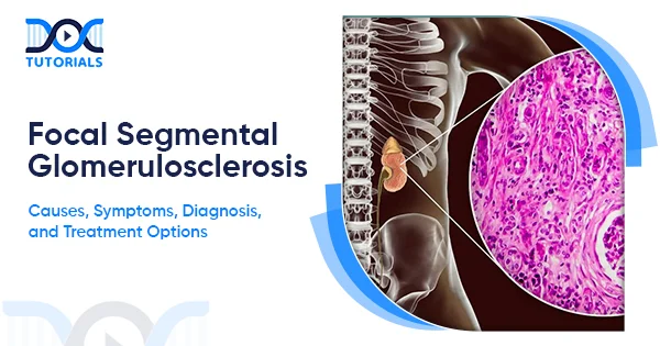
FSGS is an uncommon kidney disorder where some parts of the kidney’s tiny filters (glomeruli) become scarred, reducing their ability to clean the blood. This scarring leads to problems with waste removal, fluid balance, and overall kidney function.

FSGS is a rare kidney disease that causes scars in the glomeruli, the tiny filters of the kidneys. Each kidney contains around 1 million glomeruli. They work like a strainer, filtering out waste materials and toxins from your blood. When blood passes through the glomeruli, it filters out essential nutrients, minerals, waste products, and excess fluid.

The vital nutrients and minerals are then returned to the bloodstream, and the waste products and excess fluid are removed from the body in urine. Kidney conditions that affect the glomeruli are called glomerular diseases.

FSGS is a type of glomerular disease that scars or hardens these tiny filters (glomeruli). Here’s what the terms in the name ‘Focal Segmental Glomerulosclerosis’ mean:

  • Focal: It refers to some glomeruli in the kidneys, but not all.
  • Segmental: It refers to a few specific parts of the glomeruli that have scars. 
  • Sclerosis: It means ‘Hardening’

Thus, the name ‘Focal Segmental Glomerulosclerosis’ means hardening (Sclerosis) of some (Focal) parts of (Segmental) glomeruli.

What are the Types of Focal Segmental Glomerulosclerosis?

FSGS can develop in 4 main forms: primary (no known cause), secondary (linked to conditions or medications), genetic (due to inherited gene changes), and unknown (cause not identified even after testing).

FSGS can cause hardening of the tiny filters (glomeruli) in the kidneys, leading to kidney damage. The treatment depends on the type of FSGS you have. These include: 

  1. Primary FSGS: Also known as idiopathic FSGS, this is the most common form of FSGS, where the cause is not known or is not apparent.
  2. Secondary FSGS: This type of FSGS is caused by medications or other factors, such as infection, drug toxicity, diabetes or sickle cell disease, obesity, and other kidney diseases.

Managing the underlying cause of this FSGS can help slow ongoing kidney damage and improve kidney function over time.

  1. Genetic FSGS: Commonly known as familial FSGS, it is caused by genetic changes. It is a rare form of FSGS that is suspected if several members of a family have a history of FSGS.

It can also develop even when neither parent has a history of FSGS, but each carries a copy of an altered gene that can be passed on to the next generation.

  1. Unknown FSGS: In some instances, the underlying cause of FSGS cannot be determined even after clinical evaluation of symptoms and comprehensive laboratory testing.

What are the Causes and Risk Factors of Focal Segmental Glomerulosclerosis?

FSGS can develop due to several causes, including chronic conditions like diabetes, hypertension, obesity, lupus, certain medications, infections, and harmful drug use. Genetic changes, especially APOL1 variants, along with family history, age, and overall kidney strain, can further increase the risk of developing this condition.

Several different causes lead to FSGS, but people with primary FSGS have no apparent reason. Researchers have found that in unknown FSGS, a blood protein damages parts of the glomeruli.

Secondary FSGS is caused by heavy blood flow to the glomeruli, which can happen due to many factors, such as:

  • Diseases like sickle cell anaemia, diabetes, and lupus.
  • Certain medications, such as interferons or bisphosphonates.
  • Several drugs, like heroin or anabolic steroids.
  • Infections, including viruses like HIV (Human Immunodeficiency Virus).
  • Being obese
  • Several other kidney conditions or any congenital kidney condition.

Genetic FSGS occurs when the APOL1 (Apolipoprotein L1) gene mutates during fetal development. This is most common among people of West African ancestry. Apart from these causes, several other factors can elevate the risk of FSGS, including:

  • Certain Medical Conditions: Several chronic conditions, like hypertension, diabetes, obesity, and lupus, can increase the risk of kidney damage and FSGS.
  • Infections: Hepatitis C, HIV, and other infections, if not appropriately treated, can heighten the risk of FSGS.
  • Gene Changes: Changes in genes that control podocyte (filtering cell) integrity can increase the risk of FSGS. It is most common among people of African ancestry due to the presence of APOL1 gene variants linked to podocyte injury.

Middle-aged males are more susceptible to this disease. People with a family history of kidney disease are also at an increased risk of developing FSGS.

What are the Symptoms of Focal Segmental Glomerulosclerosis?

FSGS often develops silently, but signs like swelling, high protein in the urine, and high blood pressure may appear during checkups. When swelling, proteinuria, and low albumin occur together, it may indicate nephrotic syndrome.

FSGS does not always cause noticeable signs or symptoms that you can identify on your own. Medical practitioners identify these symptoms during a routine health examination:

  • Swelling in arms, legs, or face (Oedema)
  • High-cholestrol
  • Low albumin (a protein in the blood)
  • Proteinuria (abnormally high amounts of protein in the urine)
  • Sudden weight gain due to excess fluid in the body.
  • High blood pressure or hypertension.

If you have oedema, proteinuria, and low albumin, the healthcare professional may diagnose nephrotic syndrome, which causes the kidneys to release large amounts of protein into the urine.

What is the Diagnosis of Focal Segmental Glomerulosclerosis?

FSGS is diagnosed through tests that assess how well your kidneys are working, including blood and urine tests, GFR measurement, and, sometimes, a kidney biopsy. In some cases, genetic testing is also done to identify inherited causes and guide treatment.

There are several tests through which FSGS can be diagnosed, depending on your condition. The tests can include:

  • Blood Tests: They help to determine the levels of protein, cholesterol, and waste in your blood.
  • Urine Tests: These are done to determine the levels of protein in your urine. 
  • Glomerular Filtration Rate (GFR): It’s a blood test that helps determine how well the kidneys are functioning by measuring the rate at which the kidneys filter waste.
  • Kidney Biopsy: It involves removing a tiny sample of kidney tissue with a special needle for examination under the microscope. It helps to diagnose FSGS.
  • Genetic Testing: It helps identify whether you were born with genes that can cause kidney disease. It allows the doctor to decide on a suitable treatment option for your condition.

What is the Treatment for Focal Segmental Glomerulosclerosis? 

FSGS treatment depends on the cause and may include medications to reduce inflammation, control protein loss, and prevent swelling or complications. Lifestyle steps like staying active and maintaining a healthy weight protect kidney function.

The type of treatment for FSGS is determined based on the cause. However, the treatment usually includes:

  • Corticosteroids: These medications are considered the first-line therapy for primary FSGS. They help reduce inflammation and protein loss. The long-term use of these medicines requires careful monitoring, as they may cause side effects.
  • Immunosuppressants: These medicines are used if the steroids don’t work. They help stabilise podocytes and reduce kidney damage.
  • ACE Inhibitors or ARBs: These medicines help lower blood pressure and reduce protein loss (Proteinuria), thereby protecting kidney function.
  • Diuretics: These medications help control swelling and reduce fluid accumulation in the body.
  • Statins: These are given if you’re diagnosed with nephrotic syndrome to reduce cholesterol and triglycerides.
  • Anticoagulants: These medicines are given to patients at elevated risk of developing blood clots.

Apart from medications, the doctor will ask you to implement certain lifestyle changes to manage your symptoms:

  • Dietary Changes: The doctor may ask you to reduce sodium (salt) and protein in your diet to minimise the load on your kidneys. 
  • Healthy Habits: Avoid smoking and try to lose weight if you are obese. Contact any healthcare professional if you need help quitting smoking.  
  • Physical Exercises: Staying active will expedite your recovery. Try to devote some time to physical exercises every day.
  • Avoid Certain Medicines: Pain relievers and over-the-counter medicines like nonsteroidal anti-inflammatory drugs (NSAIDs) can cause damage to the kidneys. It is better to avoid these medicines if you’re diagnosed with FSGS or nephrotic syndrome. 

FAQs about Focal Segmental Glomerulosclerosis

  1. How serious is FSGS?

FSGS is a serious kidney condition that can permanently damage the kidneys if not treated on time. However, with proper medical care, its progression can be controlled or slowed down.

  1. Can FSGS be prevented?

There is no sure-shot way or technique to prevent FSGS. However, reducing risk factors such as high blood pressure, diabetes, and obesity can minimise the chances of developing secondary FSGS.

  1. Is FSGS the same as chronic kidney disease?

No, FSGS is a specific type of kidney disease that can progress to chronic kidney disease if not treated effectively.

  1. What is the life expectancy of a person who has FSGS?

If FSGS is diagnosed early and the person receives prompt medical care and maintains a healthy lifestyle, then they can live for decades without requiring dialysis or a transplant.

A study has found that people with FSGS who don’t respond well to treatment enter the end-stage kidney disease within six to eight years after their initial diagnosis.

  1. Which drugs can lead to FSGS?

Drugs and toxins such as interferons, bisphosphonates, anthracyclines, lithium, and calcineurin inhibitors are typically associated with FSGS.

Conclusion

FSGS is a complex kidney disorder that has no permanent cure. However, the prognosis of the diseases differs from person to person. Some people recover on their own, while some live with the disease for years without the symptoms getting worse, and some can even develop kidney failure.

Early diagnosis, identification of the underlying cause, and effective treatment options can help patients take proactive steps to slow disease progression. To understand more such complex NEET PG concepts, DocTutorials can be your study partner.

We offer crisp videos, clinical Qbank, exam-focused notes, flashcards, and mindmaps to help aspirants gain clarity on every medical concept. Join DocTutorials today and explore our NEET PG course to excel in your medical courses.

NEET PG Online Live Power Pack
DocTutorials App Download Now

Latest Blogs

Earwax Blockage: Causes, Symptoms, Risk Factors, Diagnosis, and Treatment

Earwax Blockage

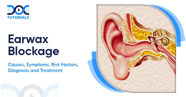
Human beings rely on five primary sense organs, and the ear is one of them. It plays a critical role in both hearing and maintaining balance. The glands in the ear canal secrete an oily wax that keeps our ears clean by trapping dirt and germs.

However, when the amount of secretion exceeds the normal level, it can block the ear canal. This leads to earwax blockage. Keep on reading to learn about earwax blockage, its causes and symptoms, and the associated treatment.

What is Earwax Blockage? 

When the earwax builds up in the ears beyond the normal amount, causing itching, ear pain, and other issues, it is known as an earwax blockage.

Medically known as “Cerumen Impaction”, earwax blockage is a condition in which the cerumen builds up in the ear beyond normal levels, blocking the ear canals and causing ear pain and itching. In severe cases, earwax blockage can cause hearing loss if left untreated.

Cerumen, or earwax, is a waxy oil that is produced by the glands present in the ear canal that help to keep the inner ear clean and protect it from infections.

Who is at Risk of Developing Earwax Blockage?

Anyone can develop an earwax blockage; however, people with certain conditions, such as hypertrichosis, developmental disabilities, eczema, misshapen ear canals, etc., are more prone to ceruminous impaction.

Earwax blockage can happen to anyone, regardless of age or gender. However, it is more commonly seen in children, affecting 10% children and 5% adults. The following section lists the people who are more prone towards developing earwax blockage:

  • People with hypertrichosis or hairy ears.
  • People who use foreign bodies, such as cotton swabs or other sharp objects, to clean their ears.
  • People above the age of 55.
  • People using hearing aids or earplugs.
  • People with developmental disabilities. 
  • People with skin conditions like Eczema. 
  • People with misshapen ear canals.

What are the Causes and Risk Factors of Earwax Blockage?

Some causes of earwax blockage include hypertrichosis, hard ceruminous debris, narrow ear canals, and the regular use of foreign bodies to clean the ear. Apart from these, risk factors such as recurrent ear infections also contribute to cerumen impaction.

The majority of people suffer from earwax blockage only because their glands naturally produce more cerumen. However, some other causes of cerumen impaction are:

  • Having hard cerumen 
  • Having hairy ears
  • Having narrow ear canals 
  • Wearing earplugs and hearing aids
  • Putting cotton swabs into the ears regularly

Apart from the causes, certain risk factors make people prone towards impacted cerumen, such as:

  • Bony growths or osteomas
  • Recurrent ear infections
  • Frequent exposure to water 
  • Males are at a higher risk than females
  • Psoriasis or dermatitis

What are the Symptoms and Diagnosis of Earwax Blockage?

The symptoms of earwax blockage include ear pain, hearing loss, ear itching, dizziness, and a feeling of fullness, which help diagnose the condition. The physician also uses an otoscope to examine the buildup of cerumen.

The primary symptom of earwax blockage is ear pain, which may or may not be associated with the following symptoms:

  • Ear ache
  • A feeling of fullness in the ears
  • Hearing loss, where the condition deteriorates over time
  • Ringing in the ears or tinnitus 
  • Itching in the ears
  • Discharge from the ear
  • Dizziness
  • Ear infections

Earwax blockage is diagnosed based on the above symptoms and a few other tests that are mentioned below:

  • Complete physical examination, including the Weber and Rinne tests.
  • An ear examination by the physician using an otoscope to check the amount of wax buildup.

What are the Treatment Options for Earwax Blockage?

Earwax blockage can be treated at home using earwax-dissolving solutions and ear irrigation, and at healthcare centres by trained staff.

Earwax blockage, if not treated in time, can cause hearing loss that worsens over time. Go through the following section to learn about the several methods to treat cerumen impaction at home:

  • Regular and Proper Cleaning of Ears: Use a washcloth to clean the outer area of your ear, and don’t forget to dry your ears after a shower properly. Don’t use sharp objects or safety pins to clean your ears.
  • Use Earwax-Dissolving Solutions: Using cerumenolytic solutions such as glycerin, mineral oil, baby oil, or saline helps soften hard impacted earwax. These solutions should be applied sparingly to the affected ear by lying on the opposite side.
  • Ear Irrigation: After softening the earwax with cerumenolytic solutions, use a syringe to rinse the ear with water or saline, then gently irrigate with a bulb syringe. 
  • In-office Earwax Removal: If the impaction is severe, visit your nearest healthcare centre, and the physician will remove it using special instruments such as a cerumen spoon, forceps, or a suction device.

What is the Prognosis of Earwax Blockage?

The prognosis for earwax blockage is good if treated promptly, but if left untreated, the person may suffer from severe infection and pain.

Once cleared, the person may again suffer from earwax blockage, and the hearing loss is temporary and partial. Hearing returns gradually after the impaction is cleared. People using hearing aids should get themselves checked every 3 to 6 months.

FAQs about Earwax Blockage

  1. How do you unblock a waxed ear?

Soften the earwax with the ceruminolytic ear drops, then clean the ear with gentle irrigation.

  1. Will the earwax blockage clear on its own?

Asymptomatic earwax blockage can clear on its own, but ceruminous impaction with clinical manifestations requires intervention by a skilled healthcare professional.

  1. How can I tell if I have an earwax blockage?

Some of the signs and symptoms of earwax blockage are:

  • Earache 
  • Ringing or noises in the ear 
  • Dizziness
  • Decreased hearing 
  • Coughing
  1. Is earwax removal painful?

No, earwax removal is not painful if performed correctly by a skilled staff member.

  1. How does a doctor remove earwax?

If the condition is severe, the doctor uses special instruments, such as a ceruminolytic spoon, suction, or forceps, to remove earwax.

Conclusion

Impacted earwax is a minor ailment that can worsen over time if left untreated. If not treated or cleared on time, the earwax blockage can cause a severe earache or infection that may lead to permanent hearing loss.

Maintain hygiene and keep your ears clean and healthy to prevent other chronic ear diseases that may affect your hearing and balance.

To understand more such complex NEET PG concepts easily, join DocTutorials today and explore our NEET PG course to excel in your career!

DocTutorials App Download Now

Latest Blogs

Hereditary Spherocytosis: Causes, Symptoms, Diagnosis, and Treatment

Hereditary

Hereditary spherocytosis is a foundational topic in Pathology, representing a unique intersection among genetics, membrane biology, and clinical presentation. It is a complex process in which a defect in the RBC membrane transforms the cell’s normal biconcave shape into a highly vulnerable, fragile sphere.

This structural transformation triggers continuous hemolysis, leading to jaundice and splenomegaly, making it both clinically significant and a very informative topic in pathology. The study of hereditary spherocytosis is necessary to enhance your knowledge of hemolytic anaemias.

Keep reading for a detailed analysis.

What is Hereditary Spherocytosis?

Hereditary spherocytosis is one of the inherited hemolytic anaemias resulting from a defective membrane protein of the RBC. Consequently, rigid and spherical red blood cells are formed. These spherocytes get trapped within the spleen and subsequently destroyed, leading to chronic extravascular hemolysis.

Hereditary spherocytosis is a genetic hemolytic anaemia due to inherited abnormalities of the RBC membrane component. Such proteins may include spectrin, ankyrin, and band 3 (4.2) protein. Structural defects weaken the RBC membrane, leading to the sequential loss of its parts.

As a result, red blood cells gradually assume a more spherical shape, becoming spherocytes that are smaller, denser, and more inflexible than normal biconcave RBCs.

Since spherocytes do not assume the normal shape, they cannot easily navigate the complex meshwork channels of the cords of Billroth, which traps them and eventually destroys them mechanically via splenic macrophages. This pathway produces chronic extravascular hemolysis as a hallmark of hereditary spherocytosis.

What are the Types of Hereditary Spherocytosis?

Hereditary spherocytosis presents clinical types based on the degree of anaemia, the amount of hemolysis, and the total number of symptoms. This classification helps inform management intervention decisions, including whether to transfuse or perform a splenectomy.

Here is the table showing the clinical classification of Hereditary Spherocytosis based on severity:

SeverityHaemoglobin (Approx.)Reticulocyte CountSymptomsSplenomegaly
Mild HS11–15 g/dLSlightly increasedOften asymptomatic or with only mild symptoms.Mild
Moderate HS8–12 g/dLMarkedly elevatedAnaemia, intermittent jaundice, and fatigue.Moderate
Severe HS<8 g/dLVery highSignificant anaemia, requiring transfusions, gallstones, and growth delay.Prominent

What are the Causes of Hereditary Spherocytosis?

Hereditary spherocytosis arises from inherited defects in RBC membrane proteins that cause membrane instability and progressive loss of surface area. The result will be the transformation of red cells into inflexible spherocytes that predominantly get destroyed in the spleen, leading to chronic hemolysis.

Hereditary spherocytosis is caused by inherited mutations of genes encoding key red blood cell membrane proteins. These proteins are responsible for strength and elasticity, thereby supporting the stability and flexibility of the RBC membrane.

The most common defects include:

  • Spectrin (α or β chains)
  • Ankyrin
  • Band 3 protein
  • Protein 4.2

These proteins attach the cytoskeletal network to the lipid bilayer. If any of them is abnormal or deficient, there will be weaker vertical connections between the two layers. This instability results in a progressive loss of membrane fragments and a reduction in the RBC’s surface area while its volume remains constant.

Hence, it changes into a spherocyte, which is typically rigid and unable to deform. Spherocytes cannot flex and extend as healthy biconcave red blood cells do, so they cannot easily pass through the splenic sinusoids. 

The spleen detects them as defective and begins removing them from the circulation, initiating extravascular hemolysis. Most cases follow an autosomal dominant inheritance, meaning a single mutated allele is sufficient to cause disease.

The remaining cases are autosomal recessive, usually producing more severe forms due to the complete loss of functional protein.

What are the Risk Factors of Hereditary Spherocytosis?

The most significant risks for hereditary spherocytosis are in families, since this disease has a strong predilection towards autosomal dominant inheritance. It is more prevalent among individuals of Northern European descent, though it can be observed worldwide and can also result from de novo mutations.

Several factors predispose one to get hereditary spherocytosis, primarily because it is a genetic disease:

  • Family History

One of the major risk factors is having a parent with the condition since most cases are autosomal dominant. Affected individuals transmit the mutation to their offspring in half of the cases. Autosomal recessive cases also occur and are identified when carrier parents, who do not show any symptoms, have an affected child.

  • Genetic Ancestry

Northern European populations show higher frequencies, although reports indicate a dominant occurrence of hereditary spherocytosis in Japanese families, North Africa, Brazil, and worldwide. In any ethnic group, the condition may manifest itself despite regional clustering.

  • Spontaneous Mutations

Around one-fourth of cases arise from new (de novo) mutations in RBC membrane protein genes, even without a family history.

  • Early Neonatal Jaundice

Babies who show frequent jaundice soon after birth, especially if it is out of proportion to physiological expectations, may be at increased risk of harbouring hereditary spherocytosis that will declare itself with recurrent hemolytic episodes later in life.

What are the Symptoms of Hereditary Spherocytosis?

Hereditary spherocytosis may present with common anaemia, jaundice, and splenomegaly since it is a chronic hemolytic process, but patients can also develop pigment gallstones or severe neonatal jaundice.

The severity of symptoms in hereditary spherocytosis ranges from asymptomatic to severe hemolytic disease, depending on the degree of membrane protein deficiency.

Common clinical symptoms include:

  • Anaemia

Patients often present with signs such as fatigue, pallor, reduced exercise capacity, and irritability in children. These symptoms may worsen during infections due to increased hemolysis.

  • Jaundice

Elevated unconjugated bilirubin from ongoing RBC breakdown leads to scleral icterus and yellowish skin discolouration. Intermittent jaundice is especially common in adolescents and adults with hereditary spherocytosis.

  • Splenomegaly

In patients with hereditary spherocytosis, the spleen enlarges and becomes soft as it continuously clears spherocytes from the circulation. Persistent splenic trapping contributes to chronic hemolysis.

  • Pigment Gallstones

Chronic bilirubin overproduction predisposes patients, often in adolescence or early adulthood, to black pigment gallstones, which may present with abdominal pain.

  • Severe Neonatal Jaundice

Newborns with hereditary spherocytosis may experience marked hyperbilirubinemia requiring phototherapy or, in rare cases, exchange transfusion.

  • Aplastic Crisis 

A sudden drop in RBC production, most frequently triggered by Parvovirus B19, can cause profound anaemia. This is a medical emergency and a classic complication in hereditary hemolytic disorders.

How is Hereditary Spherocytosis Diagnosed?

The diagnosis of hereditary spherocytosis is made based on clinical and laboratory findings, confirmed by specific tests. The findings include evidence for hemolysis, typical red cell morphology, and the exclusion of other causes.

Here is the table summarising the diagnostic tests for Hereditary spherocytosis (HS):

TestWhat it AssessesFinding in HSClinical Relevance
Peripheral SmearRBC morphologySpherocytes, polychromasiaFirst clue: visual identification of characteristic cells.
CBC IndicesRed cell parametersIncreased MCHC (Mean Corpuscular Haemoglobin Concentration), normal/low MCV (Mean Corpuscular Volume)Supports diagnosis; high MCHC is a hallmark of HS.
Reticulocyte CountBone marrow responseElevatedIndicates active hemolysis (red cell destruction).
Osmotic Fragility TestRed cell membrane stabilityIncreased fragility (hemolysis at higher saline concentration)Classical test: spherocytes lyse easily in a hypotonic solution.
EMA Binding Test (Flow Cytometry)Band 3 protein (anion exchanger)Decreased fluorescenceMost sensitive screening test; assesses deficiency of key membrane proteins.
Direct Antiglobulin Test (DAT)Presence of antibodies on RBCsNegativeDifferentiates HS from Autoimmune Hemolytic Anaemia (AIHA), where it is positive.

What are the Treatment Options for Hereditary Spherocytosis?

Treatment of hereditary spherocytosis runs from supportive care, which includes folic acid supplementation and regular monitoring, to definitive treatment, i.e., splenectomy in moderate to severe cases. Splenectomy greatly reduces hemolysis and improves symptoms.

The various treatment options for Hereditary Spherocytosis include the following:

  • Supportive Measures

Hereditary spherocytosis is initially managed with supportive therapy, as continuous red blood cell destruction increases the marrow’s need to produce new ones. Supportive therapy involves:

  • Folic acid supplementation to maintain adequate erythropoiesis in the setting of chronic hemolysis.
  • Routine monitoring for progressive anaemia, increasing jaundice, and early signs of hemolytic or aplastic crises.
  • Paediatric follow-up, especially in younger patients, to assess growth, splenic enlargement, and overall disease progression.
  • Splenectomy (Definitive Treatment)

When symptoms become significant, such as persistent anaemia, marked jaundice, or recurrent crises, splenectomy becomes the primary definitive option.

Partial splenectomy may be considered in children to reduce infection risk. Splenectomy is regarded as the first-line treatment because:

  • Reduces hemolysis by removing the organ where abnormal RBCs are predominantly destroyed.
  • Recommended for moderate to severe cases, or when complications like growth failure or frequent transfusions occur.
  • Can be performed as a total or partial splenectomy, based on age and risk–benefit considerations.
  • Cholecystectomy

Long-standing hemolysis increases the bilirubin load, predisposing patients to pigment gallstone formation.

Symptomatic stones can cause abdominal pain, jaundice, or biliary obstruction; hence, timely surgical management through cholecystectomy is important. Cholecystectomy is considered for:

  • Patients with symptomatic pigment gallstones or recurrent biliary colic.
  • Often performed simultaneously with splenectomy to minimise hospitalisation and prevent future biliary complications.
  • Blood Transfusions

Some patients may experience sudden worsening of anaemia, especially during viral illnesses such as Parvovirus B19 infection. In these situations, transfusion support becomes essential until the bone marrow recovers.

Blood transfusions are: 

  • Used in episodes of severe anaemia when haemoglobin drops rapidly.
  • Commonly required during aplastic crisis, where temporary suppression of erythropoiesis occurs.
  • Usually, a short-term measure, as needs decrease significantly after splenectomy.

What are the Complications of Hereditary Spherocytosis?

Hereditary spherocytosis, if left untreated, will eventually lead to progressive hemolytic anaemia, pigment gallstones, and a life-threatening aplastic crisis. Chronic folate deficiency may precipitate megaloblastic changes.

The lack of recognition and appropriate management of hereditary spherocytosis can result in not only the continued destruction of red blood cells (RBCs) along with metabolic strain, but also multiple short and long-term complications.

These are:

  • Extreme Hemolytic Anaemia

The ongoing destruction of RBCs may exceed the bone marrow’s capacity to compensate, leading to severe anaemia with fatigue, pallor, and limited ability to exercise.

  • Pigment Gallstones

Chronic hemolysis creates large amounts of unconjugated bilirubin, which is typically stored in the liver. This excess bilirubin can form black pigment gallstones, leading to biliary colic, cholecystitis, or obstructive jaundice.

  • Aplastic Crisis

Infection with Parvovirus B19 may transiently decrease the rate of erythropoiesis by destroying erythrocyte progenitors in the bone marrow, leading to an abrupt and life-threatening decline in haemoglobin levels.

This situation is considered to be a medical emergency in patients with hereditary hemolytic disorders.

  • A Megaloblastic Crisis

An increase in marrow activity leads to an increased demand for folate; when the demand for folate exceeds the available supply, folate deficiency will ultimately lead to megaloblastic changes in the marrow and to further deterioration of anaemia and impairment of the formation of RBCs.

  • Growth Delay in Children

Untreated through proper medical monitoring, chronic anaemia and the increased metabolic burden will typically slow down physical development and delay important developmental milestones in children.

  • Pulmonary Hypertension (Uncommon)

Prolonged episodes of hemolysis can impact endothelial cell function and result in the depletion of nitric oxide, placing certain patients at risk for the development of pulmonary hypertension in acute or chronic settings.

FAQs about Hereditary Spherocytosis

1.  Is hereditary spherocytosis always inherited?

This does not always happen. Approximately 25% of patients with hereditary spherocytosis have no familial history of this condition. The other 75% of cases are inherited from an affected parent.

2. Are spherocytes pathognomonic for hereditary spherocytosis?

Spherocytes are not exclusively pathogenic to hereditary spherocytosis but are also seen in autoimmune haemolytic anaemia. Therefore, to differentiate between hereditary spherocytosis and autoimmune haemolytic anaemia, it is essential to perform a Direct Antiglobulin test.

3.  Can hereditary spherocytosis be cured?

There is no genetic cure for hereditary spherocytosis. However, a splenectomy can substantially decrease hemolysis and improve symptoms. Although the membrane defect will remain the same.

4. Is splenectomy safe in children?

Splenectomy is usually delayed until after the child has reached 5 years of age to minimise the risk of severe infections. Immunisations and other methods to minimise infection are usually administered before and after splenectomy.

5. Why is MCHC increased in hereditary spherocytosis?

Hereditary spherocytosis causes a decreased surface area of the RBC membrane, resulting in less total intracellular fluid (H2O) within each RBC. The decrease in H2O leads to increased total Hb concentration in each RBC, thereby causing the MCHC to be elevated compared to normal erythrocytes.

Conclusion

Hereditary spherocytosis is classified as a genetic hemolytic anaemia that creates spherocyte production and red blood cell destruction within the spleen. The patient outcome can be improved with an early diagnosis, folic acid supplementation, and timely surgical intervention, through performing a splenectomy, cholecystectomy, or blood transfusions.

NEET-PG requires candidates to know the definitions of molecular defects, diagnostic tests, and clinical complications related to hereditary spherocytosis. For added guidance, DocTutorials can be your study partner.
We offer crisp videos, clinical Qbank, exam-focused notes, flashcards, and mindmaps to ensure aspirants are well-prepared and gain complete clarity over complex NEET PG concepts. Join DocTutorials today and explore our NEET PG course to excel in your medical career!

DocTutorials App Download Now

Latest Blogs

How to Perform CPR: Types, Procedure, and Essential Steps

CPR

CPR (cardiopulmonary resuscitation) is a critical technique that can restore the heartbeat by continuous compression and breathing techniques. The CPR procedure can help an individual to survive a cardiac arrest during an emergency.

Every individual, especially medical students, should learn CPR to handle critical situations until professional medical help arrives. With DocTutorials, you can cover all the aspects of CPR in detail through high-quality study materials and video recordings.

Read on to learn the steps of CPR, its guidelines, mistakes to avoid while performing CPR, and more. The well-explained CPR techniques will also help you get a brief idea about our teaching methods!

What is CPR?

Cardiopulmonary resuscitation or CPR is a process to restore the heart functioning of a person who has had a cardiac arrest. It involves compressing the chest so the heart can pump blood and return to a regular rhythm. The CPR procedure also involves breathing oxygen into the victim’s mouth to increase the oxygen level in their body and help blood blow to the crucial body organs.

You should remember the CAB order while performing CPR. Here: 

  • “C” stands for chest compression 
  • “A” represents airway; you need to open the victim’s airway 
  • “B” stands for giving rescue breaths to the patient.

How to Prepare for CPR?

Here are the steps to prepare for a CPR procedure:

  • If a person gets cardiac arrest before you and starts collapsing, ask loudly if they are alright
  • Call for medical aid immediately if the individual does not respond
  • Get an AED (automated external defibrillator) from someone who is nearby
  • Keep the person lying on their back and tilt their head back
  • Get closer to the individual and try to listen if the heart is beating
  • Check for the chest going up and down and whether the person is breathing or not
  • Touch the individual’s neck to check if there is pulse
  • If you cannot find a pulse, CPR must be performed immediately

What are the Different Types of CPR?

Following are the four main CPR types:

  • Hands-only CPR

This type is also called compression-only CPR. In this process, you don’t need to provide rescue breaths; it only involves chest compression. This technique includes continuous compression of 100-120 per minute.

  • Standard CPR

It involves combining both chest compressions and rescue breaths. In each cycle, you need to perform 2 rescue breaths after 30 chest compressions. This procedure is also known as the traditional CPR method. 

  • Hands-only CPR with an AED

In this process, AED or automated external defibrillator devices are combined with hands-on CPR to give an electric shock to the individual’s heart suffering from cardiac arrest. You can continue with the hands-on technique until the AED is ready.

  • Paediatric CPR

As the name suggests, this CPR technique is used when the victim is a child or infant. You need to provide 15 compressions after 2 rescue breaths in each cycle.

How to Perform CPR for an Adult: Steps to Follow

Cardiopulmonary resuscitation is a vital skill that can save lives in emergencies by providing essential life support. Here’s how to perform CPR effectively:

Step 1: You need to assess the scene to check if the area is safe before approaching the individual.

Step 2: Check for their response by gently tapping the patient and asking if they are okay. Look for signs of movement or response.

Step 3: If you find the person unresponsive, immediately call emergency services.

Step 4: Open the airway by tilting the patient’s head back and lifting their chin.

Step 5: Listen and feel for breaths by placing your ear near the patient’s mouth and observing chest movements.

Step 6: If the patient isn’t breathing, start compressions. Place one hand on top of the other, interlock fingers, and keep elbows straight.

Step 7: Perform 100-120 compressions per minute, depressing the chest at least 2 inches and allowing it to recoil fully between compressions.

Step 8: After 30 compressions, provide two rescue breaths. Pinch the nose, seal the mouth with yours, and give a one-second breath. Continue cycles until help arrives.

Following these steps, you can provide critical care until medical professionals arrive. Remember, CPR is a lifesaving intervention that requires practice and confidence to perform effectively.

How to Perform CPR for a Child?

The CPR procedure for a child is almost the same as that of an adult. Here are some of the essential things to consider:

  • Provide 2 rescue breaths and then 30 cycles of chest compression.
  • For small children, avoid using two hands for chest compression; only one is enough.
  • In the case of infants, use two fingers for chest compression instead of hands. Make sure the compression is 1.5 inches only and not 2 inches like adults.
  • The rescue breath process for infants is also different from that of adults. Here, you need to gently seal the infant’s nose and mouth with yours and puff air to provide rescue breath.

What are the American Heart Association (AHA) Guidelines for CPR?

The American Heart Association (AHA) updates its CPR guidelines every five years to reflect the latest scientific evidence and improve survival rates from cardiac arrest. As of the latest updates, the AHA emphasises the importance of high-quality chest compressions and minimising interruptions during CPR. 

The traditional 30:2 ratio of compressions to breaths remains standard for adults. However, there is a growing trend towards continuous chest compressions (CCC) before intubation, which may eventually replace traditional CPR with Cardio-Cerebral Resuscitation (CCR) in some settings.

Key points from recent guidelines include:

  • High-Quality Compressions: Ensure compressions are at least 2 inches deep at a rate of 100-120 per minute.
  • Minimising Interruptions: Keep pauses in chest compressions to a minimum to maintain blood circulation.
  • Technology Integration: Utilise devices like CPR feedback tools and mobile apps for real-time guidance.

What are the Mistakes to Avoid While Performing CPR?

Performing CPR effectively requires avoiding the following mistakes as they can reduce its effectiveness or even cause harm:

  • Not Calling for Help Early

If you fail to call emergency services promptly, it can delay professional medical assistance.

  • Incorrect Hand Placement

If your hand placement is not correct, the CPR will not be effective. Hands should be placed on the centre of the chest, between the nipples, with fingers interlaced.

  • Inadequate Chest Compressions

If the right sets of chest compressions are not provided, the heartbeat will not be restored even after CPR. You should give chest compressions at least 2 inches deep for adults at a rate of 100-120 per minute.

  • Interrupting Compressions

If you keep pausing between chest compressions, blood flow to and from the heart cannot be normalised. Minimise pauses between compressions to maintain continuous blood flow.

  • Not Tilting the Head

If the head of an individual who has had cardiac arrest is not tilted correctly, the airway will not open. Ensure proper head tilt to open the airway before giving rescue breaths.

FAQs About CPR (Cardiopulmonary Resuscitation)

  1. What are the advantages of providing CPR?

CPR keeps blood circulating, preventing organ damage during cardiac arrest.

  1. What needs to be done after providing CPR?

After CPR, the person is sent to a hospital. Healthcare providers assess for organ damage, determine the cardiac arrest cause, and provide necessary treatment. Many survivors initially remain in a coma.

  1. Can CPR be fatal?

CPR is beneficial even if not performed perfectly, as it helps cardiac arrest victims, potentially improving survival chances with proper technique.

  1. How will you understand whether the CPR is effective or not?

CPR is effective, even if the technique is not perfect. Look for chest rise during rescue breaths as a sign of effectiveness.

  1. Can a person’s ribs get broken due to CPR?

CPR can sometimes break ribs due to the force required to compress the chest and circulate blood throughout the body.

Conclusion

CPR is a crucial method that can save lives during emergencies like cardiac arrest. By learning the proper compression and breathing methods, you can restore the heartbeat of an individual who has had a heart attack. For healthcare professionals seeking to enhance their skills or medical students who need to learn about CPR for their MBBS syllabus, DocTutorials offers comprehensive resources. It includes expert-led videos and case-based discussions. By enrolling in our classes, you can learn more about the CPR procedure and provide critical care until medical help arrives.

NEET PG Online Live Power Pack
DocTutorials App Download Now

Latest Blogs

Cardiopulmonary Resuscitation: Definition, Types of CPR, Essential Steps, and More

Cardio Pulmonary

CPR, or cardiopulmonary resuscitation, is a vital method of saving lives by keeping the blood flowing even after the heart has stopped beating. The technique plays a critical role in maintaining the circulation of the blood after an individual has suffered from cardiac arrest.

According to a report by the Indian Institute of Emergency Medical Services (IIEMS), only 2% of the Indian population knows how to perform CPR. Thus, learning about this lifesaving mechanism is crucial not only for medical students and healthcare providers but also for other individuals to provide the necessary assistance during emergencies.

This article covers when to use CPR, its different types, a step-by-step guide to perform CPR and other crucial aspects. 

What is CPR?

The full form of CPR is cardiopulmonary resuscitation, in which the term ‘cardio’ refers to the heart, while ‘pulmonary’ denotes the lungs. Resuscitation is the procedure of ensuring a constant flow of blood and oxygen in the body, which is a vital function played by these central organs. 

CPR helps restore the functioning of an individual’s heart after a cardiac arrest. The procedure involves compressing the chest so that the heart can pump blood and come back to its regular rhythm. The CPR process involves breathing oxygen into the patient’s mouth to enhance the oxygen level in the body and help blood flow to the vital organs. 

When to Use CPR?

CPR could be required for a person who has stopped breathing as a result of several scenarios, such as:

  • Cardiac arrest or heart attack
  • Road traffic accidents
  • Choking accidents
  • Near-drowning
  • Poisoning
  • Suffocation
  • Smoke inhalation
  • Drug or alcohol overdose
  • Electrocution
  • Suspected sudden infant death syndrome

What are the Types of CPR?

Check out the various types of CPR in the section below:

  1. Standard CPR

In this technique, chest compressions are given with the addition of rescue breaths. You need to provide two rescue breaths in every cycle of 30 chest compressions. This is commonly known as traditional CPR.

  1. Hands-Only CPR

Another name for compression-only CPR, this technique is all about chest compressions and does not require rescue breaths. It includes providing continuous compressions at a rate of 100-120 per minute.

  1. Hands-Only CPR with an AED

This technique combines the use of an automated external defibrillator (AED) with manual CPR. The AED gives the heart an electric shock to restore its normal rhythm, and you keep compressing until the machine is ready.

  1. Paediatric CPR

As its name suggests, this method is used specifically on children or babies. With each cycle, you give 15 compressions and then two rescue breaths.

How to Prepare for Cardiopulmonary Resuscitation (CPR)?

Mentioned below are the common steps to consider while preparing for a CPR procedure:

  • If you see a person having a cardiac arrest and they start falling down, call out loudly to ask how they are feeling.
  • Call for medical attention immediately if the person does not respond.
  • Ask someone around you to fetch an automated external defibrillator (AED).
  • Keep the person on their back and tilt their head back slowly.
  • Lean in further to check if you hear any heartbeat.
  • Look to the chest for rising and falling to see if the individual is breathing.
  • Check for a pulse at the neck by feeling.
  • If there is no pulse, start CPR immediately.

What is the Step-by-Step Process to Perform CPR for an Adult?

Cardiopulmonary resuscitation is a medical technique that can save lives in emergencies by providing crucial life support. Here is the step-by-step guide on how to conduct CPR effectively on adults:

  1. Start by assessing the environment to make sure it is safe before reaching the person in need.
  2. Check if the individual is responsive by tapping them gently and asking if they are alright. Look for any movement or response.
  3. If the patient is not responsive, immediately call emergency services for help.
  4. Open the airway by tilting the person’s head back and lifting the chin.
  5. Listen for breathing by putting your ear beside the person’s mouth and observing for chest rises.
  6. If the person is not breathing, begin chest compressions. Place one hand on top of the other, lace your fingers together, and keep your elbows locked.
  7. Give 100-120 compressions per minute, compressing at least 2 inches into the chest and letting it recoil completely between compressions.
  8. Give two rescue breaths after 30 compressions. Pinch the nose, cover their mouth with yours, and give a one-second breath. Repeat this sequence until medical help arrives.

How to Perform CPR for a Child?

When it comes to children, the CPR procedure is almost identical to that of an adult. The following are some of the key things to consider while performing CPR for children:

  • Step 1: Check for Breathing and Signs of Life
    • Look at the chest for evidence of normal breathing or movement for no longer than 10 seconds. If the victim is not breathing or has no signs of life, then continue with CPR.
  • Step 2: Start Chest Compressions
    • Place one hand at the centre of the child’s chest and put your other hand on top, locking your elbows. Give 30 compressions to a depth of around 2 inches at 100-120 compressions per minute.
  • Step 3: Perform Rescue Breaths
    • Open the airway by lifting the chin and tilting the head back, pinching the nose. Provide two slow and gentle breaths, just enough to raise the chest.
  • Step 4: Repeat CPR Cycles
    • Continue repeating 30 chest compressions and two rescue breaths. Repeat the cycle until you notice signs of life, an AED is available, or medical help is on the scene.
  • Step 5: Infant CPR (Alternate Method)
    • For infants, use the two-finger technique for compressions on the chest, 1.5 inches deep, at a rate of 100-120 times per minute. Make sure that the chest returns to its normal position after each compression.
  • Step 6: Perform Chest Compression 
    • For younger kids, use a single hand to do chest compressions with a downward pressure of 2 inches at the same rate of 100-120 per minute. The compression should be firm enough but not too hard for their smaller chests.
  • Step 7: Deliver the Rescue Breath 
    • Carefully cover the child’s nose and mouth with your mouth and exhale a slow, gentle puff to raise the chest. 
  • Step 8: Continue Until Help Arrives
    • Keep performing CPR until there are signs of life or the EMS (Emergency Medical Services) personnel arrive. 

Consider checking out a CPR diagram to get a visual idea of how this procedure is performed. It will help you gain a more detailed idea of what exactly you need to do, which can be a lifesaver during emergencies.    

FAQs About Cardiopulmonary Resuscitation

  1. What should be done after CPR?

After CPR, the individual is sent to a hospital. Doctors analyse for organ damage, determine the cause of the cardiac arrest, and adopt necessary treatment methods. In a few cases, the survivors may also go into a coma. 

  1. How can we comprehend whether CPR is effective?

Cardiopulmonary resuscitation (CPR) can still be effective, even if the method is not performed perfectly. To check whether it is effective, you can look for the chest rise during rescue breaths.

  1. When is CPR performed?

CPR is performed in the event of an emergency when an individual’s breathing or heartbeat stops.

  1. What is the purpose of CPR in nursing?

The purpose of CPR in nursing is to assist in saving a life by maintaining oxygenation of the brain and other critical organs until advanced medical intervention is provided. 

  1. What are the limits for CPR?

The optimal duration of CPR is approximately 55–62 minutes in patients with shockable rhythms and 24–34 minutes in patients with non-shockable rhythms. 

Conclusion

Learning CPR properly and applying the same during emergencies can restore a heartbeat and save lives. As a post-graduate medical student, if you are preparing for the NEET PG exam and want to learn about CPR in further detail, DocTutorials has you covered. 

Here, we provide complete NEET PG study materials, professional video courses, a Quick Revision Program (QRP), and interactive study aids for aspiring medical students. 

NEET PG Online Live Power Pack
DocTutorials App Download Now

Latest Blogs

NEET PG Online Live Power Pack
DocTutorials App Download Now