pg-residency Archives - Page 2 of 9 -

Pediatric Dermatology for PG Residents: Decoding Skin Lesions and Vesicles 

Pediatric Dermatology

Mastering clinical genetics, dysmorphic assessments, and advanced molecular treatments like CAR T Cell Therapy is no longer optional for today’s post-graduate (PG) medical residents. This comprehensive guide connects foundational pediatric bedside examinations with the cutting-edge frontiers of gene modification and CAR T Cell Therapy. 

Get High Quality, High Yield PG Residency Notes for Free!

The Foundation of Dysmorphic Examination 

Evaluating a child for genetic syndromes begins with a meticulous general assessment and anthropometry. Understanding foundational metrics helps clinicians identify underlying genetic abnormalities before moving to advanced treatments like CAR T Cell Therapy. 

Anthropometry and Crucial Metrics 

A non-negotiable metric for any syndromic assessment is the Upper Segment to Lower Segment (US:LS) ratio. This ratio must always be correlated with age-specific norms. As a rule for all dysmorphic children, clinicians must document the US:LS ratio even if it falls within normal limits. Alongside this, the clinical checklist should include assessing consciousness, vital signs, and malnutrition staging (acute, chronic, or acute-on-chronic). A rapid screen for general signs like pallor, icterus, cyanosis, and clubbing is also essential. 

Head, Skull, and Facial Features 

Plotting the occipitofrontal circumference (OFC) on a growth chart determines whether a patient has microcephaly, macrocephaly, or normal head growth. Furthermore, analyzing the anterior fontanel for size, shape, and bulging (if open) is critical. Skull shapes vary significantly in craniosynostosis: scaphocephaly presents with increased AP diameter, while brachycephaly shows increased biparietal diameter. 

Facial features provide immense diagnostic clues. For instance, an upward lateral canthus (mongoloid slant) is associated with Down Syndrome, whereas a downward lateral canthus (antimongoloid slant) points to Noonan Syndrome. 

Below is a table summarizing key facies types and their associated conditions. 

Common Facies and Associated Syndromes 

Facies Type Key Clinical Features Associated Conditions 
Coarse Facies Thick lips, flat bridge, large tongue MPS, Cretinism 
Elfin Facies Wide mouth, smooth philtrum, pointed teeth Williams Syndrome 
Hemolytic Facies Frontal bossing, crowded teeth Thalassemia 
Other Facies Moon, Hepatic, Mask-like Cushing’s, CLD, Moebius 

The Evolution of Gene Therapy Strategies 

While physical examinations identify the phenotypic expressions of genetic disorders, the treatment landscape is rapidly shifting toward molecular interventions, laying the groundwork for complex modalities like CAR T Cell Therapy. Gene therapy strategies are broadly categorized into In Vivo and Ex Vivo mechanisms. 

In Vivo Therapies 

In Vivo gene therapy involves direct administration inside the body, where gene modification occurs internally. The primary methods for delivery include viral vectors (like AAV), nanoparticles, and RNA/Ribosomes. 

How do Antisense Oligonucleotides (ASO) function in the treatment of Duchenne Muscular Dystrophy (DMD)? 

In DMD, which is an X-linked recessive disorder characterized by deficient dystrophin, ASO therapy works via exon skipping. The ASO binds to the pre-mRNA and masks specific exons—such as skipping Exon 51 with Eteplirsen—to bypass the mutation stop codon, ultimately allowing translation into a shorter but functional dystrophin protein. 

In Vivo delivery systems heavily rely on advanced transport mechanisms, such as GalNAc conjugates which are receptor-dependent and liver-specific (administered subcutaneously), and Lipid Nanoparticles (LNP) which utilize non-receptor dependent entry via membrane fusion or endocytosis. 

In Vivo ASO Therapeutics Overview 

Disease Drug Name Gene Target Route of Administration 
Spinal Muscular Atrophy Nusinersen (Spinraza) SMN2 Gene Intrathecal 
TTR-Amyloidosis Inotersen Transthyretin (TTR) Subcutaneous 
Hyperlipidaemia Mipomersen Apolipoprotein B100 Subcutaneous 
Familial Chylomicronaemia Volanesorsen Apolipoprotein C3 Subcutaneous 

The Breakthrough of CAR T Cell Therapy (Ex Vivo) 

Ex Vivo gene therapy takes cells harvested from the patient, modifies them in a laboratory setting, and returns them to the patient. This framework is exactly how CAR T Cell Therapy operates. 

CAR T Cell Therapy is a revolutionary immunotherapy primarily utilized in pediatrics for the treatment of Acute Lymphoblastic Leukaemia (ALL). The process of CAR T Cell Therapy begins with the isolation of the patient’s own T-cells. Once isolated, these cells undergo modification ex vivo to express the Chimeric Antigen Receptor (CAR). 

After being successfully modified to become CAR T-cells, they are expanded in the laboratory and subsequently re-infused into the patient’s bloodstream. The core action of CAR T Cell Therapy is to grant specific cytotoxic activity against target cancer cells. By leveraging the body’s own immune system, CAR T Cell Therapy ensures the direct and targeted destruction of leukemia cells.  

The integration of lentiviral vectors is frequently utilized in these Ex Vivo additions to ensure functional gene expression. Thus, CAR T Cell Therapy represents a paradigm shift in how hematological malignancies are treated in pediatric patients. 

Advanced Ex Vivo Gene Editing: CRISPR/Cas9 

Beyond CAR T Cell Therapy, Ex Vivo strategies also utilize CRISPR/Cas9 technology, functioning as “molecular scissors.” A prominent example is the treatment for Sickle Cell Anaemia using Exagamglogene Autotemcel (Exa-cel/Casgevy). This mechanism involves the inactivation of the BCL11A gene, which serves as the switch that normally stops fetal hemoglobin (HbF) production. The resulting increase in HbF successfully prevents red blood cell sickling and subsequent crises. 

However, much like the stringent monitoring required after CAR T Cell Therapy, CRISPR therapies demand careful safety oversight. Clinicians must monitor for off-target effects and potential malignant transformations, as these represent permanent genetic changes compared to treatments like Hydroxyurea. 

Frequently Asked Questions (FAQs) 

1. What is the primary pediatric indication for CAR T Cell Therapy? 

The primary pediatric use for CAR T Cell Therapy is the treatment of Acute Lymphoblastic Leukaemia (ALL). 

2. How are cells modified in CAR T Cell Therapy? 

In CAR T Cell Therapy, a patient’s T-cells are isolated, modified ex vivo to express Chimeric Antigen Receptors (CAR), expanded, and then re-infused. 

3. What is the mechanism of action for CAR T Cell Therapy? 

CAR T Cell Therapy grants the modified T-cells specific cytotoxic activity directed against target cancer cells, leading to cancer cell destruction. 

4. How does CAR T Cell Therapy differ from In Vivo gene therapy? 

CAR T Cell Therapy is an Ex Vivo method, meaning cells are harvested, modified outside the body in a lab, and then returned. In Vivo therapy involves direct administration and internal modification. 

5. What is the US:LS ratio in dysmorphic examinations? 

The Upper Segment to Lower Segment (US:LS) ratio is a non-negotiable metric essential for all dysmorphic children and must correlate with age-specific norms. 

6. Which condition is indicated by an upward lateral canthus? 

An upward lateral canthus, also known as a mongoloid slant, is a feature associated with Down Syndrome. 

7. How is Spinal Muscular Atrophy (SMA) treated using gene therapy? 

SMA is treated using Nusinersen (Spinraza), an In Vivo therapy administered intrathecally that activates the inactive SMN2 gene to compensate for the missing SMN1. 

8. What vectors are used in Ex Vivo therapies like CAR T Cell Therapy? 

Ex Vivo therapies often utilize Lentiviral vectors for the laboratory integration of functional genes. 

9. How does CRISPR/Cas9 help Sickle Cell Anaemia patients? 

It inactivates the BCL11A gene, increasing Fetal Hemoglobin (HbF) production, which prevents sickling and crises. 

10. What dermatological marker indicates Tuberous Sclerosis? 

The presence of an Ash Leaf Macule (a hypopigmented spot) is a hidden dermatological marker for Tuberous Sclerosis. 

DocTutorials App Download Now

Latest Blogs

Essential Pediatric Surgery: Navigating CDH Management and Neuroblastoma Staging 

pediatric surgery

Mastering the complexities of pediatric surgery is a critical milestone for any PG Resident. The neonatal and infant physiology demands a nuanced approach, particularly when dealing with life-threatening congenital defects and early-onset malignancies. This comprehensive pediatric surgery guide delves into two high-yield topics: Congenital Diaphragmatic Hernia (CDH) and Neuroblastoma, providing factually accurate, structured insights to enhance both your theoretical knowledge and clinical acumen. 

Get High Quality, High Yield Pediatrics PG Residency Notes for Free! 

Congenital Diaphragmatic Hernia (CDH): Diagnosis and Management 

In the realm of pediatric surgery, distinguishing between causes of neonatal respiratory distress is paramount. When a newborn presents with respiratory distress alone, one must highly suspect Congenital Diaphragmatic Hernia (CDH). Conversely, if the presentation includes distress, drooling, and cyanosis, Tracheoesophageal Fistula (TEF) becomes the primary differential. 

Embryology and Presentation 

The embryological defect responsible for CDH occurs between gestational weeks 8 and 10 due to the failure of the pleuroperitoneal canal membrane to close. 

  • Bochdalek Hernia: This is the most common variant, presenting posterolaterally, and occurs more frequently on the left side than the right. 
  • Morgagni Hernia: This is a less common, anteromedial defect. 

Radiographic confirmation via chest and abdomen X-ray reveals bowel gas patterns displacing the lung, a scaphoid (empty) abdomen, and a paucity of abdominal gas. 

Pathophysiology and Pulmonary Insult 

The core issue in CDH is not merely the anatomical defect, but the resulting pulmonary insult. The diaphragmatic defect allows herniation of abdominal contents into the thorax. 

  • This causes mechanical compression, leading to pulmonary hypoplasia, which is more severe on the ipsilateral side compared to the contralateral side. 
  • Simultaneously, arteriolar remodeling occurs, characterized by muscularization and a loss of elasticity. 
  • These factors culminate in Pulmonary Hypertension, which remains the major cause of mortality in these patients. 

Antenatal prediction often relies on the Lung-Head Ratio (LHR), calculated by dividing the longest perpendicular lung measurements by the head circumference. An LHR of less than 1.0 indicates a poor prognosis due to hypoplasia and hypertension, whereas an LHR greater than 1.4 suggests a good prognosis with adequate lung volume. 

Prognostic Indicators in CDH 

Parameter Favourable Signs (Good Survival) Unfavourable Signs (High Mortality) 
Liver Position Abdominal (No herniation) Thoracic Herniation 
Onset Late gestation Early gestation 
Defect Size Small Large 
Stomach Position Intra-abdominal Thoracic 
Birth Weight > 2.5 kg Low 
Associated Anomalies None Present 

Mid-Blog Q&A Challenge 

Question: What is the most critical initial step in the management hierarchy for a newborn presenting with CDH? 

Answer: Medical Stabilization is the absolute priority to reduce Pulmonary Hypertension. In the context of pediatric surgery, CDH is treated strictly as a medical emergency first, not a surgical one; rushing to surgery is a critical warning to avoid. 

Surgical Repair and Post-Operative Safety 

Once medical stabilization is achieved, elective pediatric surgery repair is performed. For large defects where primary closure is impossible, a patch repair using synthetic mesh is required, whereas primary suture approximation is ideal for smaller defects. Laparoscopic repair is the current standard, offering reduced pain and faster recovery compared to the historical laparotomy subcostal approach. 

Post-operatively, vigilance is required for Abdominal Compartment Syndrome due to the increased pressure from returned organs. A fall in urine output to <0.5 mL/kg/hr is the earliest red flag indicator, managed by releasing muscular sutures via a Bogotá bag or delayed closure. 

Neuroblastoma: The Most Common Intra-Abdominal Malignancy 

Transitioning to oncology within pediatric surgery, Neuroblastoma is the most common intra-abdominal malignancy in newborns. It originates from embryonic Neural Crest Cells and predominantly affects infants under 1 year of age, with a median age of 22 months. The primary sites include the adrenal medulla and the sympathetic ganglia chain. 

Clinical Presentation and Diagnostics 

The clinical presentation varies wildly based on the tumor site and metastatic spread: 

  • Cervical Mass: Can present with Horner’s Syndrome (Ptosis, Miosis, Anhidrosis). 
  • Adrenal Mass: Present in 50% of cases, accompanied by pain and hypertension. 
  • Metastasis: Indicators include “Raccoon Eyes” (ecchymosis), bone pain, and anemia from marrow invasion. 
  • Spinal Involvement: Can lead to Paraplegia or Cauda Equina Syndrome. 

Diagnostic testing in pediatric surgery for Neuroblastoma includes evaluating urine markers where elevated VMA signifies a better prognosis, alongside MIBG scans, X-rays for calcification, and bone marrow aspirates. Favorable paraneoplastic syndromes include Opsoclonus-Myoclonus (OMS), known as “dancing eyes and feet,” and VIP Syndrome causing watery diarrhea. 

Staging and Histopathology 

Histopathologically, the presence of small blue round cells and Homer-Wright rosettes are characteristic, while Schwannian stroma indicates a favorable prognosis. 

The INSS Staging ranges from localized Stage 1 to distant metastasis in Stage 4. Notably, Stage 4S is a unique category for infants <1yr where spread is limited to the skin, liver, and bone marrow, carrying a remarkably better prognosis and often managed simply by observation due to spontaneous regression. 

Genetic Dashboard for Neuroblastoma 

Favorable Markers Unfavorable / Aggressive Markers 
Hyperdiploidy / Aneuploidy MYCN Amplification (Chr 2) – Rapid Progression 
TRKA Expression (Angiogenic inhibition) 1p Deletion / 17q Gain 
High Caspase-8 (Apoptosis) Diploid DNA 
 TrkB Expression 

Risk-adapted pediatric surgery and treatment modalities vary drastically. Low-risk cases may require surgery alone, while high-risk multimodal lanes involve induction chemo, surgery, local radiotherapy, stem cell rescue consolidation, and maintenance therapy using 13-cis-retinoic acid and Anti-GD2. 

Frequently Asked Questions (FAQs) 

1. What is the key radiographic diagnosis for Congenital Diaphragmatic Hernia? 

The presence of bowel loops in the thorax and a scaphoid, empty abdomen. 

2. Which type of CDH is the most common? 

The Bochdalek hernia, which is posterolateral and more common on the left side than the right. 

3. What is the primary cause of mortality in patients with CDH? 

Pulmonary Hypertension resulting from mechanical compression and arteriolar remodeling. 

4. When should surgical repair be performed in CDH cases? 

Surgery should only be performed electively after the patient has achieved medical stabilization; CDH is a medical emergency, not a surgical one. 

5. What is the earliest clinical indicator of post-operative abdominal compartment syndrome? 

A fall in urine output below <0.5 mL/kg/hr is the earliest red flag. 

6. What is the cellular origin of Neuroblastoma? 

Neuroblastoma originates from embryonic Neural Crest Cells. 

7. Which paraneoplastic syndrome in Neuroblastoma presents with “dancing eyes and feet”? 

Opsoclonus-Myoclonus Syndrome (OMS), which is considered a favourable signal. 

8. What does a Stage 4S classification mean for an infant with Neuroblastoma? 

It indicates a better prognosis where spread is limited to skin, liver, and bone marrow, often managed by observation for spontaneous regression. 

9. Which genetic marker is associated with rapid progression and an unfavorable outcome in Neuroblastoma? 

MYCN Amplification on Chromosome 2. 

10. What histology findings signify a better outcome in Neuroblastoma? 

The presence of Schwannian stroma (stroma-rich) in patients under 18 months of age. 

DocTutorials App Download Now

Latest Blogs

Mastering Pediatric Nutrition and Severe Acute Malnutrition: A Clinical Guide 

pediatric nutrition

Welcome to an essential exploration of Pediatric Nutrition. Navigating the complexities of infant and child health requires a deep understanding of nutritional foundations and the critical clinical management of Severe Acute Malnutrition. This guide is designed to equip PG residents with factually accurate protocols, developmental benchmarks, and safety guidelines essential for pediatric care.  

Get High Quality, High Yield Pediatrics PG Residency Notes for Free! 

Understanding Severe Acute Malnutrition (SAM) 

Addressing Severe Acute Malnutrition is a cornerstone of Pediatric Nutrition. According to standardized criteria for children aged 6 months to 5 years, Severe Acute Malnutrition is identified by a Weight-for-Height below -3 SD (WHO Standards), severe visible wasting, or bilateral pedal edema. Furthermore, a Mid-Upper Arm Circumference (MUAC) of less than 11.5 cm is a definitive marker for Severe Acute Malnutrition.  

When managing Severe Acute Malnutrition, residents must be vigilant about the “Silent Dangers.” Children with Severe Acute Malnutrition are effectively immunocompromised. You must prioritize the ABCs (Airway, Breathing, Circulation) first. Furthermore, clinicians often encounter an asymptomatic presentation of hypoglycemia (less than 3.0 mmol/L) where initial seizures or lethargy are absent. Infections may present without fever, and hypothermia (less than 35.5°C) is a frequent hidden sign. Beware of the dehydration trap in Severe Acute Malnutrition; skin turgor is unreliable because loose skin mimics tenting.  

The Lethal Triad 

In the realm of Pediatric Nutrition, the “Lethal Triad” in Severe Acute Malnutrition consists of Hypoglycemia, Hypothermia, and Infection. The presence of any one of these conditions mandates the immediate checking of the other two.  

Clinical Differentiation in Severe Acute Malnutrition 

Accurately diagnosing the specific type of Severe Acute Malnutrition is crucial for shaping your Pediatric Nutrition intervention strategy. 

Feature Marasmus Kwashiorkor 
Primary Deficiency Calorie Deficiency  Protein Deficiency  
Appearance Severe wasting, “Baggy pants” (loose skin)  “Sugar baby” (deceptive fat), “Flaky paint” dermatosis  
Mental Status Alert  Apathetic, miserable  
Edema Absent  Present (Generalized)  
Appetite Voracious / Good  Poor  

Both forms of Severe Acute Malnutrition may share common critical signs, including shock (cold extremities, weak pulse), specific deficiencies (like Vitamin A/D eye signs), and dermatitis.  

Why is standard electrolyte correction dangerous in patients with Severe Acute Malnutrition? 

In the management of Severe Acute Malnutrition, maintaining the electrolyte balance scale is a matter of life and death. You must actively avoid sodium overload, as a low salt diet is required to prevent a high risk of mortality. Instead, the Pediatric Nutrition focus must be on supplementing Potassium (3-4 mEq/kg/day) and Magnesium (0.8-1.2 mEq/day), starting with an initial Magnesium dose of 50% MgSO4 (8.3 mL/kg).  

Management Timeline for Severe Acute Malnutrition 

Effective Pediatric Nutrition protocols divide the treatment of Severe Acute Malnutrition into distinct phases. 

Stabilization Phase (Days 2 to 7) 

The primary goal here is to restore homeostasis.  

  • Treat hypoglycemia and hypothermia.  
  • Correct electrolytes and treat infection and dehydration.  
  • Initiate cautious feeding using F-75 formula, which is low in protein and osmolarity.  

Critical Action: Hypoglycemia Protocol Check glucose at first contact with a cutoff of less than 54 mg/dL (less than 3 mmol/L).  

  • Asymptomatic: Give 50 mL of 10% Dextrose orally, start F-75 feeds every 2 hours, and monitor glucose every 30 minutes.  
  • Symptomatic (Seizures, lethargy, apnea): Administer 5 mL/kg of 10% Dextrose IV. Transition to oral feeds once the patient is stable.  

Rehabilitation Phase (Weeks 2 to 6) 

The goal shifts to rebuilding tissue (muscle and fat).  

  • Introduce high-calorie feeding using F-100 or Ready-to-Use Therapeutic Food (RUTF), delivering 150-200 kcal/kg and 4-6 g/kg of protein.  
  • Ensure catch-up growth and provide sensory stimulation.  
  • Important: Start Iron supplementation (3 mg/kg/day) only in this phase. Do not give iron during stabilization.  

Low Birth Weight (LBW) Protocol and Feeding 

A critical sub-specialty of Pediatric Nutrition involves managing LBW infants (less than 2500 g). The primary triage decision is determining if the infant is sick or stable. If the infant meets the “Sick” criteria (Shock, Severe Sepsis, NEC, Ventilation), you must stop enteral feeds and start IV fluids. If shock is present, use an NS Bolus plus maintenance; if no shock, use maintenance only.  

For healthy/stable LBW infants, feeding methods in Pediatric Nutrition are strictly dictated by gestational age.  

Gestational Age Feeding Method & Rationale 
< 28 Weeks IV Fluids (Immature gut, inadequate suck)  
28 – 31 Weeks Tube Feeds (OGT) (No suck-swallow coordination, aspiration risk)  
32 – 34 Weeks Spoon / Paladai (Coordination begins, feed semi-upright)  
> 34 Weeks Breastfeeding (Mature suck and coordination)  

In all Pediatric Nutrition scenarios for infants, the milk choice hierarchy is: Mother’s Own Milk (Gold Standard), followed by Donor Human Milk (PDHM), then Preterm/Term Formula, while strictly avoiding Animal Milk. Target volume should reach 180 mL/kg/day.  

Anthropometry Tools and Growth Velocity 

Mastering growth tracking is non-negotiable in Pediatric Nutrition. For assessing head circumference, remember that it is typically 35 cm at birth, 40 cm at 3 months, and 45-46 cm at 1 year. Microcephaly is defined as less than -3 SD.  

To quickly estimate weight and height, use these benchmarks: 

  • Weight at 1 Year: 3x Birth Weight.  
  • Weight (1-6 years): (Age in years x 2) + 8.  
  • Height (up to 12 years): (Age x 6) + 77.  

Proper positioning for length or height checks requires aligning the Frankfurt Plane (lower orbit to auditory meatus) at a 90° angle. Finally, to confirm readiness for discharge following Severe Acute Malnutrition treatment, ensure the child has a Weight-for-Height of at least 90% median, no edema, is alert, and demonstrates a weight gain of at least 5 g/kg/day for 3 consecutive days.  

Frequently Asked Questions (FAQs) 

1. What is the standard MUAC cutoff for Severe Acute Malnutrition?  

A Mid-Upper Arm Circumference (MUAC) of less than 11.5 cm indicates Severe Acute Malnutrition in children aged 6 months to 5 years.  

2. How does the presentation of Marasmus differ from Kwashiorkor?  

Marasmus causes severe wasting without edema and is due to calorie deficiency. Kwashiorkor involves protein deficiency, characterized by generalized edema, flaky paint dermatosis, and a poor appetite.  

3. What constitutes the Lethal Triad in Severe Acute Malnutrition?  

The Lethal Triad consists of Hypoglycemia, Hypothermia, and Infection.  

4. Why is F-75 used during the stabilization phase?  

F-75 is a low-protein, low-osmolarity formula specifically designed to restore homeostasis safely without overwhelming a compromised system.  

5. When should iron supplementation begin in Pediatric Nutrition recovery?  

Iron (3 mg/kg/day) should only be started in the rehabilitation phase, never during the initial stabilization phase.  

6. How is asymptomatic hypoglycemia managed in a child with Severe Acute Malnutrition?  

Administer 50 mL of 10% Dextrose orally, begin F-75 feeds every 2 hours, and monitor blood glucose every 30 minutes.  

7. What is the empirical antibiotic strategy for Severe Acute Malnutrition?  

First line is Inj. Ampicillin (50 mg/kg QID) for 2 days, followed by Inj. Gentamicin (5-8 mg/kg/day) for 5 days.  

8. At what gestational age can a healthy LBW infant begin breastfeeding?  

Breastfeeding can typically begin at greater than 34 weeks of gestational age once mature suck and swallow coordination is established.  

9. Why are hydration signs deceptive in Severe Acute Malnutrition?  

Standard skin turgor assessments are unreliable because the child’s loose skin mimics tenting, trapping clinicians into misjudging dehydration.  

10. What are the clinical criteria for discharging a Severe Acute Malnutrition patient? 

The child must achieve a Weight-for-Height of at least 90% median, be free of edema, be alert, and show a weight gain of at least 5 g/kg/day for three consecutive days. 

DocTutorials App Download Now

Latest Blogs

A Comprehensive Guide to Pediatric Nephrology: RRT and Wilson Disease 

A Comprehensive Guide to Pediatric Nephrology

Navigating the complexities of Pediatric Nephrology requires a deep understanding of multi-systemic diseases and advanced life-sustaining interventions. This comprehensive guide provides factually accurate, high-yield insights into pediatric nephrology, focusing on the genetic and clinical spectrum of Wilson Disease, the vital involvement of the nigrostriatal pathway, and the critical mechanics of Pediatric Renal Replacement Therapy (RRT). 

Get High Quality, High Yield Pediatrics PG Residency Notes for Free! 

Understanding Wilson Disease in Pediatric Nephrology 

In the expansive domain of pediatric nephrology, systemic conditions often overlap with renal and hepatic function. Wilson Disease is a prime example of such a condition, representing an autosomal recessive disorder caused by an ATP7B mutation on chromosome 13. This mutation leads to the failure of copper transport out of liver cells, resulting in toxic accumulation.  

For a specialist in pediatric nephrology, understanding this accumulation is critical. The copper blockade leads to inflammation and oxidant damage in the liver, brain, eyes, and kidneys. The clinical expression is variable—a concept crucial in pediatric nephrology—where patients with the same genotype may exhibit vastly different phenotypes depending on their age. Childhood onset (mean 7-9 years) typically shows hepatic predominance, whereas adolescent or adult onset skews toward neurological and psychiatric predominance.  

Clinical Presentation Spectrum 

The systemic prevalence of Wilson Disease in pediatric patients presents a diagnostic challenge in pediatric nephrology. Hepatic presentations (50-60%) manifest as asymptomatic hepatomegaly, acute hepatitis-like symptoms, cirrhosis, or acute liver failure. The neurological manifestations (30-45%) are equally significant, with movement disorders being the most common, alongside dystonia, tremors, and dysautonomia.  

Psychiatric symptoms (10-20%) include personality changes, emotional lability, cognitive decline, and psychosis. These often correlate heavily with the neurological findings. Additional systemic involvements relevant to pediatric nephrology include renal complications, skeletal abnormalities, endocrine issues (like menstrual abnormalities), and rare occurrences of seizures (5-10%).  

Neurological Subtypes and the Nigrostriatal Pathway 

The Denny Brown classification divides the neurological phenotypes into distinct subtypes. The tremor-predominant subtype (70%) is characterized by a pathognomonic wing-beating tremor, elicited by proximal arm flexion, indicating basal ganglia and dentate-rubro-thalamic pathway involvement. The dystonia-predominant subtype (13-30%) is the most severe, presenting with focal, segmental, or generalized dystonia, hypomimia, or risus sardonicus. Status dystonicus is a medical emergency due to the risk of rhabdomyolysis and acute kidney injury (AKI)—a direct intersection with pediatric nephrology.  

Importantly, 11% of cases fall into the nigrostriatal pathway subtype. The nigrostriatal pathway is a critical neural circuit for voluntary movement control. When toxic copper accumulation damages the nigrostriatal pathway, it directly results in a Parkinsonian gait and posture. Pediatric nephrology patients with active involvement of the nigrostriatal pathway will frequently present with rigidity, bradykinesia, resting tremor, drooling, and significant gait difficulty. Recognizing the signs of nigrostriatal pathway dysfunction is vital for accurate subtyping and long-term systemic management in pediatric nephrology.  

Diagnostic Approaches and Management in Pediatric Nephrology Diagnostic Signs: Ocular and Neuroimaging 

Diagnostic acuity is a cornerstone of pediatric nephrology. Ophthalmologic examination may reveal Kayser-Fleischer (KF) rings—copper in Descemet’s membrane—visible in 90% of neurological Wilson Disease but only 40% of hepatic cases. Sunflower cataracts are less common but clear with chelation.  

Neuroimaging is equally distinct. MRI of the midbrain on T2/FLAIR may reveal the “Face of the Giant Panda Sign”. This consists of a dark/hypointense red nucleus representing the eyes, combined with a bright/hyperintense tegmentum. Identifying these signs expedites diagnosis in complex pediatric nephrology cases.  

In the context of Pediatric Nephrology and Renal Replacement Therapy (RRT), what is the critical equation for sizing equipment in Intermittent Hemodialysis?  

The critical equation is: Dialyzer Surface Area must be ≤ Patient Body Surface Area (BSA). The smallest available size is 0.2 m². Adhering to this is a fundamental safety note in pediatric nephrology to prevent hemodynamic collapse during the procedure.  

The Leipzig Diagnostic Calculator 

To standardize diagnosis, pediatric nephrology relies on the Leipzig Diagnostic Calculator. A score of ≤ 2 makes the diagnosis unlikely, exactly 3 means diagnosis is possible (requires genetics), and a score of ≥ 4 firmly establishes the diagnosis. The scoring matrix is detailed below:  

Diagnostic Parameter Points Awarded 
Kayser-Fleischer Rings +2  
Liver Copper (Biopsy) +2  
Neurological Symptoms +1  
Serum Ceruloplasmin (< 20 mg/dL) +1  
24hr Urine Copper (> 30 µg) +1  
Coombs-negative hemolytic anemia +1  

Management Strategy: Copper Chelation 

The goal of management in this area of pediatric nephrology is to remove excess copper and prevent re-accumulation. Level 1 involves dietary restriction combined with Zinc maintenance, which decreases gut absorption of copper and serves as a mandatory adjunct. Level 2 (First-Line) relies on D-Penicillamine as the primary chelation therapy. If the patient is intolerant to D-Penicillamine, Level 3 (Second-Line) employs Trientine.  

Pediatric Renal Replacement Therapy (RRT) 

Beyond systemic diseases, the mastery of Renal Replacement Therapy (RRT) is the pinnacle of pediatric nephrology. RRT replaces kidney function in Acute Kidney Injury (AKI) and Chronic Kidney Insufficiency (CKI), requiring collaboration between the PICU and the pediatric nephrology team.  

Foundations and Mechanisms 

RRT operates on two physical principles. Diffusion (Solute Removal) clears small solutes like urea and creatinine by moving them from an area of high concentration to low concentration. Convection (Solvent Drag) clears fluid and “little middle molecules,” driven by pressure and fluid movement. It is important to remember in pediatric nephrology that RRT does not replace metabolic or hormonal kidney functions, and the incidence of CKI after pediatric AKI is substantial at 20-30%.  

Indications for Initiation: The Non-Negotiables 

The pediatric nephrology emergency checklist for RRT initiation includes fluid overload (> 10% body weight, especially if diuretic resistant/pulmonary edema), hyperkalemia (> 6 mEq/L with EKG changes/refractory), and severe metabolic acidosis (pH < 7.2 persistent despite bicarbonate). Additionally, uremic encephalopathy (Urea > 160-200 mg/dL), emergency toxins (salicylates, phenobarbital, hyperammonemia), Tumor Lysis Syndrome, and logistic needs for nutrition/blood products in a uremic child are critical triggers.  

Deep Dive into RRT Modalities 

Pediatric nephrology practitioners must carefully select the RRT modality based on patient stability, speed requirements, and clinical presentation. The following matrix outlines these critical pediatric nephrology decisions:  

Modality Patient Status Speed & Mechanism Pros vs. Cons 
Peritoneal Dialysis (PD) Stable OR Unstable  Continuous  Pros: Simple, low cost, no vascular access.   Cons: Poor solute clearance, peritonitis risk, uncontrolled ultrafiltration.   Best For: Neonates, difficult access, low resource.  
Intermittent Hemodialysis (IHD) Stable Only  Rapid (Intermittent)  Pros: Rapid toxin and fluid removal.   Cons: Hypotension, Dialysis Disequilibrium Syndrome (DDS).   Best For: Rapid toxin clearance.  
Continuous (CRRT) Clinically Unstable  Continuous (>24hr)  Pros: Hemodynamic stability, precise fluid control.   Cons: High cost, immobilization, prolonged anticoagulation.   Best For: Sepsis, shock, multi-organ failure.  

Peritoneal Dialysis (PD) 

In pediatric nephrology, the PD cycle loop consists of three stages: Fill (10-20 mL/kg over 5-10 min), Dwell (20-30 min), and Drain (10-20 min to measure output). The standard fluid is 1.7% Dextrose, adjustable up to 2.5%, with additives like Heparin and Potassium. A core principle of PD physics in pediatric nephrology is that short dwell times yield better diffusion (solute clearance), while prolonged dwell times reach equilibrium, resulting in less clearance but more fluid removal.  

Intermittent Hemodialysis (IHD) 

IHD requires robust access, with the Right Internal Jugular being the preferred site. The pediatric nephrology prescription demands strict parameters: Blood flow (Qb) of 3-5 mL/kg/min, Dialysate flow (Qd) at twice the blood flow, and a maximum ultrafiltration of 10% of body weight or 2 mL/kg/hr. A major warning stamp in pediatric nephrology is the risk of Dialysis Disequilibrium Syndrome (DDS); therapy must start slow and short to prevent cerebral edema.  

Continuous Renal Replacement Therapy (CRRT) 

For unstable patients, CRRT is the standard of care in pediatric nephrology. The modalities scale in complexity from SCUF (fluid removal only) to CVVH/CVVHD (convection or diffusion), up to CVVHDF (Hemo-Dia-Filtration for maximum efficiency). Dosing standard is 20-25 mL/kg/hour, escalating to > 50 mL/kg/hour for hyperammonemia. Proper priming, sometimes requiring whole blood in small infants to prevent hemodilution, is an essential pediatric nephrology skill.  

Frequently Asked Questions (FAQs) 

1. What is the genetic cause of Wilson Disease in pediatric nephrology?  

It is an autosomal recessive disorder caused by a mutation in the ATP7B gene on chromosome 13, leading to copper transport failure.  

2. Which neurological circuit is associated with Parkinsonian gait in Wilson Disease? 

The nigrostriatal pathway. When damaged by copper toxicity, the nigrostriatal pathway subtype (11% of cases) presents with resting tremor, rigidity, and bradykinesia.  

3. What is the mean age of onset for pediatric Wilson Disease?  

The mean childhood onset is between 7 and 9 years, primarily showing hepatic predominance.  

4. What is a Kayser-Fleischer ring?  

It is a ring of copper deposited in Descemet’s membrane of the eye, visible in 90% of neurological Wilson Disease cases and 40% of hepatic cases.  

5. How many points does a liver biopsy with elevated copper provide on the Leipzig scale?  

A positive liver biopsy awards +2 points on the Leipzig Diagnostic Calculator. A total score of ≥ 4 establishes the diagnosis.  

6. What is the first-line chelation therapy for Wilson Disease?  

D-Penicillamine is the Level 2 primary first-line chelation therapy, while Trientine is the second-line alternative. Zinc is used as a mandatory maintenance adjunct.  

7. What is the primary indication to start emergency RRT?  

Non-negotiable indications include fluid overload (> 10% body weight), severe hyperkalemia (> 6 mEq/L with EKG changes), and severe persistent metabolic acidosis (pH < 7.2).  

8. Which RRT modality is preferred for hemodynamically unstable children?  

Continuous Renal Replacement Therapy (CRRT) is preferred for unstable patients due to its continuous, gentle nature and precise fluid control.  

9. Why is Dialyzer Surface Area critical in IHD?  

The Dialyzer Surface Area must be ≤ Patient Body Surface Area (BSA) to prevent massive fluid shifts and sudden hemodynamic collapse during dialysis.  

10. How does dwell time affect Peritoneal Dialysis outcomes?  

A short dwell time provides better diffusion and solute clearance, whereas a prolonged dwell time allows equilibrium, resulting in less clearance but increased fluid removal. 

DocTutorials App Download Now

Latest Blogs

A Comprehensive Biostatistics Guide for PG Residents 

Biostatistics Guide

Welcome to your essential biostatistics review designed specifically for PG residents. Mastering research methodology and statistical analysis is a core competency during your residency. From interpreting standard deviations to calculating outcomes using the relative risk formula, this guide breaks down high-yield concepts into digestible, factually accurate sections. 

Get High Quality, High Yield PG Residency Notes for Free! 

Understanding Observational Study Designs 

Classifying research based on the temporal relationship between exposure and outcome is fundamental.  

  • Cross-Sectional Study (Present): This design acts as a snapshot or survey of a population. It involves the simultaneous measurement of exposure and outcome to determine the prevalence percentage of a condition. An example includes measuring mean blood pressure in three communities via house visits.  
  • Case-Control Study (Past): This study starts with the outcome by looking at cases (diseased) and controls (non-diseased). It then traces back to find the exposure history. For instance, comparing DVT patients versus controls to find past risk factors.  
  • Cohort Study (Future): This study starts with exposure (e.g., smokers vs. non-smokers) and follows the groups forward to observe disease development. This design relies heavily on the relative risk formula to quantify outcomes. An example is following pregnant smokers to see if they deliver low birth weight babies.  

The 2×2 Table and the Relative Risk Formula 

Quantifying the link between exposure and disease requires utilizing the standard 2×2 table. This matrix is the foundation for applying the relative risk formula and calculating odds ratios.  

The 2×2 Matrix for Measuring Risk 

Status Diseased (Cases) Non-Diseased (Controls) 
Exposed a  b  
Non-Exposed c  d  

In case-control studies, we use the Odds Ratio (OR), calculated as OR = (a × d) / (b × c). For example, if OR = 6, it implies exposed individuals have a 6x higher chance of having the disease. 

However, in cohort studies, researchers apply the relative risk formula. The relative risk formula measures the incidence in the exposed group divided by the incidence in the non-exposed group. 

Mathematically, the relative risk formula is expressed as: RR = (a / (a + b)) / (c / (c + d)) 

By using the relative risk formula, you can determine the exact risk multiplier.  

Interpreting the Results of the Relative Risk Formula 

Once you calculate the data using the relative risk formula or the OR, interpretation follows strict rules:  

  • Value = 1: No association (Null).  
  • Value > 1: The exposure is a risk factor and increases disease likelihood.  
  • Value < 1: The exposure is a protective factor and prevents disease.  

What is the difference between “Intention to Treat” and “Per Protocol” in Randomized Control Trials? 

“Intention to Treat” includes all drop-outs in the final analysis, making it the preferred method as it reflects real-world conditions. Conversely, “Per Protocol” excludes drop-outs and evaluates the drug only under ideal conditions.  

Efficacy and Number Needed to Treat (NNT) 

In Randomized Control Trials (RCTs), researchers analyze a treatment group against a control group (placebo). The efficacy of a treatment can also be linked back to our previous risk measurements. Efficacy is calculated as 1 – Relative Risk (RR). You must use the relative risk formula accurately first to ensure your efficacy calculation is correct. 

Another vital metric is the Number Needed to Treat (NNT). NNT represents the minimum number of patients that must be treated to achieve one additional cure compared to the control group. 

The formula is: 
NNT = 1 / Absolute Risk Reduction 
NNT = 1 / (Incidence in Control – Incidence in Treated) 

If the control cure rate is 10% (0.1) and the treatment cure rate is 20% (0.2), the gap is 0.1. Therefore, NNT = 1 / 0.1 = 10, meaning you must treat 10 patients to benefit 1 extra person. 

Evidence Synthesis and Reporting Standards 

When combining data through a Meta-Analysis, systematic combinations of results from previous studies are used to reach new conclusions (data is collected from studies, not directly from patients). Reporting these findings requires strict adherence to guidelines.  

Research Study Reporting Guidelines 

Study Design / Focus Guideline Acronym 
Systematic Reviews & Meta-Analyses PRISMA (Preferred Reporting Items)  
Meta-Analyses (Quality Focus) QUOROM (Quality of Reporting)  
Randomized Control Trials (RCT) CONSORT (Consolidated Standards)  
Observational Studies STROBE (Strengthening Reporting)  
Diagnostic Accuracy Studies STARD (Standards for Reporting)  

Diagnostic Testing and Predictive Values 

Evaluating diagnostic tests is another pillar of biostatistics. 

• Sensitivity: Defined as TP / (TP + FN), it is the true positive rate used for screening. The goal is to find all diseased cases. 

• Specificity: Defined as TN / (TN + FP), it is the true negative rate used for confirmation. The goal is to rule out the non-diseased. 

After a test result is known, we look at post-test probability using Predictive Values. 

• Positive Predictive Value (PPV): TP / (TP + FP). This is the probability that a positive patient actually has the disease. 

• Negative Predictive Value (NPV): TN / (TN + FN). This is the probability that a negative patient is actually disease-free. 

Hypothesis Testing and Data Distribution 

When conducting research, understanding errors is critical. 

• Type I Error (Alpha): A false positive where you reject the null hypothesis when it is actually true. The P-Value measures the probability of committing this error. 

• Type II Error (Beta): A false negative where you accept the null hypothesis when it is false. Power is the probability of correctly finding a difference, calculated as 1 – Beta. 

Measures of Central Tendency and Skew 

Data is analyzed using the mean (sensitive to extremes), median (robust to outliers), and mode (most frequent value). In a perfectly Normal Distribution (Bell Curve), the Mean, Median, and Mode are all equal. 

The empirical rule dictates that 68% of data falls within Mean ± 1 SD, 95% within Mean ± 2 SD, and 99.7% within Mean ± 3 SD. 

However, when data is asymmetrical, it creates a skew:  

  • Right Sided (Positive) Skew: The mean is dragged by a high outlier, resulting in Mean > Median > Mode.  
  • Left Sided (Negative) Skew: The tail extends to the left, resulting in Mean < Median < Mode.  

Frequently Asked Questions (FAQs) 

1. What is the primary use of the relative risk formula?  

The relative risk formula is primarily used in cohort studies to quantify the incidence of disease in an exposed group versus a non-exposed group.  

2. How do you calculate Odds Ratio (OR)?  

Using a 2×2 table, the OR is calculated as (a × d) / (b × c), mostly utilized in case-control studies. 

3. Does the relative risk formula determine prevalence?  

No, a cross-sectional study determines prevalence through a simultaneous snapshot survey. The relative risk formula measures risk moving forward in time.  

4. What does an RR value of less than 1 indicate?  

When applying the relative risk formula, a value less than 1 indicates a protective factor, meaning the exposure helps prevent the disease.  

5. What is the formula for the Number Needed to Treat (NNT)?  

NNT is 1 divided by the absolute risk reduction, or 1 / (Incidence in Control – Incidence in Treated). 

6. Which reporting guideline is used for Randomized Control Trials?  

The CONSORT (Consolidated Standards) guideline is utilized for reporting Randomized Control Trials.  

7. When should I look at Sensitivity vs. Positive Predictive Value (PPV)?  

Use Sensitivity before a test is run to understand its screening characteristics, and use PPV after the result is known to determine patient probability.  

8. What happens during a Type I Statistical Error?  

A Type I error is a false positive where researchers incorrectly reject the null hypothesis when it is actually true.  

9. How does an outlier affect the mean and median?  

An extreme outlier will drastically drag the mean toward it, while the median remains robust and unaffected, making it a better measure for skewed data.  

10. What defines a Right Sided (Positive) Skew?  

A right-sided skew occurs when the tail extends to the right, creating a sequence where the Mean is greater than the Median, which is greater than the Mode. 

DocTutorials App Download Now

Latest Blogs

The Ultimate PG Resident’s Guide to Dysmorphic Syndromes and CAR T Cell Therapy 

Dysmorphic Exam & CAR T Cell Therapy Guide

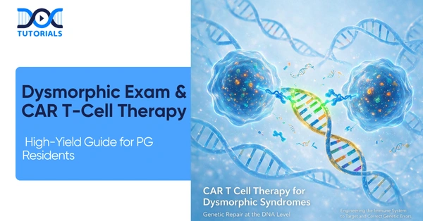
Mastering clinical genetics, dysmorphic assessments, and advanced molecular treatments like CAR T Cell Therapy is no longer optional for today’s post-graduate (PG) medical residents. This comprehensive guide connects foundational pediatric bedside examinations with the cutting-edge frontiers of gene modification and CAR T Cell Therapy. 

Get High Quality, High Yield PG Residency Notes for Free!

The Foundation of Dysmorphic Examination 

Evaluating a child for genetic syndromes begins with a meticulous general assessment and anthropometry. Understanding foundational metrics helps clinicians identify underlying genetic abnormalities before moving to advanced treatments like CAR T Cell Therapy. 

Anthropometry and Crucial Metrics 

A non-negotiable metric for any syndromic assessment is the Upper Segment to Lower Segment (US:LS) ratio. This ratio must always be correlated with age-specific norms. As a rule for all dysmorphic children, clinicians must document the US:LS ratio even if it falls within normal limits. Alongside this, the clinical checklist should include assessing consciousness, vital signs, and malnutrition staging (acute, chronic, or acute-on-chronic). A rapid screen for general signs like pallor, icterus, cyanosis, and clubbing is also essential. 

Head, Skull, and Facial Features 

Plotting the occipitofrontal circumference (OFC) on a growth chart determines whether a patient has microcephaly, macrocephaly, or normal head growth. Furthermore, analyzing the anterior fontanel for size, shape, and bulging (if open) is critical. Skull shapes vary significantly in craniosynostosis: scaphocephaly presents with increased AP diameter, while brachycephaly shows increased biparietal diameter. 

Facial features provide immense diagnostic clues. For instance, an upward lateral canthus (mongoloid slant) is associated with Down Syndrome, whereas a downward lateral canthus (antimongoloid slant) points to Noonan Syndrome. 

Below is a table summarizing key facies types and their associated conditions. 

Common Facies and Associated Syndromes 

Facies Type Key Clinical Features Associated Conditions 
Coarse Facies Thick lips, flat bridge, large tongue MPS, Cretinism 
Elfin Facies Wide mouth, smooth philtrum, pointed teeth Williams Syndrome 
Hemolytic Facies Frontal bossing, crowded teeth Thalassemia 
Other Facies Moon, Hepatic, Mask-like Cushing’s, CLD, Moebius 

The Evolution of Gene Therapy Strategies 

While physical examinations identify the phenotypic expressions of genetic disorders, the treatment landscape is rapidly shifting toward molecular interventions, laying the groundwork for complex modalities like CAR T Cell Therapy. Gene therapy strategies are broadly categorized into In Vivo and Ex Vivo mechanisms. 

In Vivo Therapies 

In Vivo gene therapy involves direct administration inside the body, where gene modification occurs internally. The primary methods for delivery include viral vectors (like AAV), nanoparticles, and RNA/Ribosomes. 

How do Antisense Oligonucleotides (ASO) function in the treatment of Duchenne Muscular Dystrophy (DMD)? 

In DMD, which is an X-linked recessive disorder characterized by deficient dystrophin, ASO therapy works via exon skipping. The ASO binds to the pre-mRNA and masks specific exons—such as skipping Exon 51 with Eteplirsen—to bypass the mutation stop codon, ultimately allowing translation into a shorter but functional dystrophin protein. 

In Vivo delivery systems heavily rely on advanced transport mechanisms, such as GalNAc conjugates which are receptor-dependent and liver-specific (administered subcutaneously), and Lipid Nanoparticles (LNP) which utilize non-receptor dependent entry via membrane fusion or endocytosis. 

In Vivo ASO Therapeutics Overview 

Disease Drug Name Gene Target Route of Administration 
Spinal Muscular Atrophy Nusinersen (Spinraza) SMN2 Gene Intrathecal 
TTR-Amyloidosis Inotersen Transthyretin (TTR) Subcutaneous 
Hyperlipidaemia Mipomersen Apolipoprotein B100 Subcutaneous 
Familial Chylomicronaemia Volanesorsen Apolipoprotein C3 Subcutaneous 

The Breakthrough of CAR T Cell Therapy (Ex Vivo) 

Ex Vivo gene therapy takes cells harvested from the patient, modifies them in a laboratory setting, and returns them to the patient. This framework is exactly how CAR T Cell Therapy operates. 

CAR T Cell Therapy is a revolutionary immunotherapy primarily utilized in pediatrics for the treatment of Acute Lymphoblastic Leukaemia (ALL). The process of CAR T Cell Therapy begins with the isolation of the patient’s own T-cells. Once isolated, these cells undergo modification ex vivo to express the Chimeric Antigen Receptor (CAR). 

After being successfully modified to become CAR T-cells, they are expanded in the laboratory and subsequently re-infused into the patient’s bloodstream. The core action of CAR T Cell Therapy is to grant specific cytotoxic activity against target cancer cells. By leveraging the body’s own immune system, CAR T Cell Therapy ensures the direct and targeted destruction of leukemia cells.  

The integration of lentiviral vectors is frequently utilized in these Ex Vivo additions to ensure functional gene expression. Thus, CAR T Cell Therapy represents a paradigm shift in how hematological malignancies are treated in pediatric patients. 

Advanced Ex Vivo Gene Editing: CRISPR/Cas9 

Beyond CAR T Cell Therapy, Ex Vivo strategies also utilize CRISPR/Cas9 technology, functioning as “molecular scissors.” A prominent example is the treatment for Sickle Cell Anaemia using Exagamglogene Autotemcel (Exa-cel/Casgevy). This mechanism involves the inactivation of the BCL11A gene, which serves as the switch that normally stops fetal hemoglobin (HbF) production. The resulting increase in HbF successfully prevents red blood cell sickling and subsequent crises. 

However, much like the stringent monitoring required after CAR T Cell Therapy, CRISPR therapies demand careful safety oversight. Clinicians must monitor for off-target effects and potential malignant transformations, as these represent permanent genetic changes compared to treatments like Hydroxyurea. 

Frequently Asked Questions (FAQs) 

1. What is the primary pediatric indication for CAR T Cell Therapy? 

The primary pediatric use for CAR T Cell Therapy is the treatment of Acute Lymphoblastic Leukaemia (ALL). 

2. How are cells modified in CAR T Cell Therapy? 

In CAR T Cell Therapy, a patient’s T-cells are isolated, modified ex vivo to express Chimeric Antigen Receptors (CAR), expanded, and then re-infused. 

3. What is the mechanism of action for CAR T Cell Therapy? 

CAR T Cell Therapy grants the modified T-cells specific cytotoxic activity directed against target cancer cells, leading to cancer cell destruction. 

4. How does CAR T Cell Therapy differ from In Vivo gene therapy? 

CAR T Cell Therapy is an Ex Vivo method, meaning cells are harvested, modified outside the body in a lab, and then returned. In Vivo therapy involves direct administration and internal modification. 

5. What is the US:LS ratio in dysmorphic examinations? 

The Upper Segment to Lower Segment (US:LS) ratio is a non-negotiable metric essential for all dysmorphic children and must correlate with age-specific norms. 

6. Which condition is indicated by an upward lateral canthus? 

An upward lateral canthus, also known as a mongoloid slant, is a feature associated with Down Syndrome. 

7. How is Spinal Muscular Atrophy (SMA) treated using gene therapy? 

SMA is treated using Nusinersen (Spinraza), an In Vivo therapy administered intrathecally that activates the inactive SMN2 gene to compensate for the missing SMN1. 

8. What vectors are used in Ex Vivo therapies like CAR T Cell Therapy? 

Ex Vivo therapies often utilize Lentiviral vectors for the laboratory integration of functional genes. 

9. How does CRISPR/Cas9 help Sickle Cell Anaemia patients? 

It inactivates the BCL11A gene, increasing Fetal Hemoglobin (HbF) production, which prevents sickling and crises. 

10. What dermatological marker indicates Tuberous Sclerosis? 

The presence of an Ash Leaf Macule (a hypopigmented spot) is a hidden dermatological marker for Tuberous Sclerosis. 

DocTutorials App Download Now

Latest Blogs

Mastering Endocrine Surgery: A High-Yield Guide for PG Residents 

Endocrine Surgery

Navigating the complexities of endocrine surgery requires a deep understanding of oncological principles, genetic syndromes, and intricate anatomical relationships. This detailed guide is meticulously crafted for PG residents aiming to master the diagnostic and operative nuances of endocrine surgery, ensuring better outcomes and robust clinical decision-making. 

Get High Quality, High Yield Endocrine Surgery PG Residency Notes for Free! 

Breast Cancer Management Protocols in Endocrine Surgery 

Understanding modern breast oncology is a fundamental component of endocrine surgery training. The evolution of breast cancer management relies heavily on precise staging and molecular profiling. The TNM prognostic staging framework categorizes the disease into Early Breast Cancer (Stage I, IIa, IIb), Locally Advanced (Stage IIIa, IIIb, IIIc), and Stage IV for metastatic spread.  

Molecular Subtype Matrix 

Systemic therapies in endocrine surgery are heavily influenced by the tumor’s receptor profile. Understanding the molecular subtype matrix is essential for guiding treatment.  

Subtype Receptor Profile Key Markers 
Luminal A ER/PR(+), HER2 (-); Low Ki-67 (Low risk)  
Luminal A (Variant) ER/PR (+), HER2 (-); High Ki-67 (High risk)  
Luminal B ER/PR(+), HER2 (+/-); High proliferation markers  
Triple Positive ER/PR(+), HER2 (+)  
HER2 Overexpressed ER/PR(-), HER2 (+)  
Triple Negative (TNBC) ER(-), PR(-), HER2 (-); Aggressive Phenotype  

Neoadjuvant Chemotherapy and Surgical Staging 

In the realm of endocrine surgery, Neoadjuvant Chemotherapy (NACT) is a powerful tool. Mandatory indications for NACT include T4 tumors with skin or chest wall infiltration, large tumors (7-10 cm), bulky axillary nodes (N2/N3), and inflammatory carcinoma. Strategic indications involve HER2 Positive, Triple Negative (TNBC), tumors greater than 2 cm or node positive, and a large tumor-to-small breast ratio to enable Breast Conservation Surgery (BCS).  

The standard chemotherapy regimen timeline involves a two-step process. Step 1 utilizes Anthracyclines (Doxorubicin + Cyclophosphamide) for 4 cycles. This is followed by Step 2 utilizing Taxanes (Paclitaxel or Docetaxel) for 4 cycles. For residual disease, HER2+ cases receive TDM-1 for 14 cycles, while TNBC cases receive Capecitabine for 6 months.  

When performing surgical management of the primary tumor, BCS requires margins with no tumor on ink for invasive disease and greater than 2mm for DCIS. Mastectomy (MRM) includes the Nipple-Areola Complex, Pectoralis Major Fascia, and Level I/II Nodes. Axillary surgical boundaries include the axillary vein inferiorly, latissimus dorsi laterally, and pectoralis major medially, with the intercostobrachial nerve passing superiorly.  

Navigating Multiple Endocrine Neoplasia (MEN) Syndromes in Endocrine Surgery 

For any resident studying endocrine surgery, the MEN syndromes represent a critical area of focus. These genetic conditions dictate aggressive screening and preemptive endocrine surgery interventions.  

MEN1 (Wermer Syndrome) Essentials 

MEN1 is linked to the MEN1 gene on Chromosome 11q13, affecting the Menin protein. It is characterized by the “3Ps”. Parathyroid disease is the most common, presenting with a prevalence greater than 95% due to multiglandular hyperplasia. Screening for parathyroid disease starts at age 8 with Calcium and PTH levels.  

Pancreatic issues (50-75% prevalence) involve gastrinomas, which have malignant potential, insulinomas, and non-functional PNETs, with screening starting at age 20. Pituitary tumors (30-35% prevalence) are primarily prolactinomas (60%) or GH-secreting tumors leading to acromegaly, with screening initiated at age 5 via prolactin and MRI. Diagnostic criteria require two or more of the three major components, or one major component alongside a family history.  

What is the most critical clinical rule to follow before performing endocrine surgery on a patient with MEN2 or MEN3?  

The pheochromocytoma must absolutely be managed first with alpha-blockade followed by beta-blockade prior to any thyroid endocrine surgery to prevent a catastrophic hypertensive crisis.  

MEN2 and MEN3 Strategies 

Endocrine surgery management for MEN2 and MEN3 hinges on the RET Proto-Oncogene mutations. These mutations dictate the timing of prophylactic thyroidectomy.  

Risk Level Mutation & Syndrome Prophylactic Thyroidectomy Surgical Deadline 
Highest Risk M918T / MEN3 (MEN2B)  Neonatal period (First months of life)  
High Risk C634 / MEN2A  Before Age 5  
Moderate Risk Various / FMTC/other  Monitor calcitonin (Operate if elevated)  

MEN2A features Medullary Thyroid Carcinoma (MTC), Pheochromocytoma in approximately 50% of cases, and Parathyroid Hyperplasia in 20-30% of cases. Conversely, MEN3 (MEN2B) presents with MTC in 100% of cases, Pheochromocytoma in 50%, Marfanoid habitus, and mucosal neuromas, notably without parathyroid disease.  

Parathyroid and Thyroid Management Strategies in Endocrine Surgery 

Primary Hyperparathyroidism and Glandular Anatomy 

In endocrine surgery, managing primary hyperparathyroidism begins with biochemical confirmation followed by localization imaging using Ultrasound and a Sestamibi Scan. If both studies concordantly localize to the same gland, a focused parathyroidectomy with intraoperative PTH (IOPTH) monitoring is indicated. If the results are discordant or negative, a bilateral neck exploration is required, which is also indicated for familial disease (MEN1) or lithium use.  

Understanding the embryological descent is vital for endocrine surgery. The superior parathyroid glands originate from the 4th pharyngeal pouch, starting high and falling posterior and deep, positioned dorsal to the recurrent laryngeal nerve (RLN). Their ectopic sites include the retro-esophageal space, para-esophageal space, and posterior mediastinum.  

The inferior glands originate from the 3rd pharyngeal pouch with the thymus, starting low and falling anterior, positioned ventral to the RLN. Ectopic sites for inferior glands include the thyrothymic ligament and anterior/superior mediastinum.  

During endocrine surgery, normal parathyroid glands exhibit a “gliding sign,” moving within fat, whereas abnormal glands do not. For multi-gland disease, endocrine surgery options include subtotal parathyroidectomy (excising 3.5 glands and leaving a 50mg vascularized remnant) or total parathyroidectomy with autotransplantation of 10-20 fragments into the brachioradialis of the non-dominant forearm.  

Thyroiditis and Pregnancy Targets 

Thyroiditis classification is crucial for decision-making in endocrine surgery. Acute infectious thyroiditis is typically bacterial (Staph/Strep) and can be hematogenous or due to a pyriform sinus fistula from the 4th branchial pouch, more commonly on the left side. Treatment requires IV antibiotics, drainage, and excision of the fistula tract.  

Subacute painful thyroiditis (Granulomatous / De Quervain’s) exhibits a toxic phase for the first 0-6 weeks with low scan uptake, a hypothyroid phase from 2-6 months, and euthyroid recovery after 6 months.  

Chronic forms include Hashimoto’s and Riedel’s. Hashimoto’s is a lymphocytic, autoimmune T-cell driven disease marked by Anti-TPO (>95%) and Anti-Tg antibodies, with histology showing lymphocytic infiltration, germinal centers, and Hurthle cells. It carries an 80x risk for B-cell lymphoma and a 30% risk for papillary carcinoma.  

Riedel’s is an IgG4-related chronic fibrosing disease presenting as a woody hard goiter fixed to strap muscles, often associated with retroperitoneal fibrosis. Endocrine surgery is hazardous in Riedel’s due to invasion, and management primarily involves tamoxifen and steroids.  

For pregnancy in the context of endocrine surgery, strict TSH upper limits are enforced. The target is strictly less than 2.5mIU/L for the 1st trimester and less than 3.0mIU/L for the 2nd and 3rd trimesters.  

Frequently Asked Questions (FAQs) 

  1. What is the primary indication for Neoadjuvant Chemotherapy in early breast cancer?  

Neoadjuvant chemotherapy is strategically indicated for HER2 Positive or Triple Negative breast cancers to facilitate breast conservation during endocrine surgery. 

  1. Which parathyroid glands are located dorsal to the recurrent laryngeal nerve?  

The superior parathyroid glands, derived from the 4th pharyngeal pouch, are positioned dorsal to the recurrent laryngeal nerve.  

  1. What is the standard radiotherapy dose for a whole breast treatment?  

The standard whole breast radiotherapy dose is 50 Gy.  

  1. When is a tumor bed boost indicated in radiotherapy?  

A tumor bed boost of +10-18 Gy is conditionally indicated for close or positive margins to reduce local recurrence.  

  1. Which genetic mutation demands a prophylactic thyroidectomy within the first months of life?  

The M918T mutation, associated with MEN3 (MEN2B), requires a prophylactic thyroidectomy in the neonatal period.  

  1. What defines the boundaries for the low-level axillary sampling area?  

The area is bounded by the intercostobrachial nerve superiorly, the latissimus dorsi laterally, the axillary vein inferiorly, and the pectoralis major medially.  

  1. How does acute infectious thyroiditis commonly present anatomically?  

Acute infectious thyroiditis, often linked to a pyriform sinus fistula, is more commonly found on the left side.  

  1. What histological findings are characteristic of Hashimoto’s thyroiditis?  

Histology typically reveals lymphocytic infiltration, germinal centers, and Hurthle cells.  

  1. In endocrine surgery for multi-gland parathyroid disease, what is autotransplanted?  

Following a total parathyroidectomy, 10-20 fragments are implanted into the brachioradialis muscle of the non-dominant forearm.  

  1. What is the TSH target for a patient in her first trimester of pregnancy?  

The non-negotiable upper limit for TSH during the first trimester is less than 2.5mIU/L. 

DocTutorials App Download Now

Latest Blogs

Understanding Caput Succedaneum: A Comprehensive Guide for Paediatrics PG Residents 

capcut succedaneum

Scalp swellings are frequent encounters in the first 1-2 days of life, making their accurate identification a cornerstone of neonatal care. Accurate diagnosis relies on identifying the specific anatomical layer involved. For PG residents pursuing their specialization in DNB Paediatrics, mastering the clinical nuances of Caput Succedaneum is an absolute necessity for daily ward rounds and board examinations.  

Get High Quality, High Yield Pediatrics PG Residency Notes for Free! 

The Anatomical Basis of Neonatal Skull Swellings in DNB Paediatrics 

When evaluating a newborn in the DNB Paediatrics curriculum, it is vital to understand the anatomical layers of the scalp to accurately diagnose conditions like Caput Succedaneum. The layers, from superficial to deep, include the Skin, Connective Tissue, Aponeurosis (Galea), Loose Areolar Tissue, Periosteum, Skull Bone, and Dura.  

There are three main pathologies that every DNB Paediatrics resident must differentiate:  

  • Caput Succedaneum: Soft tissue edema.  
  • Cephalohematoma: Subperiosteal bleeding.  
  • Subgaleal Hemorrhage: Subaponeurotic bleeding, which is a critical condition.  

Anatomically, Caput Succedaneum is located in the subcutaneous space, specifically above the periosteum. Understanding this exact location is what allows a DNB Paediatrics practitioner to confidently diagnose Caput Succedaneum over more severe hemorrhages.  

Clinical Features and Pathology of Caput Succedaneum 

In the realm of DNB Paediatrics, Caput Succedaneum is classified as a benign soft tissue swelling. The pathology behind Caput Succedaneum involves diffuse, poorly defined soft tissue edema.  

The clinical hallmark of Caput Succedaneum—and a highly tested concept in DNB Paediatrics—is that the swelling crosses the midline and suture lines (such as the sagittal suture).  

When tracking the timeline of Caput Succedaneum, DNB Paediatrics guidelines note that the swelling reaches its maximum size at birth. Typically, Caput Succedaneum resolves within 48 to 72 hours, though it can take up to one week. The etiology of Caput Succedaneum is closely associated with a normal vaginal delivery, specifically in a vertex presentation.  

How does a DNB Paediatrics resident distinguish Caput Succedaneum from a Subgaleal Hemorrhage clinically?  

While both Caput Succedaneum and Subgaleal Hemorrhage cross suture lines, Caput Succedaneum is at its maximum size at birth and resolves quickly within a week. In contrast, a Subgaleal Hemorrhage is a fluctuant mass that increases in size after birth, posing life-threatening risks like shock and requires immediate monitoring.  

Management Protocols for Caput Succedaneum 

For residents in DNB Paediatrics, the management of Caput Succedaneum is straightforward but requires strict adherence to conservative principles. The primary management for Caput Succedaneum is supportive therapy only. It is a critical rule in DNB Paediatrics that there should be no incision or drainage of Caput Succedaneum.  

Generally, there is no bleeding risk associated with Caput Succedaneum. However, DNB Paediatrics residents must remain vigilant for rare variants of Caput Succedaneum. An ‘Ecchymotic Caput’ presents a bruising and jaundice risk, while a ‘Hemorrhagic Caput’ presents a shock risk requiring transfusion.  

Feature Caput Succedaneum Cephalohematoma Subgaleal Hemorrhage 
Anatomical Location Subcutaneous (Above Periosteum)  Subperiosteal  Subaponeurotic (Loose Areolar Tissue)  
Relationship to Sutures Crosses midline & sutures  Does NOT cross sutures (Localized)  Crosses sutures (Diffuse/Spreading)  
Timing & Course Max size at birth. Resolves <1 week  Appears day 1-2. Resolves 2 weeks – 3 months  INCREASES size after birth. Resolves 2-3 weeks  
Clinical Risks Minimal (rarely hemorrhagic)  Jaundice, Linear Fracture (10-25%)  Shock, Anemia, High Mortality (CRITICAL)  

Expanding Neonatal Knowledge in DNB Paediatrics: Metabolic Bone Disease

While mastering Caput Succedaneum is essential, DNB Paediatrics training also heavily emphasizes overall neonatal bone health, specifically Metabolic Bone Disease (MBD) of Prematurity. Also known as Osteopenia of Prematurity (OOP), MBD is defined as a reduction in bone mineral content versus what is expected for gestational age.  

For a DNB Paediatrics resident, understanding the incidence is key: it affects approximately 25% of Very Low Birth Weight (VLBW, <1500g) infants and up to 50% of Extremely Low Birth Weight (ELBW, <1000g) infants. The core issue is a lag in bone mineralization driven by prematurity constraints, as 80% of rapid mineral accretion occurs during the active transport phase of the 3rd trimester.  

Clinical Presentation Timeline in DNB Paediatrics 

Unlike Caput Succedaneum, which is obvious at birth, MBD signs are late, and significant bone loss occurs before detection.  

Phase Timing Clinical Presentation 
Silent Phase Birth to 4 weeks Asymptomatic demineralization.  
Clinical Onset 4-11 Weeks (Mean 4-5 weeks) Poor postnatal growth, Craniotabes (‘ping pong ball’ skull softening), costochondral beading.  
Failure State Late Stage Fractures (Requires >80% demineralization), Respiratory failure from rib fractures.  

Biochemical Screening and Nutritional Strategies 

In DNB Paediatrics, early screening for MBD is prioritized. Screening should start at 4 weeks, or at 2 weeks if the infant is at high risk (e.g., on TPN >2 weeks or steroids). The primary screeners are Alkaline Phosphatase (ALP) >900 IU/L alongside Serum Phosphorus <5.6 mg/dL. Hypophosphatemia is the primary driver of this condition.  

Nutritional management in DNB Paediatrics aims to mimic in-utero accretion. A strict safety rule applies: never administer Calcium without Phosphate for >1-2 days, and maintain a Ca:P ratio of approximately 1.7:1 (mg/kg). Enteral dosing targets include 120-200 mg/kg/day for Calcium, 66-110 mg/kg/day for Phosphorus, and 400-700 IU/kg/day for Vitamin D.  

Frequently Asked Questions (FAQs) 

1. What exactly is Caput Succedaneum?  

In DNB Paediatrics, Caput Succedaneum is defined as a benign soft tissue swelling that presents as poorly defined edema on a newborn’s scalp.  

2. Where is Caput Succedaneum located anatomically?  

Caput Succedaneum is locate

in the subcutaneous layer, specifically above the periosteum.  

3. Does Caput Succedaneum cross suture lines?  

Yes, a defining clinical hallmark of Caput Succedaneum is that it freely crosses the midline and cranial suture lines.  

4. When does Caput Succedaneum reach its maximum size?  

Caput Succedaneum is typically at its maximum size immediately at birth.  

5. How long does Caput Succedaneum take to resolve?  

Caput Succedaneum usually resolves rapidly within 48 to 72 hours, though it can occasionally take up to one week.  

6. What is the standard DNB Paediatrics management for Caput Succedaneum?  

Management for Caput Succedaneum is strictly supportive therapy; no incision or drainage should be performed.  

7. Are there any bleeding risks associated with Caput Succedaneum?  

Usually, there are none. However, rare variants of Caput Succedaneum exist, such as ‘Ecchymotic Caput’ and ‘Hemorrhagic Caput’.  

8. What complications are associated with ‘Ecchymotic Caput Succedaneum’? 

The ‘Ecchymotic’ variant of Caput Succedaneum carries an increased risk of bruising and subsequent neonatal jaundice.  

9. How is Caput Succedaneum differentiated from Cephalohematoma in DNB Paediatrics?  

Unlike Caput Succedaneum, a Cephalohematoma is subperiosteal, presents as a well-defined tense mass, and does NOT cross suture lines.  

10. What is the primary etiology of Caput Succedaneum?  

Caput Succedaneum is most commonly associated with a normal vaginal delivery, particularly when the infant is in a vertex presentation. 

DocTutorials App Download Now

Latest Blogs

Navigating Pediatric CNS Evaluation: A Complete Guide for DNB Pediatrics Residents 

CNS Evaluation in DNB Pediatrics

The evaluation of the central nervous system (CNS) is a cornerstone of pediatric training. For residents pursuing DNB Pediatrics, mastering the intricate nuances of development, delays, and complex disorders is absolutely vital. This guide distills essential CMS Pediatric concepts to elevate your clinical acumen and board readiness. 

Get High Quality, High Yield Pediatrics PG Residency Notes for Free! 

The Core of CMS Pediatric Assessments: Understanding Milestones 

A fundamental skill in DNB Pediatrics is accurately tracking a child’s growth across specific parameters. To do this, clinicians rely on assessing five clinical domains: gross motor, fine motor, language, cognitive, and social/personal. Familiarizing yourself with a standard Child Developmental Milestones Chart is crucial for early detection of abnormalities. 

Understanding the major motor milestones timeline is a standard expectation in DNB Pediatrics. Consider these key gross motor markers: 

  • 2-3 months: Neck holding and mother recognition. 
  • 5 months: Rolling over. 
  • 7 months: Sitting with support. 
  • 9 months: Sitting without support and standing with support. 
  • 12 months: Standing without support and taking first steps. 
  • Post-12 months: Independent walking, running, and climbing stairs. 

Parental recall reliability is typically highest for major events like first crawling or sitting without support. 

Defining Delays, Stagnation, and Neurodegenerative Regression 

In DNB Pediatrics, you must sharply distinguish between normal variations and pathological states. 

  • Significant Delay: This is defined as a milestone missed by $\ge3$ months. 
  • Global Developmental Delay (GDD): This is characterized by a significant delay in $\ge2$ domains, such as motor and cognitive skills, indicating central nervous system (brain) involvement. 

A critical CMS Pediatric concept is differentiating the trajectory of a child’s skills over time. Progressive developmental delay is slow but steady. Developmental stagnation presents as a plateau and acts as a red line. However, developmental regression (a loss of previously acquired skills) is an absolute emergency requiring immediate evaluation. This is frequently seen in neurodegenerative conditions, where active damage leads to true regression. 

Clinical Scenarios in DNB Pediatrics 

When a patient presents to the CMS Pediatric clinic, isolated delays offer vital clues: 

  • Isolated Gross Motor Delay: If fine motor, language, social, and cognitive skills are normal, primary concerns include Vitamin D deficiency, bony lesions, or spasticity (e.g., Leukodystrophy). 
  • Isolated Language Delay: If a child lacks monosyllables but has normal domains, otherwise, sensory deficits like hearing impairments must be ruled out, as they can mimic delay or neurodegenerative regression. 
  • Social & Cognitive Delays: A typical Autism Spectrum Disorder (ASD) history features normal development from 0-15 months, followed by regression in language and social skills between 16-24 months. 

How should a DNB Pediatrics resident clinically differentiate between a static delay and a neurodegenerative regression? 

A static delay is characterized by an upward, albeit slower than normal, trajectory in milestone acquisition without the loss of skills. In contrast, a neurodegenerative process involves active CNS damage, resulting in true developmental regression where a child loses previously mastered abilities. It is important to note that early onset neurodegenerative diseases (occurring at <6 months) can mimic simple delay, making a meticulous CMS Pediatric history absolutely critical. 

Etiology: Uncovering the Underlying Cause 

The etiology of delays spans a broad spectrum. Perinatal causes are the most common and include asphyxia, NICU stays, ventilation, jaundice, and seizures. Genetic causes are also very common and include syndromic, chromosomal, or single-gene disorders like Down’s syndrome, Fragile X, and Angelman syndrome. 

Inborn errors of metabolism are vital CMS Pediatric topics that often present with neurodegenerative features. They are broadly divided into small and large molecule disorders. 

Metabolic Disorders in DNB Pediatrics 

Feature Small Molecule Disorders Large Molecule Disorders 
Pathology Normal development followed by a “crash” Storage accumulation over time 
Onset Early (90% <1 year; usually 1-2 years) Can be late 
Presentation Acute decompensation, regression, fever with encephalopathy Chronic baseline delay; can have regression 
Clinical Clues Abnormal ammonia, lactate, ketones Dysmorphic features (coarse facies), hepatomegaly 

Advanced Diagnostics in CMS Pediatric Care 

A thorough evaluation framework is essential. This includes taking a detailed antenatal, perinatal, developmental, family, and environmental history. Baseline routine investigations must include a CBC, vision assessment, and hearing assessment. 

Before jumping to rare neurodegenerative diseases, DNB Pediatrics protocols dictate ruling out treatable causes. This is non-negotiable for patient safety. Always check for Vitamin D deficiency (rickets), congenital hypothyroidism, and Vitamin B12 deficiency (which can cause infant tremor syndrome). 

Spinal Muscular Atrophy (SMA): A Critical DNB Pediatrics Topic 

SMA is a high-yield topic for DNB Pediatrics. The pathology involves progressive apoptosis of anterior horn cells (LMN lesion), leading to flaccid paralysis and loss of reflexes. A classic sign of severe peripheral hypotonia and SMA is the frog leg posture. 

Genetically, SMA is an autosomal recessive disorder. Normally, the SMN1 gene produces 100% functional, anti-apoptotic SMN protein. In SMA, there is a reliance on the SMN2 gene, which has a C->T mutation in Exon 7, causing a splicing suppressor to exclude Exon 7, yielding an unstable/non-functional protein. 

The therapeutic goal in this severe neurodegenerative condition is to replace SMN1 or activate SMN2

Comparison of SMA Therapies 

Therapy Target Mechanism Route & Frequency 
Nusinersen (Spinraza) SMN2 Gene Blocks suppressor (inclusion of Exon 7) Intrathecal; Loading doses then every 4 months (lifelong) 
Onasemnogene abeparvovec (Zolgensma) SMN1 Gene Gene replacement via AAV9 viral vector Intravenous; Single Dose (FDA approved 2 years) 
Risdiplam (Evrysdi) SMN2 Gene SMN2 Splicing Modifier Oral; Daily/Lifelong ( 2 months of age) 

Management of such complex neurodegenerative conditions requires a comprehensive therapy team, medical management for nutrition and comorbidities, and robust family support. 

Frequently Asked Questions (FAQs) 

1. What defines a significant developmental delay in CMS Pediatric practice? 

A significant delay is defined as a developmental milestone missed by 3 months. 

2. What are the five clinical domains assessed in developmental screening? 

The five domains are Gross Motor, Fine Motor, Language, Cognitive, and Social/Personal skills. 

3. Why is developmental regression considered a medical emergency? 

Regression indicates a loss of acquired skills, often pointing to an active neurodegenerative process or severe CNS insult requiring immediate investigation. 

4. What is the most common etiology of developmental delay? 

Perinatal and antenatal factors are the most common causes, often characterized by a history of asphyxia, NICU stay, or infections like TORCH. 

5. How does isolated language delay present, and what must be ruled out? 

It presents as a lack of monosyllables with other domains intact; sensory deficits like hearing impairment must be evaluated as they can mimic delay. 

6. What are the key clinical clues for small molecule metabolic disorders? 

These disorders typically present early (<1 year) with an acute decompensation or “crash,” regression, and abnormal ammonia, lactate, or ketones. 

7. In DNB Pediatrics, what are the absolutely “non-negotiable” treatable causes of delay to rule out? 

Residents must check for Vitamin D deficiency, thyroid dysfunction (congenital hypothyroidism), and Vitamin B12 deficiency. 

8. What is the fundamental pathology in Spinal Muscular Atrophy (SMA)? 

SMA involves the progressive apoptosis (necrosis) of Anterior Horn Cells, which are lower motor neurons (LMN), resulting in flaccid paralysis and decreased reflexes. 

9. How does Nusinersen (Spinraza) work for SMA? 

Nusinersen targets the SMN2 pre-mRNA by blocking the splicing suppressor, which allows for the inclusion of Exon 7 and the production of functional protein. 

10. What is the primary safety warning for Zolgensma gene therapy? 

Zolgensma, which replaces the SMN1 gene, carries significant safety warnings for hepatitis (which is common) and thrombocytopenia. 

DocTutorials App Download Now

Latest Blogs

A Comprehensive Clinical Guide to ADHD: Epidemiology, DSM 5 Criteria, and Management 

ADHD Clinical Guide

Attention-Deficit/Hyperactivity Disorder (ADHD) is a complex, pervasive neurodevelopmental disorder that primarily manifests in childhood and typically causes significant, lifelong impairment in cognitive, behavioral, academic, and emotional functioning. For Postgraduate (PG) psychiatric and pediatric residents, deeply understanding the DSM 5 (Diagnostic and Statistical Manual of Mental Disorders, Fifth Edition) criteria is an absolute necessity for executing an accurate clinical assessment and formulating a viable treatment plan.  

This blog details the structural brain findings, diverse etiological factors, core DSM 5 diagnostic guidelines, and evidence-based management strategies required to achieve clinical mastery in pediatric and adolescent care. 

Get High Quality, High Yield Psychiatry PG Residency Notes for Free! 

Epidemiology, Structural Findings, and Core Pathogenesis 

ADHD shares several underlying epidemiological similarities with Autism Spectrum Disorder (ASD), yet its distinct structural findings set it apart. While global brain volume changes are generally not clinically significant in ADHD (unlike the notable volume changes seen in ASD), there is an observable and measurable reduction in cortical thickness, particularly localized within the frontal lobe. Functional neuroimaging typically reveals a markedly decreased activation in both the basal ganglia and the frontal lobe regions during cognitive tasks.  

The core pathology of the disorder stems from severe neurotransmitter dysregulation, explicitly involving Dopamine and Norepinephrine systems. In patients presenting with ADHD, there is an excess reuptake of these vital neurotransmitters into presynaptic terminals, which drastically reduces their availability at postsynaptic receptors and disrupts neural signaling.  

  • Dopamine Deficit: Dopamine is fundamentally responsible for regulating learning and motor functions. A deficit in this neurotransmitter leads directly to severe learning difficulties and pronounced motor initiation issues.  
  • Norepinephrine Deficit: Norepinephrine regulates baseline arousal, sustained attention, and mood stability. A measurable deficit here translates directly into the classic clinical symptoms of inattention, hyperactivity, and persistent sleep dysregulation.  

The Multifactorial Etiology of ADHD 

The etiology of ADHD operates on a complex multifactorial inheritance model that involves intricate interactions between a patient’s genetic susceptibility and environmental triggers. The DSM 5 framework implicitly acknowledges these diverse developmental trajectories when guiding clinicians on how symptoms may present and evolve over time.  

Key Risk Factors for ADHD Development 

Risk Category Specific Identified Factors 
Genetic & Biological Complex inheritance patterns involving polymorphisms that affect neurotransmitter transport and neuronal growth, specifically including DAT gene mutations and BDNF gene (Val66Met/rs6265) alterations.  
Prenatal & Perinatal Prematurity, complications during pregnancy, parental asphyxia, maternal drug or alcohol exposure in utero, and notably high maternal sucrose intake.  
Postnatal & Environmental Epigenetic alterations and exposure to heavy metals such as Lead, Mercury, and Manganese. Routine lead screening is considered a standard clinical practice.  

The DSM 5 Diagnostic Criteria for ADHD 

The DSM 5 diagnostic criteria serve as the international gold standard for identifying and classifying ADHD in clinical practice. The DSM 5 establishes strict, objective guidelines to prevent clinical overdiagnosis and ensure diagnostic accuracy across different patient demographics. 

According to the DSM 5, the core clinical triad of ADHD consists of Hyperactivity, Impulsivity, and Inattention. To successfully meet the DSM 5 diagnostic threshold, a patient must exhibit a specific symptom count. For pediatric patients under the age of 17, the DSM 5 strongly mandates that 6 or more specific symptoms must be documented.  

  • Inattention: The clinical traits include making careless mistakes, being easily distracted by external stimuli, not listening directly when spoken to, frequently submitting incomplete homework, and exhibiting an active dislike for tasks requiring sustained mental effort, such as mathematics.  
  • Hyperactivity: The DSM 5 identifies 6 key traits in this domain, including frequently fidgeting or tapping hands, leaving one’s seat when remaining seated is expected, running or climbing at inappropriate times, being completely unable to play quietly, continuously acting as if “driven by a motor,” and talking excessively.  
  • Impulsivity: The DSM 5 outlines 3 distinct impulsive traits that further impair social and academic functioning.  

Furthermore, the DSM 5 classifies the disorder into three primary presentations: Predominantly Inattentive, Predominantly Hyperactive/Impulsive, and the Combined Type. To diagnose the Combined Type, the DSM 5 explicitly requires the patient to simultaneously meet at least 6 criteria in both the inattention domain and the hyperactivity/impulsivity domain.  

How does the DSM 5 differentiate situational inattention from clinical ADHD? 

The DSM 5 explicitly dictates that symptoms must be pervasive and present in more than one setting (for example, displaying symptoms at both home and school). This strict DSM 5 multi-setting requirement effectively rules out purely environmental triggers, ensuring that the patient’s impairment is a persistent neurodevelopmental trait rather than a localized, transient behavioral response to a specific chaotic environment.  

Timeline, Impact, and Clinical Exclusions According to the DSM 5 

The DSM 5 is exceptionally specific regarding symptom timelines to ensure developmental appropriateness. Symptoms must have a documented onset before 12 years of age and must demonstrate continuous persistence for more than 6 months; under the DSM 5, symptoms cannot be transient or episodic.  

Additionally, the DSM 5 dictates that these behaviors must cause significant, observable interference with academic, social, or occupational activities. Finally, the DSM 5 includes an exclusion criterion stating that the symptoms must be excessive for the patient’s developmental level and cannot be better explained by another overarching mental disorder.  

Comprehensive Management and Treatment Algorithm 

Adhering to a highly structured management algorithm is just as critical as correctly applying the DSM 5 diagnostic criteria. Clinical management heavily relies on the patient’s chronological age and the overall severity of the symptoms. 

ADHD Evidence-Based Management Hierarchy 

Patient Age Group Primary Interventions (First-Line) Secondary / Refractory Interventions 
Children < 6 Years Behavioral Therapy, Parent Training, and comprehensive Environmental Modification.  Early medication is strictly indicated only for severe issues like school expulsion, aggression, self-injury, or a strong family history.  
Children > 6 Years Immediate Pharmacotherapy alongside targeted Psychotherapy.  Transition to alternative drug classes depending on existing comorbidities (e.g., using Atomoxetine for heart disease, or Alpha-2 Agonists for tic disorders).  

Non-Pharmacological Interventions 

For all pediatric age groups, non-pharmacological approaches form the robust foundation of treatment, comprising up to 90% of early clinical management. Caregiver interventions involve using heavily structured daily schedules, utilizing charts and checklists, and minimizing environmental distractions by maintaining a calm room with minimal extraneous objects.  

Clinicians should coach parents on employing specific, clear communication and utilizing “calm discipline” techniques, such as time-outs, instead of yelling. Furthermore, establishing realistic and measurable goals (like consistently completing assignments) and encouraging hobbies or sports where success is highly possible are vital behavioral strategies.  

Pharmacological Interventions 

When the DSM 5 diagnostic threshold confirms a severe case requiring immediate pharmacological intervention in children over 6, the hierarchy of treatments is structured strictly by clinical efficacy and safety profiles. 

  1. Stimulants: Methylphenidate is widely considered the superior and safer first-line option, requiring a trial of 4-6 weeks. It effectively blocks DAT/NET transporters, which subsequently increases synaptic Dopamine and Norepinephrine availability. Dosing strategies typically start at 0.5mg/kg/day, targeting an eventual maintenance dose of 1-2 mg/kg/day. The drug features a peak efficacy at 1-2 hours and a half-life of 4-5 hours.  
  1. Non-Stimulants: Atomoxetine, alongside the newly approved Viloxazine (an extended-release non-stimulant approved for ages 6-17), serve as reliable second-line treatments. Atomoxetine operates as a Selective Norepinephrine Reuptake Inhibitor (SNRI) that specifically targets the prefrontal cortex. Because it does not increase dopamine levels in the Nucleus Accumbens, it possesses no major abuse potential. However, PG residents must remain vigilant regarding side effects, which include decreased appetite, mild gastrointestinal issues (vomiting/loose motions), increased blood pressure, insomnia, sleep disturbances (such as talking in sleep), weight loss, and headache. Critically, clinicians must continuously monitor for suicidal tendencies, which present in 1-2% of patients taking Atomoxetine or Viloxazine.  
  1. Alpha-2 Agonists: Medications like Clonidine and Guanfacine represent the third line of pharmacological treatment, and they are often the preferred route if the patient has a comorbid tic disorder.  

Frequently Asked Questions (FAQs) 

1. What is the core triad of ADHD under the DSM 5?  

The DSM 5 strictly defines the disorder’s core triad as Hyperactivity, Impulsivity, and Inattention.  

2. At what age must ADHD symptoms appear according to the DSM 5?  

The DSM 5 guidelines state that initial symptom onset must clearly occur before 12 years of age.  

3. How long must symptoms persist for a valid DSM 5 diagnosis?  

The DSM 5 explicitly requires that clinical symptoms persist continuously for more than 6 months.  

4. What is the minimum symptom count for children under 17 in the DSM 5?  

The DSM 5 mandates the presence of 6 or more symptoms from either the inattention or hyperactivity/impulsivity categories.  

5. Does the DSM 5 allow for diagnosis if symptoms only occur at school?  

No, the DSM 5 requires symptoms to be documented in more than one setting to effectively rule out localized environmental triggers.  

6. Which key neurotransmitters are heavily implicated in ADHD pathogenesis?  

Dopamine and Norepinephrine are the primary neurotransmitters identified as being severely dysregulated in ADHD.  

7. What is the standard first-line treatment for children under 6 years old?  

Behavioral therapy, paired with environmental modifications, is the mandatory first-line management for preschool-aged children.  

8. When should medications be utilized in children under 6?  

Medications are added early only if the child exhibits severe, unmanageable issues like aggression, active self-injury, immediate expulsion risks, or possesses a strong family history.  

9. What is the primary pharmacokinetic mechanism of Methylphenidate?  

It actively blocks DAT/NET transporters in the brain, thereby increasing available synaptic Dopamine and Norepinephrine.  

10. Why is Atomoxetine clinically considered to have a low abuse potential?  

Unlike standard stimulants, it does not artificially increase dopamine levels in the brain’s Nucleus Accumbens, thoroughly minimizing physiological abuse risks. 

DocTutorials App Download Now

Latest Blogs

NEET PG Online Live Power Pack
DocTutorials App Download Now