neet-pg Archives - Page 3 of 20 -

Layers of the Epidermis: Structure, Functions, and Disorders
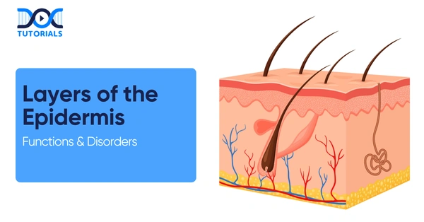
layers of the Epidermis

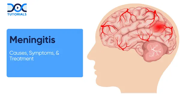
Meningitis is the swelling of the membranes (meninges) that surround the brain and the spinal cord. It is a severe disorder which may develop quickly and has to be treated immediately. It can be triggered by different organisms (bacteria, viruses, fungi, parasites) or noninfectious factors (drugs, autoimmune processes).

Acute bacterial meningitis is life-threatening, but viral meningitis is typically mild. Treatment involves a rapid diagnosis (physical examination and CSF culture) and appropriate therapy (antibiotics, antivirals, steroids). This guide addresses the aetiology and pathophysiology of meningitis relevant to NEET-PG aspirants. Keep reading to know more.

What is the Pathophysiology of Meningitis?

Meningitis is characterised by the inflammation of the subarachnoid space. Bacterial infection causes a neutrophil-rich exudate, leading to brain oedema, raised intracranial pressure, and tissue damage. Viral meningitis presents a mild lymphocytic inflammation. Cases that are not treated can cause infarction, herniation or hydrocephalus.

In bacterial meningitis, the pathogens penetrate the meninges via the bloodstream or by direct spread. They multiply in the CSF, leading to a neutrophilic reaction with purulent exudates and oedema. This causes increased intracranial pressure and disorders such as infarction, herniation and subsequent hydrocephalus.

Viral meningitis is less severe and is characterised by lymphocytic inflammation. It leads to cytokine release and mild neuronal irritation, and minimal or no pus. The course is typically subacute, with good recovery, but some viruses, such as Herpes Simplex Virus (HSV), can lead to meningoencephalitis and require specific treatment.

Fungal meningitis and tuberculosis progress gradually. Cryptococcus is prevalent in immunocompromised individuals, and tuberculosis leads to basal exudates, vasculitis, and other complications such as hydrocephalus. CSF is usually characterised by elevated leukocytes, elevated protein, and low glucose.

What are the Causes of Meningitis?

There are various causes of meningitis. Types of infections are bacterial, viral, fungal, parasitic, and tubercular. Noninfectious causes include drugs, autoimmune disorders, and cancer. Identifying the cause is key to proper treatment and prevention.

Meningitis is caused by a broad spectrum of infectious and non-infectious agents, which may vary in severity, progression, and management. The cause of meningitis varies according to region and age.

Major causes include bacterial, viral, fungal, tubercular, parasitic, and non-infectious causes, each with its own clinical and pathological characteristics. Here are the causes of meningitis:

  • Bacterial Meningitis (Acute and Life-threatening): Streptococcus pneumoniae is the most prevalent, although Neisseria meningitidis is a source of outbreaks in the youth. Vaccination has reduced Hib cases.

Listeria affects all newborns, the older generation, and those with frail immune systems, whereas Group Bacillus, Streptococcus, and Escherichia coli are prevalent among newborns. Other bacteria (such as Staphylococcus aureus and Pseudomonas) can occur after injury or surgery.

  • Viral (Aseptic) Meningitis: Mainly caused by enteroviruses. HSV (particularly HSV-2) may lead to recurrent disease. Other causes include varicella-zoster, mumps, measles, HIV, and arboviruses. Usually milder with a lymphocytic response.
  • Fungal Meningitis: Uncommon in healthy people. Cryptococcus neoformans thrives most among immunodeficient patients. Others (Histoplasma, Coccidioides, Candida) cause chronic or subacute meningitis with high protein and lymphocytes.
  • Tubercular Meningitis: As a result of Mycobacterium tuberculosis. Slow onset with basal exudates, granulomas, and infarcts. CSF shows lymphocytes, very low glucose, and high protein. Common in India; needs specific therapy.
  • Parasitic and Other Causes: Parasites (e.g. Angiostrongylus, Gnathostoma) cause eosinophilic meningitis. Naegleria fowleri causes an acute, fatal infection. Others (cysticercosis, malaria) may involve the meninges. The non- infectious causes are malignancy, autoimmune disease, drugs and sarcoidosis.

Risk Factors of Meningitis

Poor housing hygiene increases the risk of meningococcal meningitis (a bacterial infection transmitted through respiratory droplets). Risk is increased by a lack of vaccination (pneumococcal, Hib, meningococcal). Immunosuppression (HIV, chemotherapy, and splenectomy) predisposes to many forms.

Ear/sinus infections, skull fractures or neurosurgery can directly introduce bacteria into the meninges. Infants inherit risk through maternal infection (GBS, E. coli). Pregnancy predisposes to Listeria.

What are the Symptoms and Signs of Meningitis?

Meningitis is usually characterised by fever, neck stiffness and headache. Other aspects are the altered mental status, photophobia, nausea/vomiting and rash. Infants might exhibit non-specific symptoms (irritability, bulging fontanelle). 

Meningitis results in a mixture of systemic and neurological manifestations. Symptoms can build up very quickly in acute cases, but can be mild or slow in some populations, such as infants or those with chronic infections.

Typical signs and red flags must be identified early to enable prompt diagnosis and treatment:

  • Patients typically experience rapid-onset fever and severe headache.
  • Neck stiffness (nuchal rigidity) is the typical feature: patients have difficulty flexing the neck.
  • Photophobia (sensitivity to light) is frequent.
  • Meningeal irritation also causes positive Kernig’s sign (pain on extending the knee when the hip is flexed) and Brudzinski’s sign (hip/knee flexion when the neck is flexed).
  • In addition to meningitis, neurological symptoms present can be confusion, drowsiness, or coma when encephalitis is present. In the severe cases, there may be seizures.
  • A typical rash (petechial or purpuric) indicates Neisseria meningitidis.
  • Headache and photophobia are prominent in adults.
  • In children and infants, presentation is more variable: children may reject feeds, become fussy or sleepy, develop a bulging fontanelle, or cry inconsolably.
  • Fever may even be absent in neonates. Among the red flags are a sudden increase in fever, an intense headache, neck soreness, and a change in consciousness. (“Classic triad: fever, headache, neck stiffness)

Systemic signs include:

  • Tachycardia
  • Hypotension
  • Septic meningitis may be accompanied by shock.

Rapid progression, rash, and Disseminated Intravascular Coagulation (DIC) are observed in meningococcemia. Chronic or tubercular meningitis can have a subacute onset with progressive confusion, cranial nerve palsies (Most IV nerve inducing vision in both eyes), and hydrocephalus (nausea/vomiting due to increased ICP).

What is the Diagnosis of Meningitis?

Diagnosis is based on clinical features and lab tests. Suspected cases should be urgently tested by history, exam, and lumbar puncture (CSF analysis), the gold standard. Treatment should not be withheld in possible cases of bacterial meningitis.

Suspected meningitis presents with fever and signs of meningitis (stiff neck, photophobia, altered mentation). Imaging and lumbar puncture are a part of the evaluation.

In cases of focal deficits, seizures, or changes in consciousness, a CT/MRI (computed tomography/magnetic resonance imaging) scan must be performed initially to exclude increased intracranial pressure or mass lesions.

  • Lumbar Puncture (LP)

Take CSF of cell count, chemistry (protein, glucose), Gram stain, and culture. Other tests can include PCR of viruses (enterovirus, HSV), Tuberculosis Polymerase Chain Reaction (TB PCR) or acid-fast stain (in the event of suspicion of TB), and fungal antigen (cryptococcal) in immunocompromised patients.

Laboratories will then perform specific tests with CSF or blood to identify the organism. An overview of typical CSF results is as follows:

CSF ParameterBacterial MeningitisViral MeningitisTubercular Meningitis
WBC (cells/µL)100–10,000 (mostly neutrophils)<100 (mostly lymphocytes)<100 (lymphocytes 50–1000)
Neutrophils (%)Predominant (>50%)It may seem (particularly early)Predominant early (50–1000 total)
Protein>1.0 g/L0.4–1.0 g/L1.0–5.0 g/L
Glucose (ratio CSF: serum)<0.4Normal (>0.6)<0.3
Lactate (mmol/L)>2.0 (often >4.0)<2.0>2.0
Gram stain/CultureFrequently positive for bacteria.Negative (PCR for viruses)May be negative (AFB stain, culture can take weeks)

Additional tests include:

  • Blood cultures must be taken. 
  • Brain imaging (CT/MRI) can demonstrate meningeal enhancement or exclude an abscess.

Other supportive tests:

  • Complete blood count (typically presents with leukocytosis), 
  • Inflammatory markers (CRP, procalcitonin),
  • Serum chemistry (to measure dehydration, organ functionality).

Polymerase chain reaction (PCR) tests (e.g. BioFire panel) can quickly detect bacterial and viral DNA (deoxyribonucleic acid) in CSF. Nonetheless, empirical therapy should not be delayed due to PCR results.

What is the Treatment for Meningitis?

The course of treatment is based on the cause. Bacterial meningitis is a medical emergency: administer high-dose IV antibiotics and usually IV corticosteroids. Viral meningitis is largely supportive; antivirals are administered in cases of suspected herpes. Fungal meningitis is treated using suitable antifungals. In every case, supportive care is essential.

Meningitis management necessitates cause-specific urgent treatment and supportive care to minimise mortality and complications. Since delays—especially in bacterial meningitis—can be life-threatening, therapy is often started empirically based on clinical suspicion and patient factors such as age and immune status.

After the underlying cause is diagnosed, treatment is customised accordingly, with efforts to reduce inflammation, prevent complications, and support essential functions. The treatment depends on the aetiology, but in any case, the main focus is to prevent complications and minimise mortality.

  • Bacterial Meningitis: Needs broad-spectrum IV antibiotics immediately, regardless of test results, depending on age and risk factors.

The therapy is then streamlined based on culture and sensitivity reports. Close contacts of Meningococcal cases require chemoprophylaxis.

  • Role of Steroids: Corticosteroids are also administered as an adjunct to minimise inflammation and neurological destruction, especially in bacterial or tubercular meningitis. They are best initiated at an early age.
  • Viral Meningitis: Usually mild and self-limiting. The treatment is primarily supportive, and antivirals are only used in certain infections,s such as herpes viruses.
  • Tubercular Meningitis: Managed with prolonged multi-drug anti-tubercular therapy along with steroids. It is vital to initiate early treatment to avoid long-term complications.
  • Fungal Meningitis: Needs long-term antifungal treatment, usually in stages (induction and maintenance). Immunocompromised people have a more intensive management.
  • Supportive and Adjunctive Care: Involves fluid and oxygen therapies, and surveillance for seizures and complications, such as elevated intracranial pressure or hydrocephalus. In some cases, surgical operations might be necessary.

Here is a table for key treatment modalities:

Type of MeningitisImmediate ManagementDefinitive / Targeted Therapy
BacterialUrgent empiric IV antibiotics (ceftriaxone/cefotaxime + vancomycin +/- ampicillin), administer dexamethasone, fluids, and observe closely.
Adjust antibiotics in accordance with culture susceptibility (e.g. penicillin, ceftriaxone, ampicillin).Time depends on the organism (7-21 days). The close contact chemoprophylaxis in meningococcal cases.
ViralSupportive care: hydration, antipyretics, observation; hospitalise for severe and uncertain diagnosis.Antivirals only if indicated (e.g. acyclovir for HSV/VZV). The majority of cases do not require special treatment.
TubercularEarly anti-TB therapy initiation using corticosteroids; treatment of elevated ICP and complications.
Long-term ATT regimen (HRZE → HR for 12 months). Steroids were maintained during the first weeks to minimise complications.
FungalBegin antifungal treatment immediately; control intracranial pressure (e.g. repeated LPs when necessary).Amphotericin B + flucytosine as induction and fluconazole as maintenance. Long-term treatment according to immune status.
Parasitic / OtherSupportive care and stabilisation; prompt diagnosis of the underlying cause.Antiparasitic drugs, when available (e.g. miltefosine), steroids in inflammatory forms, and underlying disease (autoimmune, malignancy, etc.).

What are the Major Complications of Meningitis?

Meningitis can lead to serious, sometimes permanent complications such as hearing loss, seizures, memory and concentration problems, limb amputations (after severe sepsis), and even death. Bacterial meningitis carries the highest risk, with up to 1 in 5 survivors experiencing long-term effects.

Here are some of the key complications of meningitis based on neurological, physical and systemic classification:

Neurological Complications

  • Hearing Loss (Partial or Total): One of the most common long-term effects.
  • Vision Loss: Damage to the optic nerves or brain pathways.
  • Seizures (Epilepsy): Recurrent episodes due to brain irritation.
  • Cognitive Problems: Memory loss, poor concentration, and learning difficulties.
  • Movement and Balance Issues: Coordination problems, muscle weakness.
  • Behavioural Changes: Personality shifts, emotional instability, or developmental delays in children.

Physical Complications

  • Limb Loss/Amputation: Especially in meningococcal meningitis, where sepsis damages tissues.
  • Bone and Joint Problems: Arthritis or chronic pain due to infection spreading.
  • Kidney Damage: From septicemia or complications of treatment.

Systemic Complications

  • Sepsis: Widespread infection in the bloodstream, often fatal if untreated.
  • Increased Intracranial Pressure: Swelling of the brain leading to coma or death.
  • Hydrocephalus: Buildup of cerebrospinal fluid in the brain.
  • Blood Vessel Damage: Stroke or reduced blood supply to parts of the brain.

What is the Prevention for Meningitis?

Prevention is crucial, especially when there are complications of meningitis. The most important measures are vaccination (Hib, pneumococcal, meningococcal), prophylactic antibiotics among close contacts, and good hygiene. Avoid sharing drinks, cover coughs, and ensure proper sanitation. To minimise the risk of listeria, pregnant women must avoid unpasteurised foods.

The prevention of meningitis aims to minimise exposure to infectious agents and enhance immunity, particularly against vaccine-preventable pathogens.

The combination of immunisation, timely prophylaxis, hygiene measures, and lifestyle measures is important in reducing the risk of infection and managing outbreaks.

  • Vaccination: Hib and Streptococcus pneumoniae conjugate vaccines are commonly administered to children. At-risk groups are vaccinated against Neisseria meningitidis. These greatly reduce the incidence of bacterial meningitis.
  • Antibiotic Prophylaxis: Antibiotics (rifampin, ciprofloxacin, or ceftriaxone) should be administered to close contacts of meningococcal cases. Unvaccinated people may need prophylaxis with Hib exposure; this is not standard with pneumococcus.
  • General Hygiene: Handwashing, using separate utensils/drinks, covering coughs, and maintaining space will reduce the transmission. Masks or avoiding crowds may be advised during outbreaks.
  • Safe Food Practices: To prevent listeria, avoid unpasteurised dairy and cook meats properly, particularly in pregnant or immunocompromised people.
  • Vector Control: Prevention of some viral meningitides (arboviruses) may occur through avoidance of mosquito/tick bites, i.e., the use of insect repellent and protective clothing.

FAQs about Meningitis

  1. How quickly can meningitis become dangerous?

Bacterial meningitis may deteriorate in a matter of hours and cause serious complications or death. It is important to have early medical attention.

  1. Can meningitis spread from person to person?

Some types, particularly meningococcal meningitis, are transmitted via respiratory droplets (coughing, sneezing, close contact).

  1. Who is at the highest risk of meningitis?

People at risk are infants, elderly people, immunocompromised patients, and those who live in crowded environments.

  1. Can meningitis occur without fever?

Yes, particularly in neonates, elderly patients or immunocompromised patients where the symptoms can be atypical.

  1. Why is a lumbar puncture important in meningitis?

Lumbar puncture in meningitis assists in the diagnosis and determining the exact cause through analysis of cerebrospinal fluid.

  1. Can meningitis cause long-term disability?

Yes, survivors can have complications such as hearing loss, learning disabilities or neurological impairments.

  1. Is meningitis always contagious?

No, not every form is contagious, fungal, tubercular, or non-infectious; they do not normally pass on between individuals.

  1. What is the role of imaging in meningitis?

In some cases, CT or MRI is performed before lumbar puncture to eliminate elevated intracranial pressure or brain lesions.

  1. Can meningitis recur?

Yes, meningitis can also possibly have recurrent episodes, particularly with some viral infections or immune defects.

  1. Why is early treatment started before confirmation?

Delays in treatment, particularly in bacterial meningitis, may lead to high mortality and morbidity.

Conclusion

Meningitis is a severe disorder that entails inflammation of the meninges and has diverse etiologies. Bacterial forms result in neutrophilic exudates, and viral forms present lymphocytic inflammation. It is important to identify the symptoms early and analyse the CSF as soon as possible. Early intervention and therapy are life-saving.

For NEET-PG students, a clear understanding of the pathology and management of meningitis is crucial not only for exam preparation but also for future clinical practice. For additional guidance, DocTutorials can be your study companion. We offer crisp videos, clinical Qbank, exam-focused notes, flashcards, and mind maps. This helps ensure aspirants gain complete clarity over complex medical topics.

Join DocTutorials today and explore our NEET PG course to excel in your medical career!

NEET PG Online Live Power Pack
DocTutorials App Download Now

Latest Blogs

Meningitis: Causes, Symptoms, Diagnosis, and Treatment

Meningitis

Meningitis is the swelling of the membranes (meninges) that surround the brain and the spinal cord. It is a severe disorder which may develop quickly and has to be treated immediately. It can be triggered by different organisms (bacteria, viruses, fungi, parasites) or noninfectious factors (drugs, autoimmune processes).

Acute bacterial meningitis is life-threatening, but viral meningitis is typically mild. Treatment involves a rapid diagnosis (physical examination and CSF culture) and appropriate therapy (antibiotics, antivirals, steroids). This guide addresses the aetiology and pathophysiology of meningitis relevant to NEET-PG aspirants. Keep reading to know more.

What is the Pathophysiology of Meningitis?

Meningitis is characterised by the inflammation of the subarachnoid space. Bacterial infection causes a neutrophil-rich exudate, leading to brain oedema, raised intracranial pressure, and tissue damage. Viral meningitis presents a mild lymphocytic inflammation. Cases that are not treated can cause infarction, herniation or hydrocephalus.

In bacterial meningitis, the pathogens penetrate the meninges via the bloodstream or by direct spread. They multiply in the CSF, leading to a neutrophilic reaction with purulent exudates and oedema. This causes increased intracranial pressure and disorders such as infarction, herniation and subsequent hydrocephalus.

Viral meningitis is less severe and is characterised by lymphocytic inflammation. It leads to cytokine release and mild neuronal irritation, and minimal or no pus. The course is typically subacute, with good recovery, but some viruses, such as Herpes Simplex Virus (HSV), can lead to meningoencephalitis and require specific treatment.

Fungal meningitis and tuberculosis progress gradually. Cryptococcus is prevalent in immunocompromised individuals, and tuberculosis leads to basal exudates, vasculitis, and other complications such as hydrocephalus. CSF is usually characterised by elevated leukocytes, elevated protein, and low glucose.

What are the Causes of Meningitis?

There are various causes of meningitis. Types of infections are bacterial, viral, fungal, parasitic, and tubercular. Noninfectious causes include drugs, autoimmune disorders, and cancer. Identifying the cause is key to proper treatment and prevention.

Meningitis is caused by a broad spectrum of infectious and non-infectious agents, which may vary in severity, progression, and management. The cause of meningitis varies according to region and age.

Major causes include bacterial, viral, fungal, tubercular, parasitic, and non-infectious causes, each with its own clinical and pathological characteristics. Here are the causes of meningitis:

  • Bacterial Meningitis (Acute and Life-threatening): Streptococcus pneumoniae is the most prevalent, although Neisseria meningitidis is a source of outbreaks in the youth. Vaccination has reduced Hib cases.

Listeria affects all newborns, the older generation, and those with frail immune systems, whereas Group Bacillus, Streptococcus, and Escherichia coli are prevalent among newborns. Other bacteria (such as Staphylococcus aureus and Pseudomonas) can occur after injury or surgery.

  • Viral (Aseptic) Meningitis: Mainly caused by enteroviruses. HSV (particularly HSV-2) may lead to recurrent disease. Other causes include varicella-zoster, mumps, measles, HIV, and arboviruses. Usually milder with a lymphocytic response.
  • Fungal Meningitis: Uncommon in healthy people. Cryptococcus neoformans thrives most among immunodeficient patients. Others (Histoplasma, Coccidioides, Candida) cause chronic or subacute meningitis with high protein and lymphocytes.
  • Tubercular Meningitis: As a result of Mycobacterium tuberculosis. Slow onset with basal exudates, granulomas, and infarcts. CSF shows lymphocytes, very low glucose, and high protein. Common in India; needs specific therapy.
  • Parasitic and Other Causes: Parasites (e.g. Angiostrongylus, Gnathostoma) cause eosinophilic meningitis. Naegleria fowleri causes an acute, fatal infection. Others (cysticercosis, malaria) may involve the meninges. The non- infectious causes are malignancy, autoimmune disease, drugs and sarcoidosis.

Risk Factors of Meningitis

Poor housing hygiene increases the risk of meningococcal meningitis (a bacterial infection transmitted through respiratory droplets). Risk is increased by a lack of vaccination (pneumococcal, Hib, meningococcal). Immunosuppression (HIV, chemotherapy, and splenectomy) predisposes to many forms.

Ear/sinus infections, skull fractures or neurosurgery can directly introduce bacteria into the meninges. Infants inherit risk through maternal infection (GBS, E. coli). Pregnancy predisposes to Listeria.

What are the Symptoms and Signs of Meningitis?

Meningitis is usually characterised by fever, neck stiffness and headache. Other aspects are the altered mental status, photophobia, nausea/vomiting and rash. Infants might exhibit non-specific symptoms (irritability, bulging fontanelle). 

Meningitis results in a mixture of systemic and neurological manifestations. Symptoms can build up very quickly in acute cases, but can be mild or slow in some populations, such as infants or those with chronic infections.

Typical signs and red flags must be identified early to enable prompt diagnosis and treatment:

  • Patients typically experience rapid-onset fever and severe headache.
  • Neck stiffness (nuchal rigidity) is the typical feature: patients have difficulty flexing the neck.
  • Photophobia (sensitivity to light) is frequent.
  • Meningeal irritation also causes positive Kernig’s sign (pain on extending the knee when the hip is flexed) and Brudzinski’s sign (hip/knee flexion when the neck is flexed).
  • In addition to meningitis, neurological symptoms present can be confusion, drowsiness, or coma when encephalitis is present. In the severe cases, there may be seizures.
  • A typical rash (petechial or purpuric) indicates Neisseria meningitidis.
  • Headache and photophobia are prominent in adults.
  • In children and infants, presentation is more variable: children may reject feeds, become fussy or sleepy, develop a bulging fontanelle, or cry inconsolably.
  • Fever may even be absent in neonates. Among the red flags are a sudden increase in fever, an intense headache, neck soreness, and a change in consciousness. (“Classic triad: fever, headache, neck stiffness)

Systemic signs include:

  • Tachycardia
  • Hypotension
  • Septic meningitis may be accompanied by shock.

Rapid progression, rash, and Disseminated Intravascular Coagulation (DIC) are observed in meningococcemia. Chronic or tubercular meningitis can have a subacute onset with progressive confusion, cranial nerve palsies (Most IV nerve inducing vision in both eyes), and hydrocephalus (nausea/vomiting due to increased ICP).

What is the Diagnosis of Meningitis?

Diagnosis is based on clinical features and lab tests. Suspected cases should be urgently tested by history, exam, and lumbar puncture (CSF analysis), the gold standard. Treatment should not be withheld in possible cases of bacterial meningitis.

Suspected meningitis presents with fever and signs of meningitis (stiff neck, photophobia, altered mentation). Imaging and lumbar puncture are a part of the evaluation.

In cases of focal deficits, seizures, or changes in consciousness, a CT/MRI (computed tomography/magnetic resonance imaging) scan must be performed initially to exclude increased intracranial pressure or mass lesions.

  • Lumbar Puncture (LP)

Take CSF of cell count, chemistry (protein, glucose), Gram stain, and culture. Other tests can include PCR of viruses (enterovirus, HSV), Tuberculosis Polymerase Chain Reaction (TB PCR) or acid-fast stain (in the event of suspicion of TB), and fungal antigen (cryptococcal) in immunocompromised patients.

Laboratories will then perform specific tests with CSF or blood to identify the organism. An overview of typical CSF results is as follows:

CSF ParameterBacterial MeningitisViral MeningitisTubercular Meningitis
WBC (cells/µL)100–10,000 (mostly neutrophils)<100 (mostly lymphocytes)<100 (lymphocytes 50–1000)
Neutrophils (%)Predominant (>50%)It may seem (particularly early)Predominant early (50–1000 total)
Protein>1.0 g/L0.4–1.0 g/L1.0–5.0 g/L
Glucose (ratio CSF: serum)<0.4Normal (>0.6)<0.3
Lactate (mmol/L)>2.0 (often >4.0)<2.0>2.0
Gram stain/CultureFrequently positive for bacteria.Negative (PCR for viruses)May be negative (AFB stain, culture can take weeks)

Additional tests include:

  • Blood cultures must be taken. 
  • Brain imaging (CT/MRI) can demonstrate meningeal enhancement or exclude an abscess.

Other supportive tests:

  • Complete blood count (typically presents with leukocytosis), 
  • Inflammatory markers (CRP, procalcitonin),
  • Serum chemistry (to measure dehydration, organ functionality).

Polymerase chain reaction (PCR) tests (e.g. BioFire panel) can quickly detect bacterial and viral DNA (deoxyribonucleic acid) in CSF. Nonetheless, empirical therapy should not be delayed due to PCR results.

What is the Treatment for Meningitis?

The course of treatment is based on the cause. Bacterial meningitis is a medical emergency: administer high-dose IV antibiotics and usually IV corticosteroids. Viral meningitis is largely supportive; antivirals are administered in cases of suspected herpes. Fungal meningitis is treated using suitable antifungals. In every case, supportive care is essential.

Meningitis management necessitates cause-specific urgent treatment and supportive care to minimise mortality and complications. Since delays—especially in bacterial meningitis—can be life-threatening, therapy is often started empirically based on clinical suspicion and patient factors such as age and immune status.

After the underlying cause is diagnosed, treatment is customised accordingly, with efforts to reduce inflammation, prevent complications, and support essential functions. The treatment depends on the aetiology, but in any case, the main focus is to prevent complications and minimise mortality.

  • Bacterial Meningitis: Needs broad-spectrum IV antibiotics immediately, regardless of test results, depending on age and risk factors.

The therapy is then streamlined based on culture and sensitivity reports. Close contacts of Meningococcal cases require chemoprophylaxis.

  • Role of Steroids: Corticosteroids are also administered as an adjunct to minimise inflammation and neurological destruction, especially in bacterial or tubercular meningitis. They are best initiated at an early age.
  • Viral Meningitis: Usually mild and self-limiting. The treatment is primarily supportive, and antivirals are only used in certain infections,s such as herpes viruses.
  • Tubercular Meningitis: Managed with prolonged multi-drug anti-tubercular therapy along with steroids. It is vital to initiate early treatment to avoid long-term complications.
  • Fungal Meningitis: Needs long-term antifungal treatment, usually in stages (induction and maintenance). Immunocompromised people have a more intensive management.
  • Supportive and Adjunctive Care: Involves fluid and oxygen therapies, and surveillance for seizures and complications, such as elevated intracranial pressure or hydrocephalus. In some cases, surgical operations might be necessary.

Here is a table for key treatment modalities:

Type of MeningitisImmediate ManagementDefinitive / Targeted Therapy
BacterialUrgent empiric IV antibiotics (ceftriaxone/cefotaxime + vancomycin +/- ampicillin), administer dexamethasone, fluids, and observe closely.
Adjust antibiotics in accordance with culture susceptibility (e.g. penicillin, ceftriaxone, ampicillin).Time depends on the organism (7-21 days). The close contact chemoprophylaxis in meningococcal cases.
ViralSupportive care: hydration, antipyretics, observation; hospitalise for severe and uncertain diagnosis.Antivirals only if indicated (e.g. acyclovir for HSV/VZV). The majority of cases do not require special treatment.
TubercularEarly anti-TB therapy initiation using corticosteroids; treatment of elevated ICP and complications.
Long-term ATT regimen (HRZE → HR for 12 months). Steroids were maintained during the first weeks to minimise complications.
FungalBegin antifungal treatment immediately; control intracranial pressure (e.g. repeated LPs when necessary).Amphotericin B + flucytosine as induction and fluconazole as maintenance. Long-term treatment according to immune status.
Parasitic / OtherSupportive care and stabilisation; prompt diagnosis of the underlying cause.Antiparasitic drugs, when available (e.g. miltefosine), steroids in inflammatory forms, and underlying disease (autoimmune, malignancy, etc.).

What are the Major Complications of Meningitis?

Meningitis can lead to serious, sometimes permanent complications such as hearing loss, seizures, memory and concentration problems, limb amputations (after severe sepsis), and even death. Bacterial meningitis carries the highest risk, with up to 1 in 5 survivors experiencing long-term effects.

Here are some of the key complications of meningitis based on neurological, physical and systemic classification:

Neurological Complications

  • Hearing Loss (Partial or Total): One of the most common long-term effects.
  • Vision Loss: Damage to the optic nerves or brain pathways.
  • Seizures (Epilepsy): Recurrent episodes due to brain irritation.
  • Cognitive Problems: Memory loss, poor concentration, and learning difficulties.
  • Movement and Balance Issues: Coordination problems, muscle weakness.
  • Behavioural Changes: Personality shifts, emotional instability, or developmental delays in children.

Physical Complications

  • Limb Loss/Amputation: Especially in meningococcal meningitis, where sepsis damages tissues.
  • Bone and Joint Problems: Arthritis or chronic pain due to infection spreading.
  • Kidney Damage: From septicemia or complications of treatment.

Systemic Complications

  • Sepsis: Widespread infection in the bloodstream, often fatal if untreated.
  • Increased Intracranial Pressure: Swelling of the brain leading to coma or death.
  • Hydrocephalus: Buildup of cerebrospinal fluid in the brain.
  • Blood Vessel Damage: Stroke or reduced blood supply to parts of the brain.

What is the Prevention for Meningitis?

Prevention is crucial, especially when there are complications of meningitis. The most important measures are vaccination (Hib, pneumococcal, meningococcal), prophylactic antibiotics among close contacts, and good hygiene. Avoid sharing drinks, cover coughs, and ensure proper sanitation. To minimise the risk of listeria, pregnant women must avoid unpasteurised foods.

The prevention of meningitis aims to minimise exposure to infectious agents and enhance immunity, particularly against vaccine-preventable pathogens.

The combination of immunisation, timely prophylaxis, hygiene measures, and lifestyle measures is important in reducing the risk of infection and managing outbreaks.

  • Vaccination: Hib and Streptococcus pneumoniae conjugate vaccines are commonly administered to children. At-risk groups are vaccinated against Neisseria meningitidis. These greatly reduce the incidence of bacterial meningitis.
  • Antibiotic Prophylaxis: Antibiotics (rifampin, ciprofloxacin, or ceftriaxone) should be administered to close contacts of meningococcal cases. Unvaccinated people may need prophylaxis with Hib exposure; this is not standard with pneumococcus.
  • General Hygiene: Handwashing, using separate utensils/drinks, covering coughs, and maintaining space will reduce the transmission. Masks or avoiding crowds may be advised during outbreaks.
  • Safe Food Practices: To prevent listeria, avoid unpasteurised dairy and cook meats properly, particularly in pregnant or immunocompromised people.
  • Vector Control: Prevention of some viral meningitides (arboviruses) may occur through avoidance of mosquito/tick bites, i.e., the use of insect repellent and protective clothing.

FAQs about Meningitis

  1. How quickly can meningitis become dangerous?

Bacterial meningitis may deteriorate in a matter of hours and cause serious complications or death. It is important to have early medical attention.

  1. Can meningitis spread from person to person?

Some types, particularly meningococcal meningitis, are transmitted via respiratory droplets (coughing, sneezing, close contact).

  1. Who is at the highest risk of meningitis?

People at risk are infants, elderly people, immunocompromised patients, and those who live in crowded environments.

  1. Can meningitis occur without fever?

Yes, particularly in neonates, elderly patients or immunocompromised patients where the symptoms can be atypical.

  1. Why is a lumbar puncture important in meningitis?

Lumbar puncture in meningitis assists in the diagnosis and determining the exact cause through analysis of cerebrospinal fluid.

  1. Can meningitis cause long-term disability?

Yes, survivors can have complications such as hearing loss, learning disabilities or neurological impairments.

  1. Is meningitis always contagious?

No, not every form is contagious, fungal, tubercular, or non-infectious; they do not normally pass on between individuals.

  1. What is the role of imaging in meningitis?

In some cases, CT or MRI is performed before lumbar puncture to eliminate elevated intracranial pressure or brain lesions.

  1. Can meningitis recur?

Yes, meningitis can also possibly have recurrent episodes, particularly with some viral infections or immune defects.

  1. Why is early treatment started before confirmation?

Delays in treatment, particularly in bacterial meningitis, may lead to high mortality and morbidity.

Conclusion

Meningitis is a severe disorder that entails inflammation of the meninges and has diverse etiologies. Bacterial forms result in neutrophilic exudates, and viral forms present lymphocytic inflammation. It is important to identify the symptoms early and analyse the CSF as soon as possible. Early intervention and therapy are life-saving.

For NEET-PG students, a clear understanding of the pathology and management of meningitis is crucial not only for exam preparation but also for future clinical practice. For additional guidance, DocTutorials can be your study companion. We offer crisp videos, clinical Qbank, exam-focused notes, flashcards, and mind maps. This helps ensure aspirants gain complete clarity over complex medical topics.

Join DocTutorials today and explore our NEET PG course to excel in your medical career!

NEET PG Online Live Power Pack
DocTutorials App Download Now

Latest Blogs

Bleeding Disorders: Causes, Symptoms, and Treatment

Bleeding Disorders

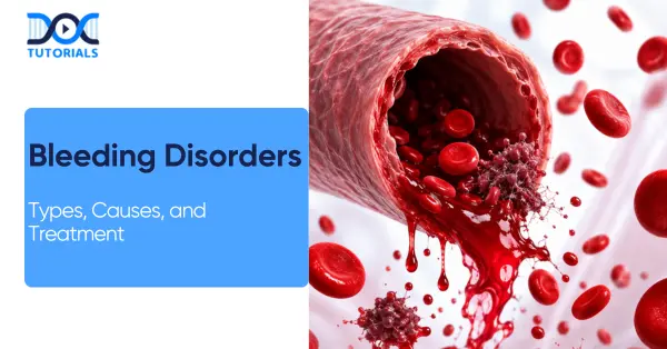
Bleeding disorders (a category of blood disorders) are a group of conditions caused by abnormalities in the body’s ability to form blood clots, leading to excessive or prolonged bleeding. These disorders can be genetic, caused by a deficiency of clotting proteins, by underlying medical conditions, or by taking certain medications.

For medical students and aspirants, bleeding disorders represent an important topic that connects theoretical knowledge with practical clinical application. Having a proper understanding of its causes, diagnostic approach, and treatment strategies is essential for effective patient management. Read on to learn about bleeding disorders in detail.

What are Blood Disorders?

Blood disorders are conditions affecting red blood cells, white blood cells, or platelets that disrupt normal blood functions and may be inherited or acquired, ranging from mild to life-threatening.

Blood disorders are medical conditions that affect the normal functioning of different components of blood, preventing them from carrying out their essential physiological roles effectively:

  • Your red blood cells are responsible for transporting oxygen from the lungs to all parts of the body.
  • White blood cells are important in the body as they help defend the body against infections by attacking harmful pathogens.
  • Your platelets are responsible for blood clotting, which helps prevent excessive bleeding after injury.

Blood disorders are broadly classified as noncancerous or cancerous. This discussion is specific to non-cancerous blood disorders. A person may either be born with a non-cancerous blood disorder due to genetic factors or develop it later in life due to underlying medical conditions that interfere with normal blood function.

Other blood disorders can be asymptomatic and may not need active treatment, whereas others can be chronic and require lifelong medical care, but do not impact life expectancy significantly.

But some blood disorders may be severe, progressive, and even life-threatening unless diagnosed and treated accordingly. Treatment of blood disorders is carried out by healthcare providers, emphasising symptom management, complication prevention, and, where feasible, treatment of the underlying cause.

What are the Various Types of Bleeding Disorders?

Bleeding disorders include haemophilia, von Willebrand disease, thrombocytopenia, platelet function disorders, DIC (Disseminated Intravascular Coagulation), factor deficiencies, and acquired haemophilia, all affecting normal blood clotting mechanisms.

There are several important types of bleeding disorders, including:

  1. Haemophilia: Haemophilia is a hereditary bleeding disorder that is mostly seen in males. It arises from a lack or complete absence of clotting factors IX and VIII, which are critical to the normal process of blood clotting. Even small injuries may result in extensive bleeding among affected individuals.
  2. Von Willebrand Disease: Von Willebrand disease is a genetic disease that occurs in both males and females. It is caused by a deficiency or dysfunction of von Willebrand factor, a major protein that helps platelets adhere to one another and form stable blood clots to prevent bleeding.
  3. Thrombocytopenia: Thrombocytopenia is a condition characterised by an abnormally low platelet count in the blood. Platelets play a crucial role in clot formation; therefore, their depletion increases the risk of easy bruising and prolonged bleeding.
  4. Platelet Function Disorders: These disorders occur when the platelet count is normal, but platelet function is defective. The impairment can be hereditary or acquired, like specific medications that influence platelet activity.
  5. Disseminated Intravascular Coagulation (DIC): DIC is a serious and life-threatening medical condition that typically occurs as a complication of infection, trauma, or other serious diseases. It is characterised by uncontrolled clot formation throughout the body, followed by excessive bleeding due to depletion of clotting factors.
  6. Factor Deficiencies: Factor deficiencies occur when the body is unable to produce adequate amounts of specific clotting factors required for normal coagulation. Such deficiencies can be either hereditary or the result of medications or underlying illnesses.
  7. Acquired Haemophilia: Acquired haemophilia is a rare autoimmune disorder in which the body produces antibodies that attack and neutralise blood-clotting factors, resulting in spontaneous and uncontrollable bleeding.

What are the Causes of Bleeding Disorders?

Bleeding disorders could be caused by either genetic mutations, liver diseases, medications such as blood thinners, autoimmune diseases, infections, blood cancers, or physical injury. These factors interfere with clotting factor production, platelet function, or normal blood coagulation pathways.

Bleeding disorders may be caused by a number of underlying factors, including:

  • Genetic Mutations: Inherited genetic defects cause conditions such as haemophilia and von Willebrand disease. This impairs the synthesis, structure, or function of clotting factors and associated proteins that are critical to normal hemostasis.
  • Medications: There are some drugs which act to prevent blood clots by interfering with the natural process, making one bleed excessively, even in cases of minor injury, such as anti-clotting drugs and anti-platelet medications.
  • Liver Disease: The liver plays a central role in the production of most clotting factors. Any hepatic injury or chronic liver disease may decrease their production, causing defective coagulation and excessive bleeding.
  • Autoimmune Disorders: Some autoimmune disorders, including systemic lupus erythematosus and rheumatoid arthritis, can lead to abnormal bleeding patterns by attacking blood vessels or clotting factors.
  • Cancer: Blood cancers such as leukaemia and lymphoma can affect bone marrow function, reducing platelet and clotting factor production, thereby increasing the risk of bleeding disorders.
  • Infections: Chronic infections like hepatitis C and HIV (Human Immunodeficiency Virus) may harm the liver or suppress the immune system, which can lead to weakened clotting mechanisms and complications of bleeding.
  • Trauma: Intensive physical trauma or surgery may interfere with the normal processes of blood clotting, causing excessive or uncontrolled bleeding, depending on the severity of the damage to the tissue.

What are Common Blood Disorder Symptoms?

Blood disorder symptoms vary based on the affected blood component and may include fatigue, weakness, pallor, and breathlessness in anaemia. Bleeding disorders commonly present with excessive bleeding, bruising, heavy periods, and signs of internal bleeding or clot formation.

Symptoms of a blood disorder depend on which blood component is affected and the degree to which normal blood function is compromised. Different blood components, including red blood cells, white blood cells, and platelets, can be affected, leading to a diverse range of clinical presentations.

For example, in anaemia, a common blood disorder, patients may experience any of the following:

  • Fatigue and generalised weakness.
  • Dizziness and lightheadedness.
  • Pale or reduced skin colouration compared to normal.
  • Increased heart rate (palpitations).
  • Shortness of breath, especially on exertion.

Bleeding Disorder Symptoms

The most common symptom of bleeding disorders includes unexplained, profuse, or extended bleeding. People with any of the following symptoms need medical advice:

  • Nosebleeds: Recurring nosebleeds that last longer than 10 minutes or more than five times a year.
  • Unusual Bleeding: Wounds that won’t stop bleeding even after 10 minutes.
  • Internal Bleeding: Could be characterised by joint pain, swelling, and stiffness due to blood leaking into joints.
  • Bruising Easily: Bruises without any cause or after experiencing a mild physical injury.
  • Bleeding after Surgery: Any surgical procedure, including dental procedures, that leads to unusual or prolonged bleeding.
  • Excessively Heavy Menstrual Bleeding: Menstrual periods that are unexplained, require regular changing of tampons/pads (every hour) and continue for more than seven days.
  • Postpartum Bleeding: Severe bleeding that persists even after delivery/miscarriage.
  • Blood in Stool: Presence of blood in stool or bleeding during defecation, which may also indicate other underlying conditions and should be evaluated by a healthcare provider.
  • Blood in Urine (Hematuria): Visible blood in urine, especially when associated with urinary urgency or discomfort, requires medical evaluation.

Blood Clotting Disorder Symptoms

People who have blood-clotting disorders may develop an unusual amount of clots in their blood vessels. These clots may form in the veins, lungs, heart, or brain. Some common symptoms include the following:

  • Swelling, pain, and tenderness of the legs: symptoms that may mean deep vein thrombosis (DVT).
  • Shortness of breath combined with chest pain, which may indicate pulmonary embolism.
  • Heart attack symptoms.
  • The symptoms of a stroke.

How are Bleeding Disorders Diagnosed?

Bleeding disorders are diagnosed using blood tests such as CBC, PT, aPTT, mixing studies, clotting factor assays, and genetic testing. These investigations help identify platelet abnormalities, clotting factor deficiencies, and underlying inherited or acquired causes.

Clinical examination, combined with special tests, is used to determine whether a person has an inherited or acquired bleeding disorder. Doctors will prescribe the following tests:

  • Complete Blood Count (CBC): This test evaluates different components of blood, including platelets. If there are few platelets in the blood, a person probably has a platelet disorder rather than a deficiency of clotting factors.
  • Partial Thromboplastin Time (PTT) / Activated PTT (aPTT): This test measures the time required for blood to clot through the intrinsic pathway and helps identify deficiencies in specific clotting factors.
  • Prothrombin Time (PT): This test measures the extrinsic clotting pathway and assesses clotting factors not measured by the PTT test, which helps in general coagulation testing.
  • Mixing Test: This test is used to identify bleeding as due to a deficiency of clotting factors or to the presence of inhibitors, such as antibodies, that disrupt the action of clotting factors.
  • Von Willebrand Factor (vWF) Tests: The tests assess the amount and activity of von Willebrand factor and are used to diagnose various forms of von Willebrand disease.
  • Tests for Clotting Factors: These tests measure the levels of certain clotting factors and may show a deficiency. Low levels of factor VIII might indicate haemophilia A.
  • Bethesda Test: The Bethesda test is performed to detect inhibitors of clotting factors, such as VIII and IX.
  • Tests for Factor XIII: The antigen and activity tests are used to check for a deficiency in Factor XIII that leads to bleeding.
  • Genetic Tests: Genetic testing allows for the diagnosis of inherited mutations that lead to bleeding disorders. In addition, patients might be referred to a genetic specialist to carry out a comprehensive investigation.

How are Bleeding Disorders Treated?

Treatment for bleeding disorders depends on the type and severity and includes iron supplements, blood transfusions, and clotting factor replacement therapy. Additional management options, like plasma transfusions or topical treatments, help control bleeding and prevent complications.

Treatment options depend on the type of bleeding disorder, its underlying cause, and the severity of symptoms. While most bleeding disorders cannot be completely cured, appropriate treatment can effectively control symptoms, prevent complications, and improve quality of life.

Here are the common treatment options:

1. Iron Supplementation

In some situations involving excessive or prolonged blood loss, doctors can prescribe iron supplements to replenish iron stores. Iron deficiency anaemia may result from low iron levels and may produce symptoms such as fatigue, weakness, dizziness, and poor physical performance.

In extreme cases, a blood transfusion might be necessary to replenish normal blood parameters when iron supplementation is inadequate.

2. Blood Transfusion

This treatment consists of replacing lost or deficient blood components with donated blood. To prevent serious blood transfusion reactions, donor blood should match the patient’s blood type. Typically, it is administered in a hospital setting under constant medical supervision.

3. Other Treatments

Depending on the type of disorder, some other forms of treatment can be employed. Localised bleeding in some bleeding disorders can be treated with topically applied agents or nasal sprays.

Other disorders like haemophilia are managed with replacement therapy, by which the lacking clotting factors are directly infused into the bloodstream to prevent or manage episodes of bleeding.

In certain cases, fresh frozen plasma transfusions are used when specific clotting factors are deficient. Fresh-frozen plasma contains important clotting factors, including factors V and VIII, which play key roles in normal blood coagulation. These transfusions are usually performed in a hospital to ensure safety and proper monitoring.

FAQs about Bleeding Disorders

  1. What are bleeding disorders?

Bleeding disorders are conditions associated with the inability of blood to clot normally due to deficiencies in clotting factors, platelets, and clotting mechanisms. People with bleeding disorders experience symptoms such as excessive bleeding, uncontrolled bleeding following an injury, or spontaneous bleeding.

  1. What are the main types of bleeding disorders?

Some common types of bleeding disorders include haemophilia, von Willebrand disease, thrombocytopenia, platelet function disorder, and clotting factor deficiencies.

  1. What are the causes of bleeding disorders?

Common causes of bleeding disorders include genetic disorders (haemophilia is an inherited disorder), low levels of platelets, liver disease, autoimmune disorders, infections, or medications such as anticoagulants.

  1. What are the symptoms of bleeding disorders?

Some common symptoms include frequent nosebleeds, easy and unexplained bruising, bleeding for a prolonged period after an injury or surgery, bleeding excessively during menstruation, bleeding gums, and even internal bleeding, which causes joint pain or swelling in severe cases.

  1. How are bleeding disorders diagnosed?

Diagnosis is done using blood investigations such as complete blood count (CBC), prothrombin time (PT), activated partial thromboplastin time (aPTT), clotting factor assays, and sometimes genetic testing to identify inherited disorders.

  1. Can bleeding disorders be cured?

Most bleeding disorders are incurable, particularly genetic ones, such as haemophilia. However, they can be effectively controlled and managed with long-term treatment and preventive care to reduce bleeding episodes.

  1. What is the treatment for bleeding disorders?

Treatment varies depending on the condition and may include clotting factor replacement therapy, iron supplements for anaemia, blood or plasma transfusions, and medications to manage bleeding. Some cases may also require preventive long-term therapy.

  1. Is haemophilia a common bleeding disorder?

Haemophilia is one of the most well-known inherited bleeding disorders, caused by a deficiency of clotting factors VIII or IX. It mostly happens in males and may cause prolonged bleeding following minor injuries.

  1. Are bleeding disorders dangerous?

Yes, there are bleeding disorders that may be serious or life-threatening, particularly when there is uncontrolled internal bleeding, significant trauma, or postoperative complications. At an early stage, it is necessary to detect and treat the disease to avoid complications.

  1. When should a person see a doctor for bleeding problems?

One should consult a doctor when experiencing frequent unexplained bleeding, bleeding longer than usual, heavy menstrual bleeding, and frequent bruising without injury because these may be signs of an underlying bleeding disorder.

Conclusion

In conclusion, bleeding disorders represent a category of diseases affecting the body’s normal mechanism of clot formation, leading to excessive bleeding, prolonged bleeding, or easy bruising. They are inherited or acquired disorders that vary in severity depending on their cause.

For NEET PG students, having a clear understanding of bleeding disorder is paramount to strengthening knowledge of clinical concepts. For additional guidance, DocTutorials can be your study companion. We offer crisp videos, clinical Qbank, exam-focused notes, flashcards, and mind maps. This helps ensure aspirants gain complete clarity over complex medical topics.

Join DocTutorials today and explore our NEET PG course to excel in your medical career!

NEET PG Online Live Power Pack
DocTutorials App Download Now

Latest Blogs

Bed Wetting: Causes, Symptoms, Diagnosis, and Treatment

Bed wetting

Bed wetting (nocturnal enuresis) is a typical disorder that is characterised by involuntary urination during sleep in a child at the age of 5 years or older. Although this is usually a normal stage of development, it can be a sign of an underlying health problem if it persists into adulthood.

Children develop bladder control skills when they are aged 2-4. Beyond the age of 5, chronic bedwetting is referred to as enuresis and should be assessed. Approximately 15% of children aged 5 continue to wet the bed, decreasing to 10% at age 7 and 5% at age 10. Keep reading to know more.

Classification of Bed Wetting

Bed-wetting, or Enuresis, refers to urinary incontinence during sleep in children older than 5 years. It is categorised by timing (nocturnal vs diurnal), symptomatology (monosymptomatic vs non-monosymptomatic), and history (primary vs secondary).

Pediatric guidelines define enuresis as the involuntary intermittent urination in a child ≥ 5 years. Until the age of 5, it is not regarded as abnormal. Enuresis can be categorised as:

  1. Nocturnal Enuresis (Bed wetting): Nighttime wetting. It is the most prevalent type.
  2. Diurnal Enuresis: Waking up with urinary incontinence.

Further classifications include:

  1. Primary vs Secondary Enuresis

Primary enuresis refers to a child who has not attained a dry period of at least 6 months. Secondary enuresis develops when a child, who has been dry at night for 6 months or more, begins to wet their bed. Secondary enuresis can usually be linked to a developing health or psychological concern (e.g., urinary tract infection, diabetes, or stress).

  1. Monosymptomatic vs Non-monosymptomatic

Monosymptomatic enuresis (MSE) is bedwetting in the absence of lower urinary tract symptoms (LUTS) in daytime. Children with MSE do not have urgency, frequency, or incontinence during the day.

Non-monosymptomatic enuresis (NMSE) is associated with nocturnal enuresis that includes other urine symptoms (daytime incontinence, urgency, frequency, voiding postponement, etc.). NMSE usually necessitates assessment of preexisting bladder or bowel issues.

Causes and Risk Factors of Bed Wetting

There are various factors that contribute to bed wetting. Common causes include genetic predisposition, slowed maturation of the bladder, deep sleep patterns and polyuria at night. Psychosocial stress and medical conditions (UTIs, diabetes, sleep apnea) can also contribute.

The aetiology of pediatric nocturnal enuresis is multifactorial with a significant genetic component, and the overall risk is approximately 44% in the presence of one affected parent and up to 77% in the presence of both affected parents.

Family history is highly penetrant, as are environmental and physiological factors. Here are the other factors:

  • Delayed Bladder Maturation: Nighttime bladder control is often delayed in many children because the bladder (detrusor muscle) and sphincter have not developed well-coordinated responses during sleep.
  • Nocturnal Polyuria: Children with enuresis have abnormal ADH (Antidiuretic Hormone) secretion, resulting in excessive urine production at night. The result is bladder overfilling during sleep without getting up to void.
  • Small Functional Bladder Capacity: A small bladder fills quickly at night, and with profound sleep or an inability to wake up, it leads to bedwetting. Capacity <65% of expected (≈30 + age × 30 mL) indicates a small bladder size.
  • Overactive Bladder/Detrusor Overactivity: Other children experience involuntary bladder contractions that result in urgency and incontinence, both day and night. In NMSE, overactive bladder can be the only problem or a component of the dysfunctional voiding.
  • Sleep Disorders: Sleep disorders such as obstructive sleep apnea predispose the risk of enuresis because they result in excessive urine and poor arousal. Children with the disorder of deep sleep cannot wake up even when the bladder is full.
  • Psychosocial Stress: Enuresis is typically an effect of stress rather than a cause, though significant life events may exacerbate symptoms or lead to secondary bedwetting.
  • Medical Comorbidities: Several health conditions may trigger or aggravate enuresis:
    • Urinary Tract Infection (UTI): Irritates the bladder, leading to urgency and accidents.
    • Diabetes Mellitus: An overload of glucose leads to polyuria (urine volume) that can fill up the bladder at night.
    • Constipation/Faecal Incontinence: A rectum that is loaded can compress the bladder, resulting in both day and night wetting. Treatment of constipation tends to lower enuresis.
    • Neurologic Disorders: The bladder control can be compromised by spina bifida, tethered cord, cerebral palsy, or other neurodegenerative diseases. A disordered stream or daytime symptoms imply a neurogenic or obstructive disorder, requiring specialist evaluation.
    • Pinworm Infection: Enterobius vermicularis can cause perianal irritation, which may result in reflex bladder contractions and enuresis (rare).
  • Deep Sleep Arousal Difficulty: Even without OSA (Obstructive Sleep Apnea), just having really good sleep can cause a child to fail to wake to bladder signals.

Signs and Symptoms of Bed Wetting

Bed wetting, or nocturnal enuresis, is characterised by involuntary urination at night, which is often unrealised by the child. It can be linked with daytime urgency or frequency (in NMSE) and psychological outcomes such as embarrassment or low self-esteem.

Nocturnal enuresis is involuntary urination at night in children who are supposed to have control over their bladders. It is often self-limiting and benign, but it can have emotional consequences, which is why early detection is crucial.

Here are the main symptoms:

  • Nighttime Incontinence: The most noticeable sign is waking up with wet pyjamas or bed linens. Frequent wet nights are commonly reported by parents without any daytime problems. The child never wakes up at the time of the episode and notices it in the morning.
  • Daytime Bladder Symptoms: Normal in monosymptomatic cases. Urgency, frequency and daytime incontinence can occur in children in NMSE. Voiding ≥ 8 times/day or <3 times/day suggests dysfunction, along with signs like squatting, leg crossing, or hesitancy.

Other associated signs and symptoms include:

  • Urinary Tract Infection (UTI) Symptoms: Painful or burning urine, hazy or bloody urine, foul smell, fever indicates UTI or other organic origin and should be evaluated immediately.
  • Faecal Soiling or Constipation: Enuresis may be accompanied by large, hard stools or faecal accidents due to pressure on the bladder caused by rectal factors.
  • Behavioural/Psychological Impact: Symptoms of anxiety, embarrassment, social withdrawal (aversion to sleepovers), and low self-esteem may be observed in a child with bedwetting. Parents and peers should be informed that enuresis is involuntary and not the fault of the child.
  • Frequency & Patterns: Frequent, daily, or intermittent (e.g., 23 nights/week). The diagnosis threshold is generally: 2 or more episodes/week over 3 months. Cases of sporadic (less than 2/month) are usually within the period of normal maturation.

Diagnosis of Bed Wetting

Pediatric enuresis is mostly diagnosed clinically. It includes ensuring the enuresis criteria (age ≥ 5, frequency ≥ 2/week for 3 months) and ruling out other causes through history, physical examination, and simple tests. The tools include a bladder diary and urinalysis.

Pediatric enuresis diagnosis is predominantly clinical and requires confirmation of meeting the criteria and ruling out underlying causes. An organised history, examination, urinalysis, and a bladder diary are most likely adequate. Here’s how the diagnosis goes:

1. Initial Assessment and History

  • Confirm Diagnostic Criteria: Make sure the child is at least 5 years old and has persistent involuntary bedwetting (at least 2 times a week) for at least 3 months.
  • Fluid Intake & Voiding Diary: Monitor fluid timing, volume and voiding patterns to determine bladder patterns, particularly evening intake and last void.
  • Family History: Ask about enuresis in the family history; also, check for a family history of diabetes, polyuria, and kidney disease.

2. Medical History and Comorbidities

  • UTI history: Dysuria, fever, frequency; recurrent UTIs can cause secondary enuresis.
  • Constipation: History of hard stools, infrequent bowel movements, or encopresis. Bladder outlet obstruction (functional) can be caused by severe constipation.
  • Diabetes Symptoms: Polydipsia (excessive thirst), polyphagia (excessive hunger), weight loss, or nocturia (getting up at night to use the restroom) should prompt a check of blood sugar levels.
  • Neurologic Symptoms: Gait abnormalities, lower-limb weakness, or spinal problems indicate neural involvement (e.g., tethered cord).
  • Psychosocial Factors: Recent stressors, behavioural issues like ADHD (Attention-Deficit/Hyperactivity Disorder), and autism that could be associated.
  • Sleep History: Snoring, sleep restlessness, daytime drowsiness, or observed apnea indicates sleep apnea.
  • Drug History: Some medications (e.g., diuretics, stimulant medications) may cause polyuria or worsen enuresis.

3. Physical Examination

  • General Exam: Growth parameters, signs of chronic illness. Check for external stigmata of spina bifida (tuft of hair, dimple) on the lower back.
  • Abdominal Exam: Palpate bladder (for retention), assess for constipation (palpable stool in colon).
  • Genitourinary Exam: Inspect for phimosis, labial adhesions, and anatomic abnormalities (e.g., ectopic ureter). Percuss the kidneys if suspicion of reflux.
  • Neurologic Exam: Strength of lower limbs, reflexes; perineal sensation, anal wink to check tethered cord or neuropathy.

4. Investigations

Most children with isolated nocturnal enuresis require only minimal testing. Key investigations include:

  • Urinalysis: Essential first step to exclude hematuria, infection (nitrites, leukocyte esterase), glucose (diabetes), or protein. Repeat if symptoms warrant.
  • Urine Culture: In case of suspected UTI (fever, dysuria, very frequent voiding) or pyuria on urinalysis, obtain a culture and treat the infection.
  • Blood Tests: Not routine, but consider blood glucose in polyuria-polydipsia; potentially renal function in a structural malady.
  • Bladder Diary/Voiding Chart: Record time and volume of each void daily within a few days. This can aid in differentiating polyuria (very large voids) and frequency (small volumes, numerous voids).

5. Imaging

  • Renal/Bladder Ultrasound: Indicated if there are recurrent UTIs, daytime incontinence, abnormal stream, or suspicion of anatomic abnormality (e.g. hydronephrosis, ureterocele).
  • VCUG (Voiding Cystourethrogram): Consider if ultrasound shows reflux or if bladder outlet obstruction is suspected.

Management and Treatment for Bed Wetting

Treatment of bedwetting in children includes education, behavioural measures, and, if needed, alarms or medications. Conservative first-line treatment includes positive reinforcement, fluid management, and treating constipation, and alarms and desmopressin are implemented in case of inadequate positive responses.

A combination of behavioural therapy and a bedwetting alarm is the most effective and safe long-term treatment. Pharmacotherapy (particularly desmopressin) can be effective to enhance the quality of life (dry nights) but often requires repetition/continuation to achieve long-term effect.

Here’s how the bedwetting treatment and management work:

1. General Principles

  • Reassurance and Education: Educate the family that enuresis is normal, mostly self-limiting, and not the child’s fault. Set realistic expectations, as treatment may take months and relapses can occur, and emphasise support over punishment.

2. Behavioural Strategies

  • Fluid Management: Limit evening fluids (particularly 12 hrs before sleep) and avoid caffeine/sodas. Encourage drinking earlier in the day.
  • Timed Voiding: Create an urge to urinate at regular times (after 2-3 hours) in the daytime and right before sleep. Other parents put reward charts for going to the bathroom prior to sleep.
  • Double Voiding: Instruct the child to urinate twice before bedtime (void, then try again a few minutes later) to ensure the bladder empties fully.
  • Constipation Management: Ensure soft bowel movements. Manage constipation (fibre, stool softeners) because a full rectum may initiate bladder contractions.
  • Protect Bedding: Use mattress protectors and easily washable bed linens. Have an extra pair of pyjamas in case of stress around accidents.

3. Lifestyle and Support

  • Engaging the child in problem-solving (seek their ideas). Provide stickers or praise for dry nights and helpful actions (changing bed clothes).
  • Avoid blaming or shaming. Do not wake children aggressively to empty (it has no benefit); use alarms to condition awakening.
  • If school concerns arise (e.g., daytime wetting), communicate with school nursing staff about toilet breaks or discreet pull-up garments, if needed.

4. Pharmacologic Therapy

Medication is reserved for when behavioural methods alone fail or for short-term situations (sleepovers, camps). Options include:

  • Desmopressin (DDAVP): A synthetic analogue of vasopressin (antidiuretic hormone). It decreases nocturnal urine production.
    • Use: First-line medication in monosymptomatic enuresis. It can be taken before going to sleep via nasal spray or oral pills.
    • Efficacy: Reduces nighttime urine output and increases the likelihood of a dry night by about 50% on average. Nevertheless, its effect subsides with discontinuation, hence it is typically intermittent or short-lasting. Not curative, but useful to manage special occasions.
    • Caveat: Risk of water intoxication and hyponatremia if too much fluid is consumed; fluid restriction (no drinking 1 hour before and after dose) is important.
  • Tricyclic Antidepressants (e.g., Imipramine): Previously used widely, now second-line because of safety issues.
    • Use: May be considered if alarms and desmopressin fail. Mechanism includes increased ADH and bladder relaxation.
    • Efficacy: Can increase dry nights by around 1 per week.
    • Caution: Risk of overdose is severe (cardiotoxicity, seizures). Contra-indicated in families where there is a risk of overdose. The use is standard with ECG before use, dose-limited, and short courses.
  • Anticholinergic Agents (e.g., Oxybutynin, Tolterodine): Indicated when symptoms of overactive bladder during the day are observed or when a small capacity of the bladder is reported. They relax the detrusor muscle.
    • Use: If the child has daytime urgency or frequency, or if an ultrasound shows a small bladder. May be added to desmopressin in refractory cases.
    • Side Effects: Dry mouth, constipation, blurred vision; should be used cautiously in children.

Prevention and Home Care for Bed Wetting

Whereas the prevention (in the sense of preventing it before it occurs) of bedwetting is somewhat constrained, since it is basically a matter of developmental timing, home care is about promoting the child’s maturation and preventing triggers.

Prevention of bedwetting is not always possible because it is a developmental complication, but with the support of home care, it can be reduced, and the control of the bladder can be improved.

Some of the important measures include:

  • Habit Training: Educate young children about proper toilet habits early (e.g., sitting posture, completing void).
  • Family Awareness: Discuss family stress and provide a supportive environment (do not make fun of the child about wetting).
  • Bowel Management: Avoid constipation by means of a high-fibre diet. A hard stool should be avoided, as it is a contributor to bladder dysfunction.
  • Sleep Hygiene: Sleep well and still; maintain good bedtime habits (use the bathroom before going to sleep; do not go to sleep right after heavy fluid consumption).
  • When to Seek Help: A medical examination is advised when bedwetting is regular, when the child is concerned about it, or when there are concerns (UTI, daytime wetting, systemic illness).

FAQs about Bed Wetting

  1. At what age is bedwetting considered normal?

Bedwetting most often occurs among younger children and is mostly normal through the age of approximately 5 years. When a child who is 5 years or over experiences frequent episodes (at least twice a week during 3 months), it is diagnosed as enuresis.

  1. What is the difference between primary and secondary enuresis?

Primary enuresis is when a child has never attained a dry spell of at least six months. Secondary enuresis is the reappearance of bedwetting in children who have been dry for six months or more.

  1. How is bedwetting diagnosed?

Clinical diagnosis of bedwetting is made on the basis of age, frequency of wetting, medical history and simple tests like urinalysis to eliminate other causes.

  1. What are the first-line treatments for enuresis?

Some of the initial treatment interventions are lifestyle and behavioural factors, which involve reducing evening fluid intake, scheduling voidings, constipation, and reward systems.

  1. When should alarm therapy be used?

Alarm therapy is recommended if initial measures do not work. It is especially effective among children and families who are eager to adhere to the regimen.

  1. What medications are used for nocturnal enuresis treatment?

The most widely used medication is desmopressin. In certain circumstances, other alternatives, such as imipramine or oxybutynin, nin can be looked into.

  1.  Which is more effective: alarms or medication?

Long-term alarm therapy is better as it trains the child, whereas drugs give short-term relief and are linked with relapse after discontinuation.

  1. Is enuresis a psychological problem?

Enuresis is more of a developmental issue, rather than a psychological disorder, though it can result in emotional distress or conditions like ADHD.

  1. Can bedwetting be prevented?

There is no guaranteed way to prevent bedwetting, but good toilet training, curing the constipation and avoiding taking fluids at night could help minimise the need to consult a doctor.

  1. When should a doctor be consulted?

Consultation with the doctor is required when the child is 5 years old or above and has frequent bedwetting, or in case of other symptoms, namely, daytime wetting, pain, or recurrence after dry days.

Conclusion

Bed wetting (pediatric enuresis) refers to a highly prevalent involuntary urination in sleep. For NEET PG, concentrate on classification, causes, and diagnosis. Behavioural measures, alarms, and medications are part of management; most children outgrow them.

 For additional guidance, DocTutorials can be your study companion. We offer crisp videos, clinical Qbank, exam-focused notes, flashcards, and mind maps. We help ensure aspirants gain complete clarity over every complex medical concept.

Join DocTutorials today and explore our NEET PG course to excel in your medical career!

NEET PG Online Live Power Pack
DocTutorials App Download Now

Latest Blogs

Perineal Tears: Symptoms, Causes, and Treatment

Perineal Tears

Not all childbirth-related complications are widely understood or openly discussed. One such condition is perineal tears. It is a type of injury that affects the area between the vagina and the rectum, commonly occurring during vaginal delivery.

A perineal tear can range in severity from minor skin involvement to deeper tissue damage and is classified into four grades. Sometimes, a surgical intervention known as an episiotomy can be performed to create a controlled opening and assist with the delivery process. Keep reading to learn about its types, causes, and treatment to improve care and recovery for the mother.

What are Perineal Tears?

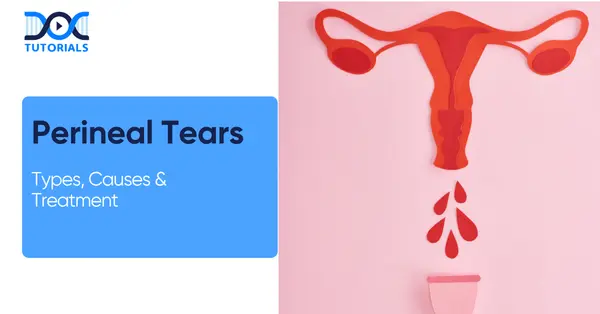
Perineal tears are injuries to the area between the vagina and anus that commonly occur during vaginal childbirth. They can range from mild skin tears to deeper tissue damage. 

A perineal tear or vaginal tear is a type of injury that may occur during birth. Also referred to as a perineal laceration, it involves a tear in the tissues surrounding the vagina and the perineum. The perineal region, commonly called the perineum, is the area located between the vaginal opening and the anus.

During a vaginal birth, the skin around the vagina naturally thickens and becomes more flexible. This process helps the baby’s head and body pass through more easily. Despite this natural adaptation, tearing is quite common. In fact, up to 90% of women who have a vaginal delivery experience some degree of tearing.

However, several factors can increase the likelihood of a vaginal tear. These include:

  • A larger-than-average baby
  • An early birth that does not give the skin time to stretch adequately
  • The use of instruments such as forceps during delivery

What are the Various Types of Perineal Tears?

Perineal tears are classified into 4 degrees: first-degree (minor skin tear), second-degree (involves skin and muscles), third-degree (extends to the anal sphincter), and fourth-degree (reaches the rectum).

Vaginal tears can be categorised into 4 grades according to severity. The size of the tear defines its type. Here are the various types of vaginal tears:

  1. First-degree Tear: This is the mildest form of tearing. It only infects the external skin around the vagina and perineum and is usually self-healing and does not require stitches.
  2. Second-degree Tear: This is the most frequently occurring type. The tear goes deeper, affecting both the skin and the underlying muscles of the vagina and perineum. Stitches are usually required for proper healing.
  3. Third-degree Tear: This type of tear extends further, reaching from the vaginal area to the anus. It involves damage not only to the skin and perineal muscles but also to the anal sphincter muscles, which control bowel movements. Stitches are necessary for recovery.
  4. Fourth-degree Tear: This is the most severe and least common form of tearing during childbirth. It extends from the vagina through the perineum, affecting the anal sphincter muscles and reaching into the rectum.

In such cases, the healthcare provider may need to perform the repair in an operating room instead of the delivery room.

What are the Causes of Perineal Tears?

Perineal tears can be caused by childbirth, sexual intercourse, shaving, waxing, vaginal dryness, and other conditions that make the skin weak and vulnerable to any type of injury. Other factors that cause perineal tears include having a large baby, quick labour, vaginal birth for the first time, or decreased skin elasticity.

Most of the cuts or tears in the vagina are commonly linked to activities such as sexual intercourse, shaving, or waxing. However, there are many other cases in which someone ends up developing a tear in and around the vagina, including pre-existing skin problems or giving birth.

Here are the primary causes involved:

  • Sexual Intercourse

Sexual intercourse is one of the common reasons why some women develop perineal tears. Lack of proper vaginal lubrication might lead to such an injury. Intense sexual activity and using sex toys might also contribute to tearing the vaginal area.

  • Shaving and Waxing Pubic Hair

Shaving and waxing pubic hair are also common factors that result in cuts on the vulva. According to research conducted in 2017, about 25.6% of those who groomed their pubic hair sustained cuts, most of which were vulvar cuts in women. In some cases, these injuries required medical treatment, such as antibiotics or even minor surgery.

  • Childbirth

About 80% of women experience perineal tearing during childbirth. A perineal tear refers to damage to the skin or muscles of the perineum, which is located between the vagina and the anus. In some cases, the tear could affect the vagina and even the anal sphincter that controls bowel movements.

Women who are giving birth for the first time are generally at a higher risk of experiencing perineal tears compared to those who have previously had a vaginal delivery.

  • Skin Conditions

Certain skin conditions can make the vaginal or vulvar area more prone to cuts and irritation. These include:

  • Eczema is a skin condition characterised by red, inflamed, and itchy skin. Symptoms of eczema include fluid exudation, crusting, and itching in the vagina and vulva.
  • Lichen Planus is an inflammatory disease that causes itchiness, burning, or a feeling of raw skin in the vulva. If the disease affects the vagina, there will be yellowish sticky discharges and tissue destruction.
  • Psoriasis, a common skin condition that is characterised by itchy and dry scales. However, in the vulvar region, Psoriasis manifests as smooth, pink spots with well-defined edges.
  • Lichen Sclerosus, an inflammatory skin disease that primarily affects the genitalia. The affected skin is easily torn or develops fissures.
  • Vulvovaginal atrophy (atrophic vaginitis), a condition that causes the vaginal tissues to become thinner, drier, and less elastic, making tears more likely.
  • Vaginal scarring or tissue damage, which may result from prior surgeries or radiation treatments in the pelvic region.
  • Vulvovaginitis, commonly caused by Candida albicans (also known as thrush), may irritate the tissues and predispose them to cuts.
  • Genital herpes (herpes simplex infection), which may develop clusters of fluid-filled blisters that may rupture and leave tiny open sores that look like cuts.
  • Other Causes

Other factors that may contribute to the formation of vaginal or vulvar cuts include:

  • Foreign body insertion into the vagina
  • Tampon insertion or extraction errors
  • Vaginal dryness
  • Vaginismus
  • Thinning of the vaginal tissue due to age
  • Steroid medication usage

What are the Symptoms of Perineal Tears?

The perineal tear symptoms typically involve pain, burning, swelling, tenderness and discomfort in the vaginal or perineal region, particularly during sitting, walking or during urination. In more serious instances, one might experience bowel or bladder control problems, emotional distress, and pain that impact daily activities and postpartum recovery.

In case of a perineal tear, a variety of symptoms can occur that disrupt everyday comfort and normal activities. It is essential to identify these symptoms to obtain the necessary treatment and aid recovery in a timely manner.

Physical symptoms may include:

  • Perineal pain or discomfort immediately after the tear, which is usually sharp, burning, or tender, particularly during motions or during use of the toilet.
  • Inability to sit, stand, or walk comfortably, which may cause problems in performing routine tasks or taking care of a newborn.
  • Bladder or bowel changes, including difficulty urinating or urgency, especially with more severe tears.

Emotional and functional effects may include:

  • Feelings of anxiety related to healing or concerns about resuming intimacy
  • Difficulty managing daily activities, including maintaining hygiene or caring for a baby
  • Uncertainty about which physical activities are safe during the recovery phase

How are Perineal Tears Diagnosed?

Perineal tears are determined by a careful physical examination whereby the medical practitioner examines the vaginal and perineal tissues to determine the extent of the injury. Reviewing the patient’s symptoms and medical history also helps identify whether the tear is new, recurring, or associated with other underlying conditions.

A healthcare provider usually conducts a physical examination and analyses your medical history to diagnose a vaginal or vulvar cut. This procedure helps them understand your symptoms and identify any underlying conditions or contributing factors that might have caused the injury.

They can also question you about the symptoms, whether they are new or recurrent, to find out whether you have had a history of similar problems. When the cuts are tiny or barely visible, the provider may use a colposcope, a specialised instrument with a bright light, to closely examine and magnify the affected area during the assessment.

Diagnostic Tests

In most situations, additional tests are not necessary to diagnose or manage vaginal or vulvar cuts, especially when the cause is clear and the injury appears minor without signs of infection.

In contrast, tests may be necessary when it is critical to establish whether there are any underlying conditions that might contribute to the injury. Tests that might be required include:

  • Culture: A sample of vaginal discharge is collected by swab and brought to the lab to determine infections such as bacterial vaginosis, yeast infections, or sexually transmitted diseases.
  • Biopsy: A small tissue sample is collected from the tear site for microscopic evaluation to determine the underlying causes of repetitive or persistent tears of the vagina.

What is a Perineal Tear Treated?

Perineal tears are treated based on their severity: mild tears usually heal on their own, whereas moderate to severe tears are treated with dissolvable stitches to repair the damaged tissue. Recovery is supported by pain-relief methods, proper hygiene, hydration, stool softeners, and cooling pads to reduce discomfort and promote faster healing.

The strategy for dealing with a vaginal or perineal tear varies according to the severity of the tear. First-degree tears do not require stitches because they can heal on their own. Second-, third-, and fourth-degree tears require stitches to repair the injured tissues and ensure proper healing.

Dissolvable sutures are used to sew these tears, and the stitches dissolve on their own after six weeks. In the case of the anal sphincter, the medical practitioner may opt for a more complex repair using dissolvable sutures. Some discomfort is expected as the tear heals.

There are some measures that you can take to ease the discomfort and promote rapid healing of the tear:

  • Thoroughly cleanse the area after going to the bathroom.
  • Pat the area dry gently using toilet paper without rubbing or wiping.
  • Maintain adequate hydration to prevent constipation, and, if necessary, use stool softeners.
  • To avoid any pain or irritation of the tear, healthcare providers can advise using cooling pads and sanitary pads after delivery.

FAQs about Perineal Tears

  1. How long does it take for a vaginal tear to heal?

Most women begin to notice relief from the discomfort caused by a vaginal tear within about two weeks. If stitches are required, they generally dissolve on their own within approximately six weeks.

In most cases, there is no need for a follow-up visit to remove stitches or for additional treatment, as healing occurs naturally.

  1. Can a woman have a vaginal delivery in a second pregnancy if she had a vaginal tear previously?

In most situations, having a tear during a previous delivery does not mean it will happen again in future childbirths. Small tears normally heal well, and they do not disrupt subsequent vaginal births.

However, if a woman has experienced a third- or fourth-degree tear before, there may be a slightly increased risk of tearing in subsequent vaginal births.

  1. When should I start pelvic floor physical therapy after experiencing a perineal tear?

Individuals may recover at an individual rate, but most people can start gentle pelvic floor therapy at 6 to 8 weeks after delivery, provided that their healthcare provider approves it.

It can also be helpful to schedule an earlier consultation to discuss recovery strategies and receive guidance on the healing process.

  1. What can I do to prepare my perineum for childbirth and potentially minimise tearing?

Preparation can typically start during the third trimester. Techniques such as perineal massage, specific strengthening exercises, and learning optimal birthing positions may help reduce the likelihood of tearing.

A trained professional can guide you through safe and effective, evidence-based methods tailored to your body’s needs.

  1. Will scar tissue from perineal tearing cause long-term problems?

Scar tissue is a normal part of the body’s healing process. With appropriate care, including physical therapy, it is usually possible to maintain flexibility and prevent tightness or discomfort, ensuring normal function over time.

  1. What are the risks of untreated severe perineal tears?

Unless properly treated, third- or fourth-degree tears can lead to complications like infection, persistent pain, urinary or faecal incontinence, and discomfort during sex. Immediate treatment and correct follow-up are important in the prevention of chronic complications.

  1. How can I manage pain after a perineal tear?

A combination of prescribed drugs, cold compresses or cooling pads, and proper hygiene can be used to manage pain. A sitz bath can also be helpful. Strain should be avoided when passing stool, loose-fitting garments should be used, and sufficient rest should be taken to aid healing.

  1. When is it safe to resume sexual activity after a perineal tear?

The majority of healthcare practitioners recommend waiting until the tear has completely healed before engaging in sexual activity, which is about six weeks following childbirth.

However, this timeline may vary depending on the severity of the tear and individual recovery, so it is best to consult your doctor before restarting sexual activity.

  1. Can perineal tears affect future pregnancies or deliveries?

In general, minor tears do not have any significant impact on future pregnancies or childbirth. Nevertheless, females with more severe tears (third and fourth degrees) might be at an increased risk during subsequent births.

Depending on your medical history, your healthcare provider will assist in identifying the safest delivery option.

  1. Are there any warning signs of infection during healing?

Yes, some symptoms can indicate an infection. These include pain, swelling, redness, abnormal or foul-smelling discharge, fever, or delayed recovery. If any of these signs appear, it is important to seek medical attention promptly to avoid further complications.

Conclusion

Perineal tears are a common childbirth condition that usually heals in a few weeks under good care. While most cases recover completely within 6 weeks of delivery, the severity can vary depending on the degree of the tear. Seeking guidance from a healthcare expert ensures better healing and reduces the risk of complications.

For additional guidance, DocTutorials can be your study companion. We provide clear videos, clinical Qbank, exam-oriented notes, and flashcards. This helps ensure aspirants gain complete clarity over complex medical topics.

Join DocTutorials today and explore our NEET PG course to excel in your medical career!

NEET PG Online Live Power Pack
DocTutorials App Download Now

Latest Blogs

Thermal Injury: Causes, Symptoms, and Treatment

Thermal injury types and cause

Thermal injuries (burns) are tissue damage caused by external heat sources, e.g., flame, hot liquids, steam, chemicals, electricity, or long-term radiation. These injuries are either superficial, first-degree burns, or deep full-thickness (third- or fourth-degree) burns, including those that affect more than one organ system when extensive.

In forensic medicine, the study of burn injuries is important in determining the cause and mode of injury or death. Keep reading to know more about its types, causes, diagnosis and treatment.

Classification of Thermal Injuries

There are 3 types of thermal injuries, classified by depth and mechanism. The depth of burns is superficial to full-thickness and deeper. Mechanisms include scalds, flame burns, contact burns, electrical burns, chemical burns, and radiation burns.

Thermal injuries are classified by the mechanism of injury and the depth (degree) of skin damage. The following table summarises the burn depth and features:

Burn DegreeSkin Layers InvolvedKey FeaturesHealing Time
First (Superficial)Epidermis onlyRed, dry, painful, no blisters; blanches on touch~5–10 days; heals without scarring
Second (Partial)Epidermis + part of dermisRed/moist with intact or ruptured blisters; very painful; blanches2–3 weeks; may scar
Third (Full)Epidermis + dermis + subcutaneousLeathery, white/brown/charred; painless (nerve loss); does not blanch>8 weeks; requires surgery

Fourth-degree burns (deep burns) penetrate all layers of the skin into muscle, bone or organs and result in charring. These are life-threatening and must always be surgically treated.

By mechanism, thermal injuries include:

  • Scalds: Caused by hot liquids or steam (common in children).
  • Flame Burns: Due to open-fire or clothing ignition.
  • Contact Burns: Through contact with hot things (e.g., metal, irons).
  • Electrical Burns: From AC (alternating current) or DC (direct current), producing serious thermal damage along the current path.
  • Chemical Burns: Caused by acids or alkalis; they chemically generate heat on tissue contact.
  • Radiation Burns: As a result of long UV (ultraviolet) exposure (sunburn) or ionising radiation.

Proper classification assists in estimating the total area burned and the treatment area. For example, the “Rule of Nines” divides the body into regions of 9% TBSA (total body surface area) each (e.g. head 9%, each arm 9%).

In children, the “Lund-Browder” chart adjusts for proportion changes. The severity of the burns is also graded (minor, moderate, major) according to TBSA, depth, age, and location.

Pathophysiology of Thermal Burns

Thermal injury causes protein denaturation and forms three zones: coagulation (necrosis), stasis (at risk), and hyperemia (reversible). Major burns cause fluid loss, shock, inflammation, and such complications as infection, respiratory damage, and organ failure.

Cellular damage from thermal energy includes protein denaturation and membrane destruction. Burns leave a characteristic three-zone pattern in the skin. They are:

  1. Zone of Coagulation: Central, irreversibly necrotic tissue.
  2. Zone of Stasis: Surrounding ischemic tissue that may survive or progress to necrosis.
  3. Zone of Hyperemia: Outer area with reversible injury.

Burns are dynamic, meaning the initial injury can worsen over a few hours or days (e.g., the stasis zone worsening). Larger size burns (>20% of body surface) result in a systemic inflammation response. It increases capillary permeability, resulting in excessive fluid loss (burn shock) and intravascular volume depletion. 

Hypotension and hypoperfusion follow if not corrected. Exposure to superheated air or combustion products may also damage the airways and lungs, leading to respiratory failure. Tissue necrosis from burns also impairs the skin’s functions: infection barrier, temperature regulation, and fluid retention.

Loss of these functions leads to a high risk of infection, hypothermia, electrolyte imbalance, and organ dysfunction. Systemically, severe burns can cause hypermetabolism and release of inflammatory mediators (cytokines, prostaglandins), which, in turn, may result in the systemic inflammatory response syndrome (SIRS) and multi-organ failure.

In forensics, extensive burns may also result in poisonous exposures: carbon monoxide (in smoke) and cyanide (in plastics) intoxication are frequent causes of death.

Causes and Risk Factors of Thermal Injury

The burn definition medical professionals use describes the injury as damage to bodily tissues caused by hot liquids, flames, hot objects, electricity, lightning, chemicals and sunlight. The risk is high with the extremes of age, work-related exposures, and substance use, as well as unsafe homes. Burn patterns are used in forensic practice to determine the cause and manner of injury, such as possible abuse or homicide.

Thermal burns arise from many sources of excessive heat. Common causes include:

  • Scalding liquids (hot water, steam, oil).
  • Open flames (house fires, explosives).
  • Hot objects (steam irons, stoves), and sunlight (ultraviolet).

Electrical injury or joule burn produces heat within the body when current passes through tissues, and there are usually entry and exit wounds. However, during a lightning strike, a filigree burns (also known as a Lichtenberg figure, arborescent burn, or feathering) is a distinctive, fern-like pattern on the skin

. Chemical burns (strong acids or alkalis) cause thermal-like injury via exothermic reactions.

Risk Factors

Certain conditions predispose to burns:

  • Age Extremes: Young children and the elderly have thinner skin and slower reflexes to avoid hot sources.
  • Occupational Hazards: Firefighters, industrial workers handling hot materials, and kitchen staff are at greater risk of exposure to flames.
  • Substance Use: The effect of intoxication by alcohol or drugs compromises judgment near fire and hot materials.
  • Domestic Risks: Lack of smoke detectors, unsafe stoves or heaters, and poor supervision of children.

The pattern and distribution of burns are also indicative of a cause in forensic practice. For example, “forced immersion” scalds (symmetrical burns on buttocks and extremities with distinct scald lines) raise suspicion of abuse in children or vulnerable adults.

Necklacing (homicidal burning method using a tyre) is a classic forensic burn scenario. The identification of these patterns aids forensic professionals in determining whether the injuries were deliberate, unintentional, or caused by others.

Clinical Features and Symptoms of Thermal Injury

Thermal burns are characterised by redness, blisters, swelling, charring, and eschar, whereas inhalation injury is characterised by facial burns, soot, and respiratory distress. Severe burns may result in oedema and renal failure.

Thermal burns have varying symptoms based on depth and extent. In partial-thickness (nerve endings are exposed) burns, pain is intense, but in full-thickness burns (nerve destruction), pain can be surprisingly weak. Common signs include:

  • Redness (erythema)
  • Blistering
  • Swelling
  • Charring
  • Eschar formation

In inhalation injuries, one may see:

  • Facial burns
  • Singed nasal hairs
  • Soot in the mouth or sputum
  • Hoarseness
  • Respiratory distress

Fluid shifts cause generalised oedema and often oliguric renal failure in severe cases. In forensic examination of a burned body, several findings distinguish antemortem from postmortem burns:

FeatureAntemortem BurnPostmortem Burn
Skin rednessPresent (due to vasodilation)Absent (no circulation)
BlistersPresent: fluid rich in albumin and chlorideMay occur; contain air/thin fluid
Soot in airwaysCarbon particles in the trachea/lungsAbsent
Carboxyhemoglobin (CO-Hb)Elevated (>10%) from smoke inhalationNormal
Haemorrhage at the injury sitePresent (capillaries intact)Absent (no vital reaction)
Curling’s ulcer (duodenum)Common in fatal burns (stress ulcers)Absent

Other notable forensic findings include “pugilistic attitude” – a flexed posture of burned bodies due to muscle contraction at >60°C – and heat fractures of bones (bones split due to dehydration, often with a distinctive pattern that does not cross suture lines).

Burned bodies also exhibit thermolysis of soft tissues and “parchment” skin. The cause of death in fire victims is often inhalation injury (asphyxia from smoke, CO or cyanide poisoning) rather than cutaneous burns.

Findings in forensic reports can include lung weight, cherry-red tissue discolouration (CO poisoning), and cyanide atomic absorption.

Medical Diagnosis of Thermal Injury

Medical diagnosis of thermal injury involves both forensic and clinical evaluation. Forensic methods help determine the cause, severity, and identity, while clinicians assess burn size, depth, and related complications, such as inhalation injury.

Forensic-medical diagnosis of thermal injuries involves thorough external and internal examination. Investigations may include:

  • Autopsy: Close dissection to determine depth of burns, internal organ injuries, and smoke inhalation.
  • Toxicology: Determining the quantity of carboxyhemoglobin, cyanide, and other poisonous gases formed during combustion is essential.
  • Histopathology: Skin biopsies can confirm the vitality of burns (e.g. neutrophils, red blood cells), extravasation in antemortem burns, and estimate the age of lesions.
  • Imaging: Post-mortem CT (computed tomography) scans of the body could reveal heat fractures or retained foreign material.
  • Forensic Odontology/Anthropology: Dental or skeletal analysis can help identify severely burned remains.

In clinical diagnosis, the healthcare practitioners measure the size and depth of the burns. The “Rule of Nines” or Lund-Browder chart quantifies TBSA. The depth is assessed by appearance, capillary refill (blanching), pain, and sensation. Patients with burns should be evaluated for associated injuries (inhalation, trauma) and for burn severity.

Management and Treatment of Thermal Injury

Thermal injuries are initially managed using the ABC principles (immediately after injury): airway protection, fluid resuscitation using the Parkland formula, and analgesics. Proper wound care, infection prevention, and tetanus prophylaxis are essential. Severe burns are surgically managed and strictly supported, with nutrition and observation of complications.

Initial management of thermal injuries follows trauma protocols (ABCs). Key steps include:

  • Airway and Breathing: Check for inhalation injury. Early intubation in case of respiratory distress or facial burns.
  • Circulation: Start large-bore IV access promptly. Remove constricting clothing/jewellery.
  • Fluid Resuscitation: For major burns, aggressive IV fluid resuscitation is required. The Parkland formula is a standard guideline:

4 mL × body weight (kg) × % TBSA (second/third-degree only) of lactated Ringer’s over 24 hours, with half in the first 8 hours. Adjust fluid to maintain urine output (0.5–1 mL/kg/h in adults).

  • Pain Control: Give analgesics (when necessary- opioids) as burns are painful.
  • Burn Care: Cool the burn with lukewarm (not ice-cold) water to limit depth. Gently clean and debride wounds. Use topical antimicrobials (e.g. silver sulfadiazine) to avoid infection. Cover with a sterile dressing.
  • Tetanus Prophylaxis: Update tetanus immunisation, as burns are prone to tetanus.
  • Wound Management: Superficial burns heal spontaneously. Partially-thickness burns can need debridement and specialised dressings. Full-thickness burns require surgical treatment (excision and grafting).
  • Supportive Care: Monitor for shock (IV fluids, hemodynamic monitoring), provide nutritional support (high-calorie diet), and prevent hypothermia (warm ambient temperature).

Admit to a burn centre if criteria are met (e.g. >20% TBSA, face/hands/genitals, electrical/chemical burns).

Rehabilitative therapy, scar management (pressure garments), and reconstructive surgery are treatment options that may be used on a long-term basis.

Prevention of Thermal Injury

Thermal injury prevention involves fire control, safe kitchen operations, controlled water temperature, and workplace safety precautions. Social consciousness and safety laws help reduce the incidence of burns, although forensic vigilance is important for identifying abuse or arson.

Preventing thermal injuries is critical, especially in high-risk settings. Safety measures include:

  • Fire Safety: Install working smoke detectors and keep fire extinguishers accessible. Practice fire escape plans.
  • Kitchen Safety: Use hot liquids with caution; handle pot handles carefully; be cautious around stoves. Never leave cooking unattended.
  • Water Temperature: Set water heaters to less than 120 F (49 C) to avoid scalds; check bath water temperature.
  • Workplace Precautions: Use protective equipment (gloves, face shields) when handling hot materials or chemicals. Follow electrical safety protocols.
  • Education: Educate children on fire safety, do not play with matches, lighters or fireworks.
  • Sun Protection: Wear sunscreen and protect yourself to avoid radiation burns (sunburn).

FAQs about Thermal Injuries

  1. How are burns classified by degree?

Burns can be categorised as deep: first-degree (epidermis, only, red, painful), second-degree (partial-thickness, blisters, very painful), and third-degree (full, charred, often painless). The fourth-degree burns extend to muscle or bone.

  1. What is the Rule of Nines?

The Rule of Nines is a quick method to estimate burn surface area. In adults: head/neck = 9%, each arm = 9%, anterior trunk = 18%, posterior trunk = 18%, each leg = 18%, perineum = 1%.

  1. How can you tell if burns occurred before death?

Antemortem burns show vital reactions – redness, blisters with protein fluid, haemorrhage at edges, and soot in airways. Blood tests reveal high carboxyhemoglobin and cyanide. Postmortem burns do not have these signs (there is no inflammation or inhaled soot).

  1. What is “pugilistic attitude”?

It is a typical flexed posture of a burned body due to muscle shrinkage in heat (> 60 °C). It resembles a boxing stance and results from heat-stiffened muscles.

  1. How are burns managed initially?

Follow trauma ABCs: secure airway (intubate if needed), support breathing, then start IV fluids. Cool the burn, provide analgesics, clean/debride wounds and place sterile dressings. Use formulas such as the Parkland formula (4 mL × kg × % TBSA) to guide fluid resuscitation.

  1. What is the Parkland formula?

A guideline for fluid resuscitation in major burns: 4 mL per kg of body weight per cent of TBSA burned (2nd/3rd-degree burns) of lactated Ringer’s in the first 24 hours. Half of that volume is given in the first 8 hours after the burn.

  1. Can electric injuries be considered thermal burns?

Electrical injuries cause serious thermal damage along current paths, but they also cause internal organ injury (arrhythmias, muscle damage). When providing a forensic classification, an electrical burn is frequently mentioned in contrast to a thermal burn.

  1. How do chemical burns differ from thermal burns?

Chemical burns occur due to an exothermic reaction with acid or alkali and can extend the damage to tissue until the agent is eliminated. They are treated by copious irrigation and neutralisation, unlike simple cooling for thermal burns.

  1. Why is burn depth painful initially but not in full-thickness burns?

Partial thickness burns reveal nerve ends, resulting in severe pain. Full-thickness burns destroy the nerves within the affected area; the victim might not experience any pain within the burnt area.

  1. What are some key forensic signs of burn-related death?

In addition to vital reactions, there is soot in the airways (from inhalation), thermal fractures of the bone, black lung (from inhaled carbon), and internal signs of systemic poisoning (e.g., cardiac carbonisation, cerebral infarcts due to hypoxia).

Conclusion

Burns can be thermal, chemical, radiation-induced, or electrical, each with distinct forensic and medical consequences. Medical students and forensic trainees must be able to understand burn classification, pathophysiology, and related factors.

In NEET PG forensic medicine, the Rule of Nines, burn depth features, and vital reactions in burns are high-yield topics. For additional guidance, DocTutorials can be your study companion. We offer crisp videos, clinical Qbank, exam-focused notes, flashcards, and mind maps.

Join DocTutorials today and explore our NEET PG course to excel in your medical career!

NEET PG Online Live Power Pack
DocTutorials App Download Now

Latest Blogs

Sarcoidosis: Causes, Symptoms, Diagnosis, and Treatment

sarcoidosis.

Sarcoidosis is a systemic inflammatory disease which is characterised by the formation of noncaseating granulomas in various organs. Typical presentations are bilateral hilar lymphadenopathy and changes in the interstitial lung, although sarcoidosis may occur in the skin, eyes, heart, and other organs.

This NEET-PG pathology review covers sarcoidosis aetiology, clinical features, diagnostic criteria, management, and complications, emphasising information relevant for medical students. Keep reading to know more.

What are the Causes of Sarcoidosis?

The exact cause of sarcoidosis is unknown. It is thought to have an exaggerated response of the immune system to unknown antigens. Risk is genetically predisposed, affected by race and environment.

There is no single known cause of sarcoidosis, and its formation is associated with a mixture of genetic and environmental factors, as well as immune-related factors:

  • Unknown Aetiology: No specific trigger is found. Sarcoidosis is likely an autoimmune response (primarily Th1-mediated) to microbial or environmental antigens. Exposures (beryllium, insecticides, mould), infections (mycobacteria, Propionibacterium acnes) are investigated, but none of them can explain all the cases.
  • Genetic Factors: Genetic predisposition is indicated by familial clustering and HLA (Human Leukocyte Antigen). There are also specific HLA-DR alleles and ACE (Angiotensin-Converting Enzyme) gene variants associated with susceptibility, which point to heritable immune hyperreactivity.
  • Demographics: Most prevalent among adults between 20 and 40 years. It occurs more in African Americans than in whites, and the morbidity among African American females is the highest. Women tend to experience extrapulmonary disease, whereas men tend to exhibit cardiac involvement.
  • Geographic/Seasonal Factors: Seen worldwide but more common in northern latitudes and urban areas. There is some seasonal variation, but there are no definite prevention measures.
  • Immune Factors: Patients have an exaggerated cellular immune response at disease sites, characterised by CD4+ T-helper cell accumulation and inverted CD4/CD8 ratios in affected tissues.

High concentrations of cytokines (IL-2, IL-6, TNF-α, IFN-γ) favour the development of granuloma. These are immunologic pathways which play a central role in the pathogenesis of sarcoidosis.

Pathogenesis and Histopathology of Sarcoidosis

The pathology of sarcoidosis is characterised by an over-response of the immune system, which results in the production of compact noncaseating granuloma in tissues. Granulomas are composed of epithelioid histiocytes and giant cells.

The pathology of sarcoidosis is characterised by the dysregulated immune response, resulting in the appearance of distinct noncaseating granulomas, which are the key features of the disease and may be localised to various organs.

  • Granuloma Formation: The characteristic lesion is the noncaseating epithelioid granuloma, with tight clusters of activated macrophages and Langhans-type giant cells. These do not have central necrosis, although there may be rare fibrinoid necrosis.

The presence of surrounding lymphocytes (primarily CD4+ T cells) and fibroblasts may ultimately result in fibrosis.

  • Cellular Details: Epithelioid histiocytes release enzymes and cytokines, such as ACE, lysozyme, and calcitriol. The overproduction of calcitriol may lead to hypercalciuria and hypercalcemia.

Giant cells are the result of fusion and may include Schaumann or asteroid bodies- nonspecific but supportive findings.

  • Organ Involvement: Granulomas can affect almost any organ, most commonly the lungs and intrathoracic lymph nodes (up to 90%). They may also affect skin, eyes, heart, liver, spleen, bone marrow, and nervous system, affecting normal tissue (e.g., fibrosis in lungs, arrhythmias in heart).
  • Pathological Variants: Granulomas appear in skin lesions (e.g., lupus pernio), but acute sarcoidosis (Löfgrens syndrome) may have erythema nodosum without granulomas. Fibrosis is present in chronic disease, and granulomas in neural tissue in neurosarcoidosis.
  • Non-Specific Findings: Granulomas can increase the ACE levels and lead to hypercalcemia. Pulmonary granulomas and lymphadenopathy tend to be seen with imaging.

Histopathology of sarcoidosis must be interpreted with caution. Since granulomas can be caused by a large number of diseases (e.g. tuberculosis, fungal infections, lymphoma), the most important diagnostic measure is to rule out other causes.

Special stains for acid-fast bacilli and fungi should be negative in sarcoidosis, confirming the granulomas are noncaseating and not due to infection.

Clinical Manifestations or Symptoms of Sarcoidosis

Sarcoidosis symptoms can vary widely depending on the organ affected, and most patients have no symptoms. Respiratory (dry cough, dyspnea), skin (erythema nodosum, lupus pernio), eye (uveitis), and constitutional (fatigue, fever) symptoms are common. Multisystem disease may cause weight loss and lymphadenopathy.

Patients with sarcoidosis may present with any combination of the following features:

  • Respiratory: Mostly impacts the lungs with a result of dry cough, chest pain, and increasing dyspnea. The test can be normal, but bilateral hilar lymphadenopathy is usually seen on imaging. Chronic disease may lead to fibrosis and respiratory failure.
  • Constitutional: Other non-specific symptoms are fatigue, low-grade fever, night sweats, and weight loss. Fever, hilar adenopathy, erythema nodosum, and polyarthritis (Löfgren syndrome) have a good prognosis and generally resolve.
  • Skin: Prevalence of approximately 10 -35%. Involves erythema nodosum and lupus pernio, together with papules or nodules, which can have granuloma on biopsy.
  • Eye: Observed in as many as 50% cases. Involves uveitis, conjunctivitis and copious tears, in which the eyes exhibit signs of redness, pain, and vision alterations. The untreated cases may result in cataracts or glaucoma.
  • Lymphatic: Lymphadenopathy usually occurs, including hilar and peripheral nodes. In systemic disease, splenomegaly and hepatomegaly could be present.
  • Musculoskeletal: It may involve arthralgia or arthritis (usually ankle) and bone lesions. Sarcoid arthritis is generally acute and migratory. Lytic lesions may be rarely caused by granulomas in bone.
  • Cardiac: Cardiac sarcoidosis can either present as palpitations, syncope or heart failure because of granulomatous infiltration of either the myocardium or conduction system. Arrhythmias and heart block are deadly complications; severe cases of these situations require an implantable defibrillator.
  • Nervous System (Neurosarcoidosis): Neurologic involvement may include cranial neuropathies (facial nerve palsy in particular), aseptic meningitis, seizures, or hypothalamic/pituitary dysfunction (resulting in diabetes insipidus). There is also a report of psychiatric symptoms (depression, anxiety).
  • Kidneys/Metabolic: Granulomas may produce excessive 1,25-dihydroxyvitamin D, leading to hypercalcemia and hypercalciuria. Patients can get kidney stones or nephrocalcinosis. Renal sarcoidosis (granulomatous nephritis) is rarer.
  • Others: Parotid gland enlargement (Heerfordt’s syndrome), central diabetes insipidus, and other organ-specific symptoms occur. Nonspecific constitutional features (malaise, fever of unknown origin) are seen in some patients.

Risk Factors and Epidemiology of Sarcoidosis

The incidence of sarcoidosis depends on the population. African Americans and individuals of Scandinavian or Irish ancestry have the greatest risk. The average age of onset is 20-50 years. The involvement of organs varies in women and men. There are no lifestyle factors that are conclusively shown to cause sarcoidosis; thus, prevention cannot be done.

Sarcoidosis shows distinct patterns across populations, with risk influenced by demographic and genetic factors rather than modifiable lifestyle behaviours. This makes prevention challenging, as highlighted in the key risk factors summarised below:

FactorEffect on Sarcoidosis Risk
Age (20–40 years)High risk (peak incidence)
GenderSlight female predominance
RaceMost common in African Americans; Caucasians have lower prevalence; high prevalence in Scandinavia/Japan
GeneticsFamilial clustering; HLA associations (e.g. DR alleles)
Environmental ExposuresImplicated (beryllium, mould), but not proven causes
Lifestyle (Smoking)Not protective; no lifestyle prevention known

Diagnosis of Sarcoidosis

The diagnosis involves a compatible clinical and radiological image and histological findings of noncaseating granulomas, and ruling out other etiologies. The most important are chest imaging (bilateral hilar lymphadenopathy or interstitial disease), supportive laboratory findings (elevated ACE, calcium), and confirmatory biopsy (granulomas without infection).

Diagnosis of sarcoidosis is based on 3 criteria: 

  1. Clinical and radiologic findings consistent with sarcoidosis, 
  2. Histopathologic evidence of noncaseating granulomas in at least one organ, and 
  3. Exclusion of alternative diagnoses (especially infections and malignancies).

The workup typically involves:

1. Imaging Studies

  • Chest X-ray: The classical observation is bilateral hilar lymphadenopathy (BHL), which is evident in most patients. Interstitial lung infiltrates (reticular or nodular patterns) may also be found. Scadding’s stages of sarcoidosis (0–IV) describe CXR progression.

The following tables describe the stages based on X-ray findings:

StageChest X-ray Findings
IBilateral hilar adenopathy only
IIBilateral hilar adenopathy + diffuse lung opacities (interstitial infiltrates)
IIIDiffuse lung opacities alone (hilar nodes regressing)
IVAdvanced fibrosis (reticular/fibrotic changes)
  • High-Resolution CT (HRCT): Gives additional detail; presents micronodules along bronchovascular bundles, fibrosis, and predominance in the upper lobes. In the case of a normal chest X-ray, HRCT is able to identify parenchymal disease.
  • Other Imaging: In case of particular organ involvement, imaging is guided: MRI (brain MRI or heart MRI in case of CNS/cardiac sarcoid), PET scans or Gallium scans to identify some occult inflammation, and so on.
  • Radiographic Staging (Scadding System): The appearances of the chest X-ray are classified into stages that represent the severity of the disease. The staging has a role in prognosis and management.

2. Pulmonary Function Tests (PFTs) 

Most pulmonary sarcoidosis produces a restrictive pattern (reduced total lung capacity, normal or elevated FEV₁/FVC ratio) and reduced diffusing capacity (DLCO). Approximately 10% may show an obstructive component. Reduction in DLCO and exercise desaturation can be used to assess pulmonary hypertension.

3. Laboratory Tests

  • Serum Angiotensin-Converting Enzyme (ACE): Increased in most sarcoidosis patients, indicating granuloma burden. However, ACE has limited sensitivity/specificity. It may serve to aid diagnosis or track activity.
  • Calcium and Vitamin D: Granulomatous production causes hypercalcemia (2-7% of patients) and increases 1,25-dihydroxyvitamin D. Hypercalciuria is more common. The routine check of serum calcium, creatinine, and urinalysis is performed.
  • Other Markers: An increase in alkaline phosphatase could be indicative of hepatic granulomas. Non-specific markers of inflammation (ESR, CRP) can be raised. Serologic tests like ANA or RF may be done to exclude other connective tissue diseases.
  • Tuberculin (PPD) Test: Often negative in sarcoidosis due to anergy. A positive PPD suggests latent TB (and may coexist or mimic sarcoid).

4. Biopsy
A tissue biopsy demonstrating noncaseating granulomas is essential for a definitive diagnosis. The biopsy site is chosen based on accessibility and clinical involvement:

  • Lymph Node Biopsy: In case of peripheral lymphadenopathy, an excisional node biopsy is performed. Transbronchial biopsy or mediastinoscopy can be used to sample mediastinal nodes.
  • Lung Biopsy: Granulomas in pulmonary sarcoidosis are frequently found on transbronchial lung biopsy (via bronchoscopy). Surgical lung biopsy (video-assisted thoracoscopic biopsy) is rarely needed if other sites are uninformative.
  • Skin Biopsy: Easy for cutaneous lesions (especially papules or plaques). The conjunctival or lacrimal gland biopsy can also be done in case of ocular involvement.
  • Other Sites: If these organs show abnormalities, liver, spleen, or nerve biopsies can be performed.

The following table summarises the key pathological findings and important differential diagnoses to consider when evaluating suspected sarcoidosis:

CategoryCondition / FindingKey FeaturesHow to Differentiate / Rule Out
Key Pathology FindingNoncaseating epithelioid granulomasMacrophages (epithelioid histiocytes), giant cells and lymphocytes; no necrosis; no organisms on stain.Confirm with biopsy; ensure absence of infection using special stains (AFB, PAS, GMS)
Infectious CausesTuberculosis & atypical mycobacteriaCaseating granulomas are more common; systemic symptoms like fever and weight loss are also present.AFB stain, culture, and PCR for mycobacteria
Fungal infections (Histoplasmosis, Coccidioidomycosis)Sarcoidosis may look like granulomas; an endemic history of exposure.PAS, GMS stains; fungal cultures/serology
Occupational DiseaseBerylliosisSimilar granulomas; history of beryllium exposure (industry-related)Exposure history, beryllium lymphocyte proliferation test
MalignancyLymphoma / metastatic cancerLymphadenopathy, systemic symptomsBiopsy with malignant cells (not granulomas)
Autoimmune / VasculitisGranulomatosis with polyangiitisNecrotising granulomas, vasculitis, renal involvementANCA testing, biopsy showing vasculitis
Interstitial Lung DiseasesHypersensitivity pneumonitisExposure-related, diffuse lung involvementHistory of antigen exposure, imaging patterns
Idiopathic pulmonary fibrosisProgressive fibrosis without granulomasHRCT pattern (UIP), no granulomas.
Other CausesCat-scratch diseaseInfectious granulomas, lymphadenopathySerology for Bartonella
Foreign body reactionGranulomas around foreign materialHistory, biopsy showing foreign material

Treatment and Management of Sarcoidosis

Many cases of sarcoidosis are mild and self-limited, requiring only observation. Corticosteroids are used as the main therapy to treat symptomatic or progressive disease. Chronic or refractory cases are treated with steroid-sparing agents (methotrexate, azathioprine).

The sarcoidosis treatment depends on disease severity, organ involvement, and symptoms. These principles are mainly to treat patients with organ-threatening/severe disease and monitor those with mild, asymptomatic involvement. Key points include:

  • Observation: In asymptomatic or limited disease (e.g., Stage I), observation without treatment is appropriate. Approximately two-thirds spontaneously remit in 2-3 years. Follow-up is frequent, which involves clinical examination, chest X-rays, and pulmonary function tests.
  • Corticosteroids: Systemic corticosteroids (e.g., prednisone) can be used as first-line treatment of active disease. Recommended in symptomatic pulmonary disease or severe extrapulmonary progression (ocular, cardiac, CNS, hypercalcemia, renal).
  1. Dosage: Typically starts at 20–40 mg prednisone daily (≈ 0.3 — 0.6 mg/kg). In 4-6 weeks, the response is again checked, and the dose is reduced to maintenance (10-20 mg/day). Depending on the response, treatment can last from 6 months to 1–2 years.
  2. Response and Monitoring: Signs of improvement include relief of symptoms, improved imaging, and normalised labs. Tapering frequently results in relapses, and treatment is balanced to control the disease and reduce steroid toxicity.
  • Steroid-Sparing Agents: In cases of intolerance to steroids or in the case of chronic treatment, other immunosuppressive agents are used:
  1. Methotrexate: A folate antagonist that suppresses immune activity. The most typical dose ranges between 10 and 20 mg per week. Methotrexate is prescribed to treat lung, skin, and joint disease and is commonly the initial steroid-free treatment option.
  2. Azathioprine: A substitute antimetabolite used in some cases where methotrexate is contraindicated.
  3. Leflunomide: A less commonly used alternative to methotrexate.
  4. Hydroxychloroquine: Useful in hypercalcemia, skin lesions, arthritis and mild disease.
  5. Mycophenolate mofetil: An additional alternative in refractory cases.
  • Biologic Agents: Biologic therapies with TNF-α have been tried in refractory or life-threatening instances (e.g. severe cardiac or neurosarcoidosis):
    1. Anti-TNF Therapy: Infliximab (IV) and adalimumab (SQ) block tumour necrosis factor-α, a cytokine central to granuloma formation. They have demonstrated their use in chronic sarcoidosis, in which the conventional agents are not effective.
    2. Others: New treatments encompass monoclonal antibodies against IL-6 or IL-12/23, which are experimental.
  • Organ-Specific Management:
    1. Ocular: Uveitis and ocular inflammation are treated with topical (eye drops) and/or systemic steroids to avoid loss of vision. Consultation in ophthalmology is necessary.
    2. Cardiac: Systemic steroids are initiated in case of cardiac involvement; pacemaker or defibrillator implantation may be required in arrhythmias.
    3. Neurosarcoid: Methotrexate or infliximab may be used in combination with high-dose steroids. Involvement of neurology is typically required.
    4. Hypercalcemia: Hydration and steroids (or antimalarial drugs) to reduce vitamin D activation and lower calcium.
  • Supportive Care:
    1. Pulmonary Rehabilitation: Exercise can help manage breathlessness.
    2. Eye and Heart Monitoring: It is recommended that regular ECGs and ophthalmologic exams, even in asymptomatic patients, be conducted because of the risk of silent involvement.
    3. Lifestyle: Patients are to avoid tobacco and sun exposure (to reduce calcium photosynthesis). Immunosuppressive therapy requires vaccinations (flu, pneumococcal).

Complications and Prognosis of Sarcoidosis

Sarcoidosis has a favourable prognosis; lots of patients experience spontaneous remission. Chronic disease, however, may lead to serious problems. The most dreaded are pulmonary fibrosis and hypertension, loss of sight, heart block, and brain impairments. In untreated or severe cases, mortality ranges from 3-5%.

Sarcoidosis is usually prognostically favourable, depending on the severity of the disease, the organ involvement, and timely treatment, with a probability of chronic complications in some patients:

  • Outcomes: Approximately 60-70% achieve remission in 2-5 years, particularly in Löfgren syndrome. Beyond 2-3 years, the disease is more likely to be chronic.

The majority of chronic cases result in mild-moderate disability, with permanent damage, which occurs in 10-12% of cases. Life expectancy is typically normal with low mortality (1-5%).

  • Pulmonary Complications: Chronic disease can lead to lung fibrosis, bronchiectasis and progressive respiratory failure. Pulmonary hypertension may develop, and end-stage disease may need lung transplants.
  • Cardiac: Additional concerns include myocardial involvement, cardiomyopathy, arrhythmias, and heart block, followed by a risk of sudden cardiac death.
  • Ocular: Unchecked chronic uveitis can lead to glaucoma, cataracts or vision loss.
  • Neurologic: Neurosarcoidosis may lead to meningitis, seizures, hydrocephalus or persistent cranial nerve palsies with potential irreversible deficits.
  • Other Complications: May involve the liver, hypercalcemia-induced kidney stones, and steroid-induced diseases such as osteoporosis, diabetes, and infections.
  • Mortality: Sarcoidosis rarely causes death, with the total mortality in untreated patients approximated at 3–5%. When it happens, it is normally the result of respiratory failure, cardiac complications or immunosuppressed infections.

FAQs sbout Sarcoidosis

  1. What is sarcoidosis?

Sarcoidosis is an inflammatory systemic disease that is characterised by noncaseating granulomas. It is most likely to hit the lungs (bilateral hilar lymphadenopathy) and can include the skin, eyes, heart, and other organs. Its cause is not known, and it has no definite cure.

  1. What causes granuloma formation?

Granulomas are the consequence of a hyper-immune reaction consisting of T cells and macrophages. This is driven by cytokines (TNF-α, IFN-γ, IL-2). Possible triggers include environmental antigens, infections, and genetics, though none are proven.

  1. How is sarcoidosis diagnosed?

Diagnosis involves biopsy of noncaseating granulomas without necrosis or infection (negative AFB/fungal stains) and clinical and radiological correlation to exclude others.

  1. What is Löfgren’s syndrome?

A mild acute sarcoidosis is characterised by a fever, bilateral hilar lymphadenopathy, erythema nodosum, and usually ankle arthritis. It prognoses very well and normally heals in 6-24 months.

  1. What lab abnormalities are seen?

There are increased ACE, hypercalcemia, and occasionally dysfunctional liver enzymes. Blood samples can be associated with mild anaemia or leukopenia.

  1. What are the main treatments?

Corticosteroids (e.g., prednisone) are used as the first-line treatment. In severe cases or cases resistant to treatment, immunosuppressants (methotrexate, azathioprine) or biologics (e.g., infliximab) are added. Mild cases might only require observation.

  1. What is the prognosis?

In mild and acute cases, about 60-70% achieve remission. Chronic disease may cause organ damage in 10–20%. The overall mortality is low (1 to 5%), and cardiac or neurologic involvement aggravates the outcomes.

  1. Can sarcoidosis be prevented?

No. The reason is not known, and this is impossible to prevent. General lung health might be improved by avoiding irritants such as smoke or dust, but it cannot prevent disease.

  1. What tests and imaging are used?

Primary tools are chest X-ray and CT. PET scans, MRI (heart/brain), pulmonary function, etc., assist in the determination of the extent and severity. Biopsy remains confirmatory.

  1. How often is monitoring needed?

Follow-up (after 3 to 6 months) is recommended with clinical examination, imaging, and lung tests. Eye tests and ECGs (electrocardiograms) are recommended due to potential silent involvement.

Conclusion

Sarcoidosis is a systemic inflammatory disease which is based on noncaseating granuloma and has an ambiguous aetiology. The topic is significant to NEET-PG because of its unique pathology, diverse manifestations, and a clear approach to diagnosis and treatment (corticosteroids and immunosuppressants).

DocTutorials help students use advanced technology and learning methods to grasp the syllabus effectively. Our online video lectures, Qbanks, exam-focused edition notes, flashcards, and mind maps help them practice strategically and prepare for exams.

Join DocTutorials today and explore our NEET PG course to excel in your medical career.

NEET PG Online Live Power Pack
DocTutorials App Download Now

Latest Blogs

Intermittent Explosive Disorder: Causes, Symptoms, and Treatment

Intermittent_Explosive

Intermittent Explosive Disorder (IED) is an impulse-control disorder characterised by unexpected aggressive outbursts (verbal or physical). Episodes can include yelling, throwing, or physical assault, usually accompanied by relief or remorse, and can interfere with social and occupational functioning.

It typically starts during childhood or adolescence (age ≥6) and can improve over time. The behaviour is unplanned, disproportionate and impulsive; it is not due to a different disorder. Keep reading to know more about IED.

What are the Causes of Intermittent Explosive Disorder?

IED is probably caused by the interaction between genetically susceptible personality, particularly brain differences with serotonin, and environmental stressors such as maltreatment. The combination of all these factors can lead to the inability to control anger and aggression.

The aetiology of IED is multifactorial, involving genetic, neurobiological, and environmental factors. It is argued that inherited traits and brain chemistry are key factors:

  • Genetic Factors: A strong genetic component is exhibited by family and twin studies. Impulsive aggression is approximately 45-50% hereditary, and a first-degree relative with IED is approximately a threefold risk factor. The overall genetic contribution is between 28-45% (verbal aggression and aggression towards others).
  • Neurobiological Factors: IED reveals chemical and brain changes. Impulsive aggression has been associated with low serotonin (5-HT), fronto-limbic grey matter and increased amygdala reactivity. Serotonin dysfunction is the most consistent finding.
  • Environmental Factors: Childhood experiences play a significant role in the risk of IED. The probability of subsequent abuse, trauma, violence, chronic stress or family conflict is enhanced by exposure to abuse, trauma, violence, chronic stress or family conflict.

What are the Risk Factors of Intermittent Explosive Disorder?

Intermittent Explosive Disorder (IED) has risk factors that predispose it. It affects more men and normally starts in childhood or adolescence, with few new cases after 40. Risk factors include family history, exposure to violence in childhood, exposure to violence, co-morbid conditions, and other factors.

Causes refer to aetiology, whereas risk factors are those attributes that relate to an increased risk of developing IED. Major risk factors are:

  • Gender: IED rates are higher among males. Impulsive acts of aggression are more prevalent in men and boys than in females.
  • Age: Onset often occurs in childhood or adolescence; symptoms peak in the second decade of life. After 40 years, new cases are uncommon.
  • Family History: Family history of IED or impulse-control disorders increases risk.
  • Childhood Trauma: A history of childhood abuse (physical/verbal) or witnessing violence strongly predisposes one to IED.
  • Psychiatric Comorbidity: Comorbid mental disorders, particularly impulsivity or aggressive ones, are risk factors. These include:
  • Attention-Deficit/Hyperactivity Disorder (ADHD).
  • Conduct Disorder or Oppositional Defiant Disorder (particularly in youth).
  • Personality Disorders (e.g. antisocial, borderline, narcissistic).
  • Substance use disorders (alcohol, drugs) – both a risk and a common co-occurrence.
  • Personality Traits: Traits like impulsivity, hostility, and poor anger control (even without a formal disorder) predispose to IED.

What are the Symptoms of Intermittent Explosive Disorder?

IED patients experience unpredictable, brief flashes of intense anger or violence, which may lack obvious causes. Outbursts are disproportionate and can involve yelling, aggression, or property damage, accompanied by physical arousal such as a racing heart. Between episodes, they appear normal.

The 7 signs of intermittent explosive disorder include:

  1. Aggressive Outbursts: Verbal (shouting, insults, threats) or physical (hitting, slapping) aggressive behaviour.
  2. Impulse-driven: The aggression is impulsive, not pre-planned or used for secondary gain. Patients frequently report experiencing an accumulation of tension that bursts out.
  3. Destructive Behaviour: The episodes may lead to property damage or assault.
  4. Ouburst Duration: The outbursts are usually less than 30 minutes. Frequency can vary: some people experience multiple episodes within a week; others have months between episodes.
  5. Emotional Cycle: Patients usually experience irritability, tension or an energy surge just before an outburst.
  6. Physical Symptoms: The physical symptoms are rapid heart rate, shaking, heat flushes or tightness in the chest.
  7. Repentance after Outburst: They often feel remorse, guilt or embarrassment.

Examples of behaviours (episodes) include:

  • Yelling/screaming (often profanely)
  • Intense arguments and temper tantrums
  • Threatening gestures or menacing postures
  • Road rage, violence toward strangers or family members
  • Punching walls, breaking dishes or furniture
  • Slapping, pushing, or kicking others
  • Threatening weapons or verbally assaulting others
  • Physical fights or sexual/ domestic assaults
  • Sexual or romantic jealousy outbursts (less common)

Physical/emotional signs during outbursts:

  • Sudden increase in energy or adrenaline (“rush”)
  • Racing heart, palpitations, chest tightness
  • Trembling or shaking hands
  • Headache
  • Tingling or hot flush sensation
  • Feeling out of control or detached

Feelings of relief and exhaustion are common after an episode, but guilt or remorse are also common. Most individuals who have IED are aware of their excessive response and might apologise when they calm down. Yet, they can do little to prevent the next outburst without assistance.

In NEET PG Psychiatry, keep in mind that IED disorder episodes are brief, impulsive, and remorseful. This assists in differentiating between IED and planned aggression or between chronic anger disorders. The presence of remorse and episodic nature are characteristic.

What is the Diagnosis of Intermittent Explosive Disorder?

Intermittent Explosive Disorder (IED) is a clinically diagnosed disorder under DSM-5 or ICD-11 criteria, as a recurrent, disproportionate, impulsive, explosive outburst. No lab tests are available; diagnosis is based on history, pattern of episodes, and ruling out medical, substance-related, or other psychiatric etiologies.

The diagnosis of IED disorder is carried out according to clinical criteria (DSM-5 or ICD-11) and episode pattern. There are no laboratory tests; it relies on a thorough history and ruling out other causes.

Key DSM-5 criteria include recurrent impulsive aggressive outbursts (verbal or physical) that are grossly out of proportion to triggers. Specifically:

Either,
A. Minor Outbursts: Verbal or short-term physical aggression at least 2 times a week during 3 months, causing no serious harm, or,
B. Major Outbursts: 3 outbursts in a year, resulting in property damage or physical assault on others.

  • The aggression is impulsive or anger-based, not premeditated for gain.
  • Outbursts result in significant distress or social/occupational dysfunction.
  • The patient is at least 6 years old (or at the developmental level).
  • The behaviours cannot be attributed to other psychiatric disorders (e.g. not just in bipolar mania or substance withdrawal).

ICD-11 criteria involve recurrent short spells of verbal or physical aggression and lack of impulse control and outbursts that are evidently disproportional to provocation.

IED is not diagnosed when aggression is attributed to other disorders (e.g. adjustment disorder, bipolar disorder, ASD or conduct/ODD in youth).

Diagnostic Workup

  • Clinical Interview: Evaluate frequency, nature, and circumstances of outbursts. Take patient history, family/friend history.
  • Medical Evaluation: Eliminate neurological or medical conditions (e.g. brain tumours, head injury, dementia, thyroid problems, hypoglycemia) that may result in aggression.
  • Substance Use: Screen for intoxication/withdrawal from alcohol or stimulants, as these can mimic or exacerbate IED.
  • Psychiatric Differential: Rule out mood disorders (particularly bipolar), PTSD, personality disorders, ADHD, conduct disorder, ODD and substance use disorders. These may co-exist with IED, but the diagnosis of aggression must be excessive compared to that of the other disorder.
  • Severity Assessment: Aggression level can be determined using tools such as the Life History of Aggression Scale or anger questionnaires (primarily in research).

The table below shows the major differences in the DSM-5 and ICD-11 diagnostic criteria of Intermittent Explosive Disorder (IED):

Criterion / AspectDSM-5 (312.34)ICD-11 (6C73)
Core DefinitionFrequent outbursts of aggression are caused by the inability to suppress.Frequent short-term verbal or physical aggression or property damage as a result of impulsiveness.
Frequency Threshold≥2 minor outbursts/week for ≥3 months OR ≥3 major outbursts/year.None based on a particular numerical limit; characterised by a pattern of repeated outbursts.
IntentAnger-driven/impulsive (no external gain, such as money or power).Episodes of anger that are grossly out of proportion to stress.
ExclusionsFailure to be better explained by some other disorder or substances; age 6 or older.None of the other mental/neurodevelopmental disorders explains it; it is not included in the chronic anger pattern.

What is the Treatment for Intermittent Explosive Disorder?

Successful management of IED often incorporates medications and psychotherapy. The aim is to decrease outbursts and severity and enhance impulse control. The first-line treatment is Cognitive-Behavioural Therapy (CBT) and other psychotherapies that are typically used in combination with pharmacotherapy to normalise mood and aggression.

IED is managed through a mixture of psychotherapy and medications to help decrease the impulsive aggression and enhance emotional control. Here are the treatment options for IED disorder:

1. Non-pharmacological Treatment

  • Cognitive-Behavioural Therapy (CBT): CBT for anger management is the cornerstone. It assists the patients in identifying triggers, developing coping skills and altering hostile thinking. 

Research demonstrates that anger, hostility, and impulsivity can be reduced within minutes and over about 12 weeks with the aid of relaxation, cognitive restructuring, coping, and relapse prevention.

  • Anger Management Programs: These are planned programs (often conducted in groups) that concentrate on impulse control, communication skills, and conflict management.
  • Family Therapy or Education: The use of family involvement can help to enhance the dynamics and support. Incidents can be minimised by teaching communication and de-escalation techniques.
  • Stress Reduction and Lifestyle: Regular physical activity, sleep, and stress management (e.g. meditation) can help to decrease impulsivity. Treatment may be facilitated by lifestyle changes such as a balanced diet, good sleep hygiene, and substance avoidance, but these are all adjunctive.

2. Pharmacotherapy

There are no FDA-approved, specifically IED-reducing drugs, though many medications are used off-label to minimise aggression:

  • SSRIs (Selective Serotonin Reuptake Inhibitors): First-line medication class. Fluoxetine is best supported: A placebo-controlled trial showed that it significantly reduced the number of aggressive outbursts.

Other SSRIs (citalopram, sertraline) are also prescribed. The way SSRIs work is by raising brain serotonin, which is normally low in IED.

  • Mood Stabilisers / Anticonvulsants: These are particularly applied where mood swings or impulsivity are high. Options include:
    • Lithium: Evidence of decreasing aggression.
    • Carbamazepine & Oxcarbazepine: Are capable of decreasing impulsive aggression. Oxcarbazepine, e.g., was identified as decreasing total, verbal, and object-directed aggression.
    • Valproate (divalproex): It was previously in use, but it was found to be ineffective in the case of IED with some side effects.
    • Phenytoin: Minimal evidence indicates that it could potentially decrease impulsivity (used in some studies).
  • Antipsychotics: A typical antipsychotic (e.g. risperidone) can be used in cases of severe aggression, though this is less convincingly supported. They do not have a direct role in IED alone.
  • Anxiolytics (e.g. benzodiazepines) are not recommended because they can lead to dependence and may cause disinhibition. The extreme cases may include short-term use.

There are some reports of buspirone or beta-blockers being used to help with anger, although they are not routine. The following table describes a probable prescribed medication:

Medication ClassExamplesNotes/Effects
SSRIs (Antidepressants)Fluoxetine, Sertraline, EscitalopramMost researched class; enhances serotonin, and impulsive aggression diminishes. It can take approximately 2-3 months to take effect.
Mood stabilisers / AnticonvulsantsLithiumCarbamazepine, OxcarbazepinePhenytoinLithium is effective at decreasing aggression (observed levels); oxcarbazepine decreases aggressive acts; valproate is ineffective.
AntipsychoticsRisperidone, QuetiapineConsider for refractory cases with severe aggression; evidence is limited.
AnxiolyticsNone routinely recommended(Minimal relaxation of anxiety but disinhibition and dependency risk).

What are the Prevention Strategies for Intermittent Explosive Disorder?

There is no established way of totally preventing IEDs. Nevertheless, severity and better results can be alleviated by early management and targeted control of risk factors. Strategies concentrate on education, early therapy and control of related conditions.

IED disorder prevention is aimed at early treatment and not necessarily a cure. Upon the detection of symptoms or comorbidities, timely assessment and therapies such as CBT (Cognitive Behavioural Therapy) may reduce the risk and avoid progression, particularly among young people.

  • Early Identification: Careful identification of potentially at-risk youth (e.g., having ADHD or conduct problems, abused in the past) and the administration of interventions (anger management training, counselling) could aid in avoiding the transition to the full-blown IED level.
  • Treatment of Comorbidities: The presence of aggressive or impulsive symptoms should not be overlooked; early intervention in conditions such as ADHD or substance abuse can decrease the risk of IED.
  • Family Support: Families with a history of violence or IED should be offered support, parenting classes, and stress management, as this can make the situation more stable.
  • Anger Management Education: Impulse control could be enhanced by teaching children and adolescents frustration-coping skills (e.g., deep breathing, time-outs, communication).
  • Avoiding Substance Abuse: Avoiding alcohol/drug abuse in individuals at risk is significant, as these substances may provoke or aggravate outbursts.
  • Community Programs: In high-risk populations, school or community interventions that educate on emotional regulation may be beneficial.

What are the Complications of Intermittent Explosive Disorder?

IED may contribute to severe psychosocial and legal issues. Recurring aggressive behaviours not only decrease quality of life but also may harm health, relationships, education and career. It is important to understand these complications to appreciate the importance of treatment.

Major complications include:

  • Psychosocial Impairment: Individuals with IED generally lack good social functioning as they have bonding difficulties and poor family life. This impacts work and school performance, exposure to job loss or discipline, and reduced quality of life.
  • Legal Problems: Impulsive aggression might result in criminal offences, such as assault, restraining order, or road rage DUI (Driving Under the Influence). The repeated outbursts can lead to imprisonment or a lawsuit due to property damage.
  • Physical Injuries: Outbursts can lead to self- or other-directed destructive behaviours, including cuts, bruises, or worse. Long-term anger may also lead to hypertension and other diseases related to stress.
  • Co-occurring Disorders: IED is frequently associated with conditions such as depression, anxiety, substance abuse, and other impulse-control disorders. It is also associated with increased prevalence of physical health conditions such as headache, heart disease and diabetes.
  • Suicidality: Self-harm and suicide risks are high in people with IED, so it is important to monitor suicidal intentions.
  • Long-term Consequences: IED may be chronic when left unattended. Although outbursts can decrease in the later years, long-term consequences such as legal matters and broken relationships can still persist.

Since IED disorder creates severe impairment, comparable to or greater than other psychiatric disorders, it is not taken lightly. Treatment not only seeks to prevent the outbursts, but also to avert these extreme implications in life.

FAQs about Intermittent Explosive Disorder

  1. What differentiates IED from normal anger?

IED includes repetitive, severe, and impulsive violence that is disproportionate to provocations, in contrast to common anger that is more restrained and context-specific.

  1. How common is IED?

The prevalence of IED disorder is relatively high; approximately 4-7% of individuals experience it at some point in their lives, more so in males and often in youth.

  1. At what age is IED diagnosed?

IED is diagnosed in people 6 years of age or older because small children can experience developmentally normal tantrums.

  1. What are the main symptoms of IED?

The main symptoms comprise abrupt verbal or physical outbursts, loss of control, and disproportionate reactions, with subsequent regret.

  1. What causes IED?

IED is caused by a set of genetic, neurobiological (e.g. serotonin malfunction), and environmental (such as childhood trauma) factors.

  1. How is IED diagnosed?

It is clinically diagnosed, focusing on recurrent impulsive aggression and excluding other causes, on the basis of DSM-5 or ICD-11.

  1. What is the first-line treatment for IED?

The first-line treatment is Cognitive-Behavioural Therapy (CBT) that may be used in combination with medication, such as SSRIs, to control the symptoms.

  1. How is IED different from ODD or conduct disorder?

IED is impulsive and episodic in nature, whereas ODD (Oppositional Defiant Disorder) and conduct disorder display consistent behavioural patterns, and they may involve planned aggression.

  1. Can IED be prevented?

Prevention is not certain; however, early intervention, treatment, and control of risk factors can diminish the severity.

  1. What are the key exam points about IED?

Key points to remember include impulsive, disproportionate aggression, diagnosis only in individuals aged 6 years or older, and the need to rule out other disorders. The initial treatment is Cognitive-Behavioral Therapy (CBT).

Conclusion

Intermittent Explosive Disorder is a chronic psychiatric disorder characterised by impulsive aggression outbursts. It is important for NEET-PG Psychiatry due to clear DSM-5/ICD-11 criteria, identifiable risk factors, and defined management (CBT and SSRIs).

To strengthen your understanding of topics like Intermittent Explosive Disorder in Psychiatry, DocTutorials can support your preparation. We offer structured exams, high-quality video lectures, Qbank, and clinical case questions in specific formats to suit NEET PG aspirants.

Join DocTutorials today and explore our NEET PG course to stay ahead in your medical career.

NEET PG Online Live Power Pack
DocTutorials App Download Now

Latest Blogs

Proctitis: Causes, Symptoms, Diagnosis, and Treatment

Proctitis Causes Symptoms

Proctitis is a disease caused by inflammation of the inner lining of the rectum, the last part of the large intestine. It is typically characterised by symptoms like pain in the rectum, bleeding, discharge, and an urge to pass stools that is often painful and disruptive to normal life.

The disorder can develop as a result of numerous causes, such as infections, inflammatory bowel diseases, radiation therapy, or autoimmune disorders. For medical students and aspirants, proctitis is an important topic that bridges clinical understanding with real-world patient management. Read on to learn in detail about this condition.

What is Proctitis?

Proctitis is inflammation of the rectal lining that disrupts normal bowel function and leads to symptoms such as pain, bleeding, and urgency. It commonly occurs due to infections, inflammatory bowel disease, or radiation exposure.

Proctitis refers to inflammation of the rectum, the final portion of the large intestine just before the anus. The term itself combines “procto,” meaning rectum, and “itis,” meaning inflammation.

The condition occurs when the inner lining of the rectum is irritated by infections, inflammatory bowel disease (IBD), radiation, or other chemical or bacterial triggers. The rectum helps pass stool, so any inflammation can significantly affect normal bowel function.

The symptoms of proctitis may include rectal pain, diarrhoea, bleeding, rectal discharge, and a persistent urge to pass stool. The symptoms can be either short-lived or chronic.

It is usually found in people with inflammatory bowel disease, including ulcerative colitis or Crohn’s disease, and can also be a side effect of radiation therapy or as a result of sexually transmitted infections.

What are the Causes of Proctitis?

Proctitis can result from infections, IBD such as ulcerative colitis, radiation therapy, or antibiotic-induced gut imbalance. Other causes include surgical changes, food protein sensitivity in infants, and eosinophilic infiltration.

A variety of conditions, medical treatments, and external factors can lead to the development of proctitis. These include:

  • Inflammatory Bowel Disease (IBD): Rectal inflammation is observed in almost 30% of patients with IBD. It is seen more frequently in people with ulcerative colitis compared to those with Crohn’s disease.
  • Infections: Sexually transmitted infections (STIs) are one of the most frequent causes, particularly among people who have anal intercourse. These are gonorrhoea, genital herpes, monkeypox, and chlamydia. Also, Salmonella, Shigella, and Campylobacter are possible foodborne infections associated with proctitis.
  • Antibiotic Use: Some antibiotics used to treat infections may disrupt healthy gut bacteria. This could result in the proliferation of harmful bacteria, such as Clostridioides difficile, in the colon and rectum, leading to inflammation.
  • Surgical Procedures: Diversion proctitis may develop after specific surgeries involving the colon or small intestine. In such cases, stool is rerouted away from the rectum to an opening created surgically, known as a stoma.
  • Food Proteins: Proctitis in infants may result from an allergy to proteins present in cow milk or soy-based formula. Breastfed infants may also be affected if the mother consumes dairy products.
  • White Blood Cell Accumulation: Eosinophilic proctitis occurs when eosinophils, a type of white blood cell, build up in the rectal lining. This condition is more commonly observed in children under the age of two.

What are the Risk Factors of Proctitis?

Risk increases with unsafe sexual practices, multiple partners, and lack of protection during intercourse. Patients undergoing pelvic radiation therapy are also at higher risk of developing rectal inflammation.

Certain factors can increase the likelihood of developing proctitis:

  • Sexual Activity: Proctitis is also prone to being triggered by sexual activity that increases the risk of STIs. This includes having multiple sexual partners, practising anal intercourse, not using protection such as condoms, or having a partner diagnosed with an STI.
  • Radiation Therapy for Cancer: Radiation around the rectum or prostate can cause rectal inflammation. Proctitis caused by radiation can start during therapy and last weeks or months later, or it can manifest years later.

What are the Symptoms of Proctitis?

The common symptom of proctitis is tenesmus, a constant urge to pass stool even after evacuation. Patients may also experience rectal pain, bleeding, mucus discharge, diarrhoea, or constipation.

The symptoms of proctitis can vary slightly depending on the underlying cause. The most common symptom is tenesmus, which refers to a constant and uncomfortable urge to defecate. This sensation often continues even after a bowel movement.

Other proctitis symptoms may include:

  • Blood present in the stool or bleeding from the anus
  • Pain in the rectum, abdomen, or anal region
  • Discomfort or pain during bowel movements
  • Mucus or discharge from the anus
  • Loose and watery stools or diarrhoea
  • Constipation
  • Swollen lymph nodes in the groin

Anyone experiencing symptoms such as anal discharge, bleeding, or persistent pain should consult a doctor without delay.

How is Proctitis Diagnosed?

Diagnosis involves a clinical history, physical examination, and rectal evaluation using digital exam or anoscopy. Confirmatory tests include stool studies, blood tests, and endoscopic procedures like sigmoidoscopy or proctoscopy.

Healthcare professionals begin by inquiring about your symptoms and subsequently conduct a physical exam of the rectum to examine evidence of proctitis. This check-up typically involves rectal examination and anal inspection.

In case of suspected proctitis, the provider will ask additional questions to learn more about the potential cause and the type of condition. These may include:

  • Medications you have taken recently.
  • Whether you have a family history of autoimmune diseases.
  • Whether you have had unprotected sex or contact with unscreened partners.
  • Whether you have recently travelled abroad.

Based on your answers and the initial findings, the provider will decide which further tests are necessary. Tests may include:

  • Blood Test: A blood test can help identify infections or other conditions that can cause proctitis. It can also exhibit indicators of blood loss or increased white blood cell counts.
  • Stool Test: A stool sample can be tested to confirm an infection, bleeding, or an excess of white blood cells.
  • Rectal Culture: In this test, the healthcare provider collects a sample from the inside of the rectum using a cotton swab. This sample is then cultured to identify bacteria or viruses.
  • Proctoscopy: This is done when the inside of the rectum is examined with a short and rigid endoscope. The proctoscope consists of a hollow tube that is fitted with a light and a camera. If required, instruments can be passed through it to collect a tissue sample for biopsy.
  • Flexible Sigmoidoscopy: A slightly longer, flexible scope may be used to examine the rectum and the lower part of the colon (sigmoid colon).

This helps determine colon involvement, which may indicate inflammatory bowel disease or a gastrointestinal infection. The procedure typically requires bowel preparation beforehand.

What are the Treatment Options for Proctitis?

Treatment for proctitis depends on the underlying cause and may include antibiotics, antivirals, or anti-inflammatory drugs. Chronic cases like IBD require long-term immunomodulators, corticosteroids, and lifestyle modifications.

The most appropriate treatment for proctitis largely depends on its underlying cause. In acute inflammation, targeted therapies may directly resolve the condition. Nonetheless, where proctitis is associated with a chronic disease, treatment will tend to be a mix of drugs as well as long-term changes to both the diet and lifestyle.

Infections

When an infection is identified as the cause, treatment focuses on eliminating the specific pathogen. Medications are usually prescribed by doctors based on the kind of infection. Antibiotics are used to treat bacterial infections, including sexually transmitted infections (STIs), and antiviral medications are used to treat viral infections, such as genital herpes.

Injury

In cases of proctitis caused by physical irritation like anal intercourse or other related activities, it is important to avoid the activity that causes the irritation. Medications may also be recommended to alleviate or manage pain and treat symptoms such as diarrhoea.

Radiation Therapy

Proctitis associated with radiation therapy is relatively common. Research from 2015 indicates that nearly 75% of individuals receiving pelvic radiation develop acute symptoms, while about 20% may experience long-term effects. The treatment depends on the severity. Mild cases can go away on their own.

In cases of severe symptoms, doctors can prescribe corticosteroid enemas to reduce inflammation. Additional medications like sucralfate, commonly used for treating stomach ulcers, may also help alleviate symptoms of radiation-induced proctitis.

Inflammatory Bowel Disease

In cases of proctitis associated with inflammatory bowel disease (IBD), it needs to be treated continuously to manage the symptoms. The main objective should be to minimise inflammation, avoid exacerbations, and sustain remission since no cure has been developed yet.

To achieve this, doctors may prescribe various categories of medications, including:

1. Corticosteroids

These medicines suppress the immune system and reduce inflammation in the treated area. Common options include:

  • Hydrocortisone
  • Prednisone
  • Methylprednisolone
  • Budesonide

2. Immunomodulators

The medications help control the immune system, which contributes to chronic inflammatory diseases like Crohn’s disease. They are commonly used to manage and reduce the severity of symptoms. Examples include:

  • Methotrexate
  • Cyclosporine
  • 6-mercaptopurine
  • Azathioprine

3. Aminosalicylates

Also known as 5-ASA drugs, these medications are commonly used to control inflammation. Examples include:

  • Mesalamine
  • Balsalazide
  • Olsalazine
  • Sulfasalazine

4. Ulcerative Colitis

Ulcerative colitis (UC) is a chronic type of IBD that causes inflammation in the lining of the colon and rectum. In severe cases, ulcers or open sores may form. Although there is no cure, a combination of therapies can effectively manage symptoms, reduce flare-ups, and maintain remission.

Medications

If a drug causes inflammation, such as some antibiotics, it is crucial to stop taking the medication. In these situations, the physician can prescribe appropriate substitutes to ensure that one can still undergo the required treatment and the rectal lining is given time to heal.

Medications used to treat UC will likely act on the immune response, as it is an immune-mediated disease. These may include:

  • Anti-integrin drugs, e.g. vedolizumab (Entyvio)
  • Interleukin 12/23 inhibitors, such as ustekinumab (Stelara)
  • Immunomodulators, e.g. methotrexate
  • TNF-α inhibitors, such as adalimumab or infliximab
  • Janus kinase (JAK) inhibitors, such as upadacitinib or tofacitinib

Other treatments, such as aminosalicylates or corticosteroids, might also be prescribed by the doctors.

Lifestyle Changes

Although no specific diet can cure UC, certain foods may worsen symptoms. For instance, high-fibre or spicy foods can trigger discomfort in some individuals. A balanced diet that reduces symptoms and flare-ups and promotes healing is frequently advised.

Surgery

Surgery might be required in situations when medications and other treatment methods are not effective. A review indicates that about 20-30% of patients with UC may require surgery. Surgery may involve:

  • Ileostomy, where waste is emptied into a pouch outside the body attached to the small intestine.
  • Colectomy, removal of the colon.
  • Ileoanal pouch surgery, in which an internal pouch is made to join the small intestine to the anal muscles.

FAQs about Proctitis

  1. What are the common causes of proctitis?

Proctitis is a condition caused by inflammation of the rectal lining and may result from various factors. Most prevalent are infections (particularly sexually transmitted infections), inflammatory bowel diseases like ulcerative colitis, and radiation. In other cases, it can also be caused by antibiotic exposure, surgery, or immunologically related disorders.

  1. What symptoms should raise suspicion of proctitis?

The most common symptoms are pain in the rectum, blood, mucus, diarrhoea, and constant need to defecate (tenesmus). There are also those who might have abdominal pain or discomfort during bowel movements, which can greatly impact their day-to-day life.

  1. What is the clinical diagnosis of proctitis?

A physical examination typically initiates diagnosis, such as a digital rectal examination and anoscopy. This can be followed by stool tests, blood tests and such procedures as proctoscopy or flexible sigmoidoscopy to determine the precise cause and extent of inflammation.

  1. What are the treatment options for proctitis?

The treatment varies according to the underlying cause. Bacterial infections are treated with antibiotics, whereas viral infections can be treated with antiviral medications.

Anti-inflammatory drugs, corticosteroids or immunomodulators are often used in conjunction with lifestyle changes in situations associated with inflammatory bowel disease.

  1. Can proctitis be prevented?

Although risk cannot be avoided in all cases, it can be minimised through the implementation of safe sexual practices, treatment of comorbidities such as inflammatory bowel disease and avoidance of unnecessary use of antibiotics. Risk can also be minimised by carefully monitoring radiation therapy.

  1. How long does it take to recover from proctitis?

The recovery time depends on the cause and severity. Symptoms may be managed over time in all forms of chronic cases, although in most mild and acute cases, they can be treated and relieved within a period of 4 to 6 weeks.

  1. What are some of the factors or triggers of proctitis?

Apart from infections and inflammatory diseases, proctitis can also be triggered by physical injury to the rectum. This can be irritation caused by the insertion of objects, exposure to specific chemicals or trauma to the anorectal area.

  1. Is there a risk of cancer in proctitis?

There is no direct association between proctitis and colorectal cancer. Nevertheless, the healing process of severe or chronic inflammation can be delayed and must be assessed and treated properly to prevent complications.

  1. Is proctitis contagious?

Proctitis is not contagious. But in the event that it is triggered by sexually transmitted infections like gonorrhoea, herpes or chlamydia, the infections can be passed on during sexual intercourse. Early detection and notification of partners are important in the successful control and prevention of transmission.

  1. Does diet and lifestyle have an influence on the symptoms of proctitis?

No, diet and lifestyle can influence symptom intensity, particularly in inflammatory bowel disease. Flare-ups can be reduced by avoiding trigger foods (spicy or high-fibre foods may exacerbate symptoms), staying hydrated, and managing stress, which in turn facilitates recovery.

Conclusion

Proctitis is a condition characterised by inflammation of the rectal lining, which can lead to discomfort and disturbances in normal bowel habits. It is commonly associated with sexually transmitted diseases, but other causes such as inflammatory bowel disease, infections, or radiation exposure may also contribute.

For additional guidance, DocTutorials can be your study companion. We offer crisp videos, clinical Qbank, exam-focused notes, flashcards, and mind maps. This helps ensure aspirants gain complete clarity over complex medical topics.

Join DocTutorials today and explore our NEET PG course to excel in your medical career!

NEET PG Online Live Power Pack
DocTutorials App Download Now

Latest Blogs

Hamstring Injury: Symptoms, Diagnosis, and Treatment

Hamstring Injury

A hamstring injury is a strain in any of the three large muscles in the back part of the thigh or their tendons close to the knee or pelvis. These injuries are particularly frequent among athletes, accounting for nearly 12% to 29% of all sports-related injuries, with recurrence rates reported to be over 30%.

Due to their high incidence and tendency to recur, they are a key focus area in the NEET PG exam and clinical practice. Keep reading to learn about the causes, recognise early symptoms, and understand the right diagnostic and treatment approaches for hamstring injuries.

What is a Hamstring Injury?

A hamstring injury is a strain or tear of the muscles on the back of the thigh, and is common in activities that require running and jumping. The severity depends on the extent of muscle damage, with higher-grade injuries causing more intense symptoms and functional limitations.

A hamstring injury is a strain or tear of the muscles in the back of the thigh. It is one of the most commonly observed sports-related injuries. These tears are commonly referred to as hamstring strains, pulled hamstrings or torn hamstrings.

The hamstrings consist of three major muscles in each leg and are among the largest muscles in the thigh. They play a crucial role in almost all leg movements. Activities such as running, jumping, and squatting place high stress and strain on these muscles, which are highly prone to injury compared to most other muscles.

What are the Causes of Hamstring Injury?

Hamstring injuries can be brought about by a mixture of both mechanical stress and other internal or external factors. These are environmental factors, genetic factors, and lifestyle habits like lack of flexibility or proper warm-up.

Hamstring injuries can result from a combination of physical strain and contributing factors. Here are the key causes of hamstring injury:

  • Infectious/Environmental Causes

Hamstring injuries are typically mechanical, although some environmental factors can increase the risk. For example, exercising or playing sports on uneven or overly hard surfaces may increase the risk of injury. But there is no evidence that infections themselves cause hamstring injuries.

  • Genetic/Autoimmune Causes

There is some evidence suggesting that genetic factors may influence the likelihood of hamstring injuries. Certain individuals may inherit muscle or tendon traits that make them more prone to strains. Also, autoimmune diseases affecting muscle health may indirectly increase injury risk.

  • Lifestyle and Dietary Factors

Habits are important factors in hamstring injury. Muscle strain may be caused by poor warm-up habits, lack of flexibility, and inadequate strength training. Moreover, inadequate nutrition may delay muscle recovery and increase the chances of injury.

What are the Risk Factors of Hamstring Injury?

Several factors increase the likelihood of hamstring injuries, including high-intensity sports, previous injuries, and muscle imbalances. Age, fatigue, and poor flexibility further raise the risk by reducing the muscle’s ability to handle physical stress.

Some of the things that may make one susceptible to a hamstring injury include:

  • Sports: Activities involving sprinting, rapid running, or intense stretching, such as dancing, can increase the risk.
  • Prior Hamstring Injury: Hamstring injuries happen more frequently in people who have suffered a previous hamstring injury, particularly in those who resume activity sooner than the time of injury.
  • Muscle Fatigue, Weakness, and Poor Flexibility: Weak or tired muscles are more prone to injury, and muscles that lack flexibility may struggle to handle physical stress.
  • Muscle Imbalance: When the quadriceps (front thigh muscles) are stronger than the hamstrings, the likelihood of injury increases.
  • Age: It is more likely that hamstring injuries increase with age.

What are the Symptoms of Hamstring Injury?

Hamstring injuries are accompanied by sudden pain in the back of the thigh, swelling, stiffness, and limited mobility. Extreme instances can include bruising, muscle weakness, and a pop sensation during injury.

The most common symptoms of a hamstring muscle injury include:

  • A noticeable lump or tight knot in the muscle that can be seen or felt.
  • Bruising around the affected area.
  • A popping feeling or sound during the time of the injury (more prevalent in severe cases).
  • Muscle spasms.
  • Stiffness in the muscle or tightness.
  • Pain in the back of the thigh, especially during movement or activity.
  • Pain near the area where the hamstring connects to the buttocks, particularly while sitting (relief may occur when shifting weight away from that side).
  • Swelling.
  • Tenderness when touched.
  • Difficulty moving the leg properly.
  • Pain may radiate from the back of the thigh to the groin, buttocks, or other parts of the leg.

More severe (higher-grade) strains lead to stronger and more obvious symptoms. Grade 2 and Grade 3 injuries often result in reduced strength in the affected hamstring.

How is a Hamstring Injury Diagnosed?

Diagnosis is done through clinical examination with the help of imaging methods to determine the amount of muscle damage. Proper assessment helps differentiate hamstring injuries from similar conditions and ensures accurate treatment planning.

To diagnose the injury, it must be evaluated correctly to determine its severity. The diagnosis of a hamstring injury is done as follows:

  • Clinical Evaluation

A hamstring injury is typically diagnosed with a clinical examination. A healthcare provider will review the patient’s history, including how the injury occurred, the symptoms experienced, and any prior injuries.

Then, a physical examination is performed to determine movement, muscle strength, and tender points.

  • Diagnostic Tests

To confirm the diagnosis and determine the depth of the injury, other tests may be necessary:

  • Imaging Tests: MRI (magnetic resonance imaging) is the most common test used to identify soft-tissue injuries, including muscle tears. The injury can also be assessed in real time using ultrasound.
  • X-rays: Although X-rays cannot show soft tissue damage, they are useful in ruling out fractures or other bone-related conditions.
  • Differential Diagnosis

Certain conditions may have symptoms similar to a hamstring injury, such as:

  • Sciatica: The pain that runs along the leg to the lower back as a result of compression of the nerves.
  • Muscle Cramps: Spasmodic contractions of the muscle, which cause pain.
  • Tendonitis: Inflammation of tendons around the hip or knee.

These conditions should be differentiated by a proper evaluation to provide the appropriate treatment approach.

What are the Treatment Options for Hamstring Injury?

The treatment aims to relieve pain, reduce swelling, and restore muscle strength through rest, medication, and rehabilitation. Most cases are treated conservatively; however, severe injuries may require advanced treatment or surgery.

The primary aim of treatment is to ease pain and control swelling. The following can be prescribed by a medical practitioner:

  • Avoid intense physical activities to give the muscle time to recover.
  • Use ice packs a few times per day to help decrease the swelling and pain.
  • Apply a compression bandage or wear compression shorts to control swelling.
  • When possible, elevate the leg above heart level to reduce swelling.
  • Take over-the-counter painkillers such as ibuprofen (Advil, Motrin IB, and others) or acetaminophen (Tylenol, others).

1. Physical Therapy

A medical practitioner or physical therapist can lead you through some light hamstring stretching and strengthening exercises. As pain and swelling resolve, more exercises can be added to restore strength and flexibility slowly.

2. Surgery or Other Procedures

The majority of the partial hamstring ruptures heal without surgery and respond well to physical therapy. Nevertheless, once the tendons have lost their attachment to the pelvis or shinbone, they can be repaired. Severe muscle tears may also necessitate surgery.

A more recent treatment method is platelet-rich plasma (PRP) therapy. This involves drawing a sample of your blood, separating the platelets and healing components, and injecting them into the injured area. Despite ongoing research, PRP therapy has proven promising for enhancing muscle injury recovery.

FAQs about Hamstring Injury

  1. What are the common causes of hamstring injuries?

Hamstring injuries are commonly a result of abrupt and vigorous actions like running or jumping, which overstretch the muscles. The risk can be enhanced by other conditions such as lack of flexibility, inability to warm up properly, and muscle imbalance.

  1. What does a hamstring injury feel like?

A hamstring injury is mostly characterised by sharp and acute pain in the back of the thigh. Some individuals may feel or hear a “pop” or tearing sensation at the time of injury.

The swelling and tenderness usually occur within a few hours and may be accompanied by the presence of bruising or skin discolouration on the back of the leg.

  1. How is a hamstring injury diagnosed and treated?

Imaging tests such as MRI can be used to confirm a suspected hamstring tear. Management is generally non-surgical and involves rest, ice, compression, and elevation (RICE), along with nonsteroidal anti-inflammatory drugs (NSAIDs) like ibuprofen to help manage pain and inflammation.

  1. How long does it take to recover from a hamstring injury?

Depending on the injury, the recovery time varies. Mild strains may heal within a few weeks, whereas more severe tears can take several months. Following a structured rehabilitation plan can help speed up recovery.

  1. What should I avoid with a hamstring injury?

Do not overstrain or resume physical activity too soon. Straining the muscle prior to complete healing may result in reinjury or a more serious tear. It’s important to consult a healthcare provider before resuming normal activities.

  1. When should I see a doctor for a hamstring injury?

You should seek medical attention in case of severe pain, excessive swelling, or inability to walk. Seeing a doctor is also recommended in case the symptoms are not alleviated by home care.

  1. Are there any long-term effects of hamstring injuries?

Without proper treatment, hamstring injuries can lead to persistent pain, loss of muscle strength, and an increased risk of subsequent injuries. Early treatment and proper rehabilitation are essential for full recovery.

  1. Can a hamstring heal without surgery?

The majority of partial hamstring injuries can be treated through rest and physical therapy, without requiring surgery. However, if the tendons detach from the bone, surgical repair may be required. Severe tears can also be subject to operative care.

  1. What role does physical therapy play in recovery?

Physical therapy is important in recovery as it enhances strength, flexibility, and functional performance. To ensure effective healing, a therapist may develop a custom rehabilitation program to help in safe healing.

  1. Are there any specific exercises to strengthen the hamstrings?

Yes, exercises like hamstring curls, deadlifts, and bridges can help strengthen these muscles. It is advisable to consult a healthcare provider or physical therapist before beginning any new exercise routine.

Conclusion

Hamstring injuries are common, painful disorders that can interfere with daily life and sports performance unless addressed effectively. Although they might be minor at first, lack of rest or returning to activity too soon may slow down the healing process and lead to the risk of recurrence.

For NEET PG aspirants, a clear understanding of topics such as hamstring injuries is essential for both the exam and clinical practice. For additional guidance, DocTutorials can be your study companion. We offer crisp videos, clinical Qbank, exam-focused notes, flashcards, and mind maps. This helps ensure aspirants gain complete clarity over complex medical topics.

Join DocTutorials today and explore our NEET PG course to excel in your medical career!

NEET PG Online Live Power Pack
DocTutorials App Download Now

Latest Blogs

NEET PG Online Live Power Pack
DocTutorials App Download Now