neet pg exam Archives -

NEET PG Books for 2027: Your Essential Resources to Crack the Exam
NEET PG Books | DocTutorials

Preparing for NEET PG? The right NEET PG books can make all the difference between an average score and a top rank. NEET PG books play a crucial role in exam preparation. The quality of a candidate’s preparation largely depends on the study materials they use, including books and notes.

Selecting the best options from a wide range becomes a challenge for many students. Whether you are aiming to strengthen your basics or master high-yield topics, the right study material can streamline your preparation. In this guide, you will learn about the best NEET PG books for each subject, how to use them effectively, and key strategies to enhance your preparation.

Which Are the Best Books for NEET PG Preparation?

The following is a list of the best books to prepare for the NEET PG exam 2027:

SubjectsBooks nameAuthor Name
PharmacologyReview of PharmacologyGobind Rai Garg, Sparsh Gupta, Ranjan Patel
AnatomyENT for the entrance examRajesh Kaushal 
ENTSelf-assessment and review of basic anatomy and forensic medicineManisha Sinha and Sachin Budhiraja
Forensic MedicineSelf-assessment and review of BiochemistryFinal Edge Image-Based
BiochemistrySelf assessment and review of BiochemistryRebecca James
MicrobiologySelf-assessment and review of obstetrics & GynecologyRachna Chaurasia / Apurba Sastry / Ananthanarayan 
MedicineComplete review of medicine for NBEDeepak Marwah or Mudit Khanna
PathologyReview of Pathology and GeneticsSparsh Gupta & Devesh Mishra
PediatricsReview of Microbiology and ImmunologyTaruna Mehra or OP Ghai
PhysiologyReview of PhysiologyDr. Soumen Manna Dr. Krishna Kumar
PSMCommunity medicine (PSM)Vivek Jain
SurgerySurgery essencePritesh Singh SRB
OBS and GynaeFinal Edge Image-BasedSakshi Arora
RadiologyReview of RadiologyRajat Jain
OphthalmologyComprehensive OphthalmologyAK Khurana Ruchi Rai
OrthopedicsOrthopedics quick reviewApurv Mehra
PsychiatryReview of PsychiatryPraveen Tripathi
AnesthesiaPROAFS Anesthesia for NBEProfs by Vivek Jain
DermatologyReview of DermatologySaurabh Jindal
Image-BasedFinal Edge Image BasedDeepak Marwah

How to Use NEET PG Books Effectively for Exam Preparation?

To ensure complete coverage of the syllabus, you must use your NEET PG study materials wisely. Referring to the right resources minimises the chances of missing out on important topics. A well-structured study plan and the best books for NEET PG can help streamline your preparation for the NEET PG exam 2027.

  1. Choose the Right Books

Selecting books by reputed authors is crucial for comprehensive preparation. Look for study materials that cover a major portion of the NEET PG syllabus. Some of the recommended books include Review of Physiology,  Review of Radiology, and Review of Pathology and Genetics. Adopting a subject-wise approach is the most effective strategy. You must focus on one subject at a time, strengthen your understanding, and then move on to the next.

  1. Follow a Subject-Wise Plan

Organising your preparation subject-wise ensures that you give equal attention to all topics. Research each topic thoroughly and take notes, as they will be invaluable for revision. Having structured notes makes it easier to retain key concepts and revise efficiently.

  1. Practice MCQs Regularly

Once you are confident about your concepts, start solving multiple-choice questions. Practicing from a book for NEET PG MCQ will help you get familiar with the exam pattern and improve problem-solving skills. Additionally, solving questions from the PYQ NEET PG book will give you insight into frequently asked questions and help you assess your performance.

  1. Focus on Concept-Based Learning

Instead of rote memorisation, focus on understanding the core concepts. Superficial reading will not help you tackle complex questions. A strong conceptual foundation allows you to answer confidently, no matter how the questions are framed.

  1. Prioritise Thorough Revision

Revision is just as important as learning new topics. After completing each subject, review it multiple times before the exam. You must aim to revise every subject at least three to four times, ensuring that you retain crucial information and reinforce your understanding. A well-planned revision strategy can make a significant difference in your final performance.

What Are the Benefits of the NEET PG Books for 2027?

NEET PG books for 2027 play a crucial role in exam preparation by offering comprehensive knowledge, concept clarity, and ample practice opportunities. Here are the key benefits of using them:

  • Complete Syllabus Coverage: NEET PG books thoroughly cover the entire syllabus, ensuring you gain all the essential information required for the exam.
  • In-Depth Concept Explanations: These books provide well-structured explanations, often supplemented with diagrams, tables, and illustrations, making it easier to understand and retain information.
  • Extensive Practice Material: With multiple-choice questions (MCQs), as well as short and long-answer questions, these books offer plenty of practice exercises to evaluate your understanding and pinpoint weak areas.
  • Regular Updates: The content is frequently revised to align with the latest syllabus and exam pattern changes, ensuring you always study with up-to-date material.

Preparation Strategy for NEET PG 2027 Exam

The following are some key tips to help you prepare efficiently and stay on track for the exam:

  1. Effective Time Management

Start by analysing how you currently spend your time and then create a structured daily schedule. Use a calendar and set reminders for important tasks, allocating specific time slots for each activity. Staying consistent with your routine is key to maintaining a steady study pace.

  • Keep a journal and a calendar
  • Set priorities
  • Follow a proper schedule
  • Take care of your health
  • Establish a disciplined routine
  1. Practice with Sample Papers

Incorporating NEET PG sample papers into your study plan is crucial. Aim to solve at least one mock test or a previous year’s paper daily. Choosing the best book for NEET PG previous year question papers can give you an edge by helping you understand question patterns, assess your performance, and get familiar with the difficulty level of the exam.

  1. Understand the Exam Pattern

Your study strategy should align with the NEET PG exam format and marking scheme. While answering questions, stay calm and focused to avoid unnecessary mistakes that could affect your final score.

  1. Go Through the Syllabus Thoroughly

A thorough understanding of the NEET PG syllabus is the foundation of your preparation. Simply skimming through topic names is not enough—you need to grasp the key subjects and subtopics covered in the exam. Identify high-weightage topics and focus more on them while ensuring you do not neglect less emphasised sections.

NEET PG Exam Pattern 2027

Having a clear understanding of the NEET PG exam pattern is essential for effective preparation. Here is a detailed breakdown of the exam structure:

Exam ComponentDetails
Mode of ExaminationComputer-based (Online Mode)
Language of ExaminationEnglish only (No regional languages)
Exam Duration3.5 hours (9:00 AM – 12:30 PM)
Types of QuestionsObjective-type (Multiple Choice Questions)
Total Marks720
Total Number of Questions186
Marking Scheme+4 for each correct answer, -1 for each incorrect answer
Number of Questions Per SectionPart A: 50 questions
Part B: 100 questions
Part C: 150 questions

FAQs on NEET PG Books

  1. What is the top book for Pharmacology?

For Pharmacology, Review of Pharmacology by Gobind Rai Garg, Sparsh Gupta, and Ranjan Patel is widely regarded as the best choice.

  1. Which anatomy book is the best for NEET PG?

Among the numerous anatomy books for NEET PG, Self-Assessment and Review of Anatomy by Rajesh Kaushal stands out as the most recommended book for Anatomy.

  1. How many books are necessary for NEET PG preparation?

Instead of focusing on the number of books, you should prioritise the quality of study materials. As long as you have a thorough understanding of the concepts, the number of books they use becomes less important.

  1. What is the mode of NEET PG 2026 counselling?

The NEET PG 2026 counselling process will take place online. To participate, candidates must first register and then select their preferred colleges and specialties for admission to postgraduate courses.

  1. Do top NEET PG books contain practice questions?

The majority of NEET PG books provide sample test papers for practice.

  1. How many hours should I study daily for NEET PG?

For NEET PG, it is not necessary to study for long hours. Quality is preferred over quantity. One should ideally study for 6-8 hours a day. This will help you be consistent and stay on track with your studies. It is ideal to have 45-50 minutes of in-depth study followed by short breaks in between.

  1. What is the best way to revise for NEET PG?

It is necessary to revise for NEET PG regularly. Revisions should be done in a scheduled manner and strictly followed. This will help you cover all your subjects well before the exam.

  1. Is solving MCQs important for NEET PG preparation?

Yes, it is necessary to solve MCQs for NEET PG. This will help you get familiar with the exam and gain confidence to face it well.

  1. Can I buy all NEET PG books online in one place?

Yes, you can buy almost all NEET PG preparation books, including complete sets, subject-wise guides, and previous year question banks (PYQs) from a single source using trusted online platforms.

  1. Which subject book should I buy first for NEET PG preparation?

For NEET PG, it’s best to begin with high-yield, foundational subjects to build a strong base. Pathology (using Review of Pathology by Sparsh Gupta or Devesh Mishra) and Anatomy (using Review of Anatomy by Rajesh Kaushal or DAMS) are ideal starting points, as they are core subjects with high exam weightage.

Conclusion 

The NEET PG 2027 books serve as essential resources to strengthen your concepts, enhance problem-solving skills, and streamline your preparation. By selecting the right books, following a structured study plan, and consistently revising, you can improve your chances of securing a top rank. Understanding the exam pattern, practising MCQs, and managing time effectively will further boost your confidence. With the right strategy and dedication, you can stay ahead in your preparation and achieve success in NEET PG 2027.

With DocTutorials, you gain access to a comprehensive question bank, previous years’ questions, high-yield study materials, and expert-led practice sessions tailored to the latest exam trends. Their structured approach ensures you stay ahead in your preparation and maximise your performance.

Start your journey with DocTutorials today and take a step closer to achieving your NEET PG 2027 goals.


NEET PG Online Live Power Pack
DocTutorials App Download Now

Latest Blogs

NBEMS NEET PG 2026: What the NBEMS Portal Offers, Login Process, and Key Features Explained 

NBEMS NEET pg 2026

NEET PG 2026 is considered one of the most crucial medical entrance exams in India for students who hold an MBBS degree and want to enrol in post-graduation courses like MD, MS, PG Diploma, DNB, and DrNB. The exam is conducted by the National Board of Examinations in Medical Sciences (NBEMS), and is the sole entrance to post-graduate medical education in the country. With more than 2.42 lakh candidates appearing for the examination in recent years, the competition continues to grow each year. 

To simplify application, examination and result-related processes, NBEMS has a dedicated portal as a central hub for all candidates. Understanding how the NBEMS portal works, its login process, and its key features can help aspirants navigate the NEET PG journey more efficiently. Continue reading to learn about NBEMS NEET PG 2026 and its role in assisting candidates during the admission process.

About NEET PG 2026

The NEET PG 2026 is a mandatory national-level computer-based entrance examination conducted for MBBS graduates aspiring to secure admission into MD, MS, PG Diploma, and DNB courses across India. It serves as the single gateway for 50% of All India Quota seats, private medical colleges, and armed forces institutions, excluding premier Institutes of National Importance like AIIMS and JIPMER.

The National Eligibility cum Entrance Test for Postgraduate courses (NEET PG) is a national-level entrance examination conducted for admission to various MD, MS, PG Diploma, Post DNB, and NBEMS Diploma programmes across India.

Qualifying the NEET PG 2026 examination is mandatory for candidates seeking admission to MCI/NMC-recognised government and private medical colleges offering postgraduate medical courses, except for Institutes of National Importance (INIs). Admissions to INI institutions are conducted separately through the INI-CET examination. Besides, if foreign nationals want to take admission to a PG Diploma or MD/MS program in India, they will also have to pass NEET PG.

NEET PG 2026 serves as the gateway for admission to the following:

  • All India Quota seats are available at 50% in the States and UTs.
  • Private medical colleges and medical institutes across the country (deemed universities).
  • Medical institutions under the Armed Forces Medical Services.
  • Post-MBBS DNB courses with NBEMS Diploma programmes.

Admissions to the following institutions are not conducted through NEET PG:

  • AIIMS, New Delhi and other AIIMS institutions
  • PGIMER, Chandigarh
  • JIPMER, Puducherry
  • NIMHANS, Bengaluru
  • SCTIMST, Thiruvananthapuram

NEET PG 2026 Highlights

To prepare for the entrance exam for the postgraduate medical program, candidates must have an understanding of the important information and test format. The NEET PG 2026 application form can be submitted online through the official NBEMS portal.

The table below provides an overview of the examination:

ParticularsDetails
ExaminationNEET PG – National Eligibility cum Entrance Test for Postgraduate Courses
Conducting AuthorityNational Board of Examinations in Medical Sciences (NBEMS)
Exam LevelNational Level
FrequencyOnce Every Year
Mode of ExaminationComputer-Based Test (CBT)
LanguageEnglish
Duration3 Hours 30 Minutes
Number of Questions180
Maximum Marks720
Marking Scheme+4 marks for each correct answer, -1 mark for each incorrect answer, and 0 marks for unanswered questions
Negative Marking25% negative marking, i.e., -1 mark per incorrect response

Conducting Body of NEET PG

The National Board of Examinations in Medical Sciences (NBEMS) was set up in 1975 as the official body to ensure uniformity in postgraduate medical education and conduct the NEET PG exam. Beyond managing student results and merit lists, NBEMS oversees several other critical medical screening and certification tests nationwide, including NEET-MDS, NEET-SS, and FMGE.

The National Board of Examinations in Medical Sciences (NBEMS) was established by the Government of India in 1975 with the objective of enhancing the quality of medical education and maintaining uniform standards in postgraduate medical examinations throughout the country. The organisation also strives to maximise the use of the existing healthcare infrastructure to increase medical training and assessment.

NBEMS is the conducting body of the NEET PG exam, the declaration of the NEET PG result and providing the NEET PG merit list to the designated counselling authorities for seat allotment.

NBEMS organises several medical examinations at the national level apart from NEET PG, such as:

  • NEET-MDS
  • NEET-SS
  • DNB-PDCET (Post Diploma Centralised Entrance Test)
  • Fellowship Entrance Test (FET)
  • Foreign Medical Graduate Examination (FMGE)
  • Foreign Dental Screening Test (FDST)
  • MRE/DRE
  • Fellowship Exit Examination (FEE)
  • DNB/DrNB Final Examinations (Theory and Practical)

The examinations are used by NBEMS to set a national standard for the assessment of the knowledge, skills and competency of medical graduates and postgraduate candidates in different medical domains.

How to Complete the NEET PG 2026 Application Through the NBEMS Portal

To apply online, candidates must register on the official portal and provide all the personal, academic, and internship details and then generate the Applicant ID. To complete the submission successfully, applicants must select their preferred exam city on a first-come, first-served basis, upload properly formatted documents, and pay the requisite registration fee online.

The NEET PG 2026 application process is conducted entirely online through the NBEMS portal. An official notification and information bulletin by the National Board of Examinations in Medical Sciences (NBEMS) will be issued that will include important dates, criteria and detailed instructions for applying. The process for the application, based on the admission cycle in the last year, is expected to be as follows:

  1. Register on the NBEMS Portal

Candidates need to refer to the official NBEMS examination website and link the NEET PG 2026 application after it gets activated. Applicants will need to fill in basic details at the time of registration, including their name, date of birth, mobile number, email address and other personal information.

Once successful registration is completed, a unique Applicant ID and Applicant password are generated. These credentials are necessary for future logins, application updates, downloading of the admit card and other activities related to the result.

  1. Complete the NEET PG 2026 Application Form

After logging in, candidates need to fill out the online application form with accurate information. The typical form will request information on:

  • Personal information
  • MBBS qualification details
  • Medical college and university information
  • Internship commencement and completion dates
  • Medical registration details
  • Category information (General, OBC, SC, ST, EWS, etc.)
  • Preferred examination cities

Applicants should carefully verify all entries before submission. Information submitted in the application form is considered official and may be used throughout the admission and counselling process.

  1. Upload Required Documents

The NBEMS portal requires candidates to upload scanned copies of specific documents according to prescribed guidelines. These generally include:

  • Recent passport-size photograph with a plain background
  • Scanned signature
  • Thumb impression image
  • Additional supporting documents, if required

Depending on the notification, candidates may also need to provide documents such as:

  • MBBS degree or provisional certificate
  • Internship completion certificate
  • Category or reservation certificates

NBEMS specifies file size, dimensions, and format requirements in the information bulletin. Adhering to these guidelines will help avoid rejection of your application because of image problems.

  1. Choose Your Preferred Exam City

Candidates need to opt for their choice of city in which they wish to appear for the examination from the list of available test centres in India. City allotment is typically on a first-come, first-served basis.

Since exam city selection often follows a first-come, first-served approach, early registration increases the chances of securing a preferred location. When the number of applicants for a given city exceeds the capacity, they will be required to choose a different city.

  1. Pay the Examination Fee Online

Once the candidate has filled out the application form, they need to pay the examination fee using the online payment gateway by the candidate through any of the following methods:

  • Credit Card
  • Debit Card
  • Internet Banking
  • Other approved digital payment methods

Based on past years, the fee structure is anticipated to be as follows:

  • ₹3,500 for GEN/OBC/EWS candidates
  • ₹2,500 for SC, ST & PWD candidates

Additional taxes and transaction fees may apply. Before moving on, candidates must ensure that the payment is confirmed as successful.

  1. Download the Confirmation Page

Once completed the application form and payment process are completed, candidates need to download the application/confirmation page.

The confirmation page consists of the following information:

  • Application number
  • Personal details of the candidate
  • Exam city preferences
  • Payment confirmation

It is advised to maintain a copy of the application/confirmation page for future reference, especially during the counselling and verification of documents.

  1. Use the NBEMS Correction Window if Needed

The correction facility is usually provided by NBEMS after the application’s closing. During this period, candidates can modify certain editable details in their submitted applications.

A separate window for correcting images is also provided to candidates whose uploaded photo/signature/thumb impression does not meet the prescribed criteria.

  1. Download NEET PG 2026 Admit Card

The admit card can be downloaded by candidates whose applications get approved before taking the examination.

The admit card consists of the following details:

  • Date and timing of examination and shift
  • Time of reporting at the centre
  • Exam centre detail
  • Instructions to the candidates

It is the responsibility of the candidates to download, print and bring the admit card along with a valid photo ID proof to the exam centre.

  • Important Guidelines for Using the NBEMS Portal

The candidates are strictly advised to follow the official NBEMS application process and notifications while filling out the application form. Don’t click on unofficial websites, agents or third-party services that promise admissions or help with applications.

Always double-check website addresses and directions printed in the official information bulletin. Using the official NBEMS portal ensures accurate information, secure application submission, and timely updates regarding NEET PG 2026.

Key Features and Services Offered on the NBEMS Portal

The NBEMS portal is a single multi-user platform that acts as a digital single window solution offering login gateways for institutes, trainees and faculty directly involved in training and academic records management. It also provides separate dedicated portals for institutional accreditation, examination schedules, grievance redressal, and post-qualification degree or convocation services.

The NBEMS portal serves as an integrated platform for all medical graduates, trainees, faculty and institutions affiliated with NBEMS. It simplifies access to exams, accreditation services, training management and other vital academic and administrative processes.

The following are the main sections and services within the NBEMS portal:

  • Institute Login

The Institute Login section is meant to enable NBEMS-accredited institutions to carry out their official online activities. Institutions can update records, submit necessary documents and manage trainees’ information through this portal, and ensure regulatory compliance. It serves as a communication centre between NBEMS and institutions.

  • Trainees Login

The Trainees Login portal provides access to all important academic and training-related information for post-graduate trainees. Users have the ability to monitor progress in their training, submit required reports, update personal information and access notifications for their NBEMS training. It assists trainees in effectively navigating their learning process.

  • Faculty Login

The Faculty Login section is for the faculty members of institutions affiliated with NBEMS. It allows them to carry out teaching activities, check trainee activities, provide academic information and engage in various NBEMS-related activities. This helps to ensure transparency and proper academic supervision.

  • Accreditation Section

The Accreditation Section offers resources and services to institutions that are applying for accreditation or renewal of NBEMS. Institutions can submit applications, upload compliance documents, monitor accreditation status and view guidelines. This section ensures that educational facilities are fit for the training of postgraduate medical students.

  • Exams Conducted by NBEMS

This section gives information about all the medical entrance/certification exams conducted by NBEMS, such as NEET PG, DNB, DrNB and more. Notices, application form, admit card, exam schedule, result notification, etc. are made available on the website for the candidates. It gives the primary source of information for examinations.

  • Grievance Redressal Portal

The Grievance Redressal Portal is a platform where candidates, trainees, faculty and institutions can raise their concerns and ask for resolutions on their issues. Through the portal, users can submit complaints, track complaints, and get updates.

  • Training & Monitoring

The Training & Monitoring section supports NBEMS in monitoring and ensuring the quality of and progress in all postgraduate medical education programs. It provides an easy means of submitting and reviewing training records, assessments and compliance reports.

  • Degree & Convocation Services

This section contains information about the issuance of degrees, convocation ceremonies and certification of degrees provided by NBEMS. Qualified candidates can get updates on degree distribution, eligibility and important convocation announcements.

FAQs About NBEMS NEET PG 2026

  1. Who conducts NEET PG 2026 and NEET MDS 2026?

Both NEET PG 2026 and NEET MDS 2026 are being organised by the National Board of Examinations in Medical Sciences (NBEMS). The organisation is responsible for managing the examination process, releasing results, and coordinating with counselling authorities.

  1. What is the role of the NBEMS portal for NEET PG aspirants?

The NBEMS portal serves as the official online platform for NEET PG-related activities, including registration, application submission, admit card download, result access, and official notifications. It acts as a central hub throughout the admission process.

  1. Is qualifying NEET PG compulsory for PG admissions in India?

Yes, it is mandatory to clear NEET PG to enrol in MD, MS, PG Diploma, DNB and DrNB courses provided by various medical institutions in India.

  1. How can candidates register on the NBEMS portal?

The candidates can register on the official NBEMS website using their personal information, email address, and mobile number. Upon successful registration, an Applicant ID and password are generated for future logins.

  1. What documents are generally required while filling out the NEET PG application form?

Typically, candidates are required to submit a photo, their signature, a thumbprint and other required documents (as detailed in the information bulletin). All files must be of the required size and format.

  1. Can candidates edit their NEET PG application form after submission?

Yes, in general, NBEMS offers an application correction window where the applicant can change some editable information after the due date for application submission. Discrepancies related to the image can also be corrected separately.

  1. How are exam cities allotted for NEET PG 2026?

Exam city allocation typically depends on the availability of the exam city when the application is submitted. Candidates who have completed the application forms early will have better chances of getting their desired examination city.

  1. What services are available under the Trainees Login section of the NBEMS portal?

The Trainees Login section allows post-graduate trainees to keep track of training progress, upload academic records, update personal details and receive pertinent notifications regarding their NBEMS training programmes.

  1. How does the Grievance Redressal Portal help candidates?

The Grievance Redressal Portal allows candidates, trainees, faculty and institutions to file their complaints and queries. Users can also track the progress and resolution status of their grievances online.

  1. What is the function of the Training & Monitoring section?

The Training & Monitoring section supports NBEMS to assess the quality and progress of postgraduate medical training programmes. It facilitates institutions and trainees to submit training records, assessments and compliance reports.

Conclusion

The NBEMS portal plays a crucial role in simplifying the entire NEET PG 2026 journey, from registration and application submission to admit card download, result access, and post-examination services. It is the official digital platform of NBEMS where candidates, trainees, faculty members and institutions can have a central system to handle the important academic and administrative processes. The knowledge of the portal’s login procedure, application process, and essential features can guide aspirants to avoid common pitfalls and remain informed during the entire admission process.

NEET PG Online Live Power Pack
DocTutorials App Download Now

Latest Blogs

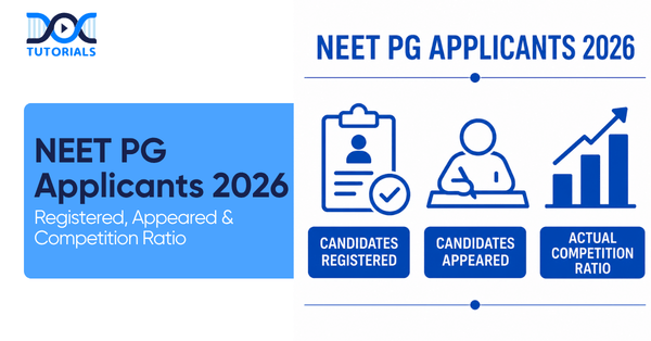
NEET PG Applicants 2026: How many candidates registered, appeared, and the actual competition ratio 

NEET PG Applicants

Lakhs of candidates appear for NEET PG annually to get admissions in MD/MS, PG diploma, and DNB courses. One such question often asked by candidates preparing for NEET PG exams is the following: How many candidates apply for the NEET PG exam, and what is the actual degree of competition among those who appear in the NEET PG?

Although the numbers of registrations and appearances were lower in previous years, the official data for the most recent cycle shows a clear picture of rising competition.

Discover how many candidates took NEET PG in the last 5 years, find out the total PG seats available in each state, and see which specialities are trending! Click below to grab the PDF now!

What Is NEET PG?

NEET PG is a national-level entrance examination organised by NBEMS for getting admission in postgraduate medical programmes. It acts as an entry test for MD/MS, PG Diploma, and DNB courses. 

This exam is computer-based, and according to the latest pattern, there are 180 multiple-choice questions with a total of 720 marks. 

How Many Students Registered for NEET PG?

Over the last few years, the number of candidates applying for NEET PG has been continuously above two lakh.

YearRegistered Candidates
20242,28,540
20252,42,493
20262,73,096

The record number of 2,73,096 applicants in 2026 reflects the increasing interest of medical graduates in opting for specializations in postgraduate courses. 

How Many Students Actually Appeared?

Not all applicants appear in the examination. But their participation ratio is always very high.

YearAppeared Candidates
20242,16,136
20252,30,114
20262,65,980

In NEET PG 2026, a massive 2,65,980 candidates appeared for the examination out of 2,73,096 registrations. This means that over 97 percent of registered candidates ultimately took the exam. 

NEET PG 2027: Expected Number of Candidates

As of now, NBEMS has not officially provided any statistics regarding the exact number of candidates for the upcoming NEET PG 2027. However, looking at the consistent upward trend, multiple reports estimate that the total number of competitors in the examination will easily exceed 2.8 lakh candidates next year.

The exact number will only become known when NBEMS shares the final examination statistics.

What Is the Actual Competition Ratio in NEET PG?

The competition in NEET PG becomes clearer when the number of candidates is compared with the available postgraduate seats.

According to the latest 2026 seat matrix data, there are around: 

  • 42,936 government PG medical seats 
  • 86,360 total PG medical seats in government medical colleges, private institutes, deemed universities, and DNB hospitals. 

Considering the overall numbers from the latest 2026 data: 

  • Number of candidates appeared: 2,65,980 
  • Total number of PG seats: 86,360 This makes up to one seat for approximately three candidates overall. 

Considering only the highly competitive government PG medical seats: 

  • Number of candidates appeared: 2,65,980 
  • Number of government PG seats: 42,936 It means that one government seat is shared among roughly six applicants. 

These numbers prove NEET PG to be one of the most competitive examinations for admission to PG colleges among all medical entrance tests held in India.

Why the Competition Is So High?

Two main reasons determine the level of competition:

1. Candidate Pool Size

Over two lakh MBBS graduates participate in NEET PG every year. Each year, the size of the candidate pool grows with the increasing number of medical colleges producing MBBS graduates.

2. Limited Number of PG Medical Seats

Even though the number of PG medical seats keeps growing every year, there are still fewer places than the number of candidates registering to appear in the NEET PG examination.

What This Means for Aspirants

Statistics clearly demonstrate that securing a postgraduate medical seat requires much more than simply passing the exam. You will be competing against top MBBS graduates nationwide for a highly limited number of clinical seats. 

Looking at the most recent NEET PG 2026 data: 

  • 2,73,096 registered candidates 
  • 2,65,980 appeared as candidates 
  • Over 2.8 lakh candidates are expected to apply for NEET PG 2027 
  • Approximately 86,360 total PG seats are available across India 

Therefore, it is incredibly important that future applicants prepare properly to secure their desired medical specialization and dream college. 

Frequently Asked Questions

1. How many students will register for NEET PG 2027?

While the official numbers for NEET PG 2027 will only be revealed closer to the exam date, current trends indicate that well over 2.8 lakh candidates are expected to register next year.

2. How many students appeared for NEET PG 2026?

According to the latest official exam statistics, exactly 2,65,980 students appeared for the NEET PG 2026 exam out of the 2,73,096 candidates who successfully registered.

3. How many candidates appeared for NEET PG 2025?

During the 2025 cycle, a total of 2,30,114 candidates appeared for the examination, while 2,42,493 had officially registered.

4. What is the actual competition ratio in NEET PG?

When looking at the latest 2026 figures, over 2.65 lakh candidates competed for approximately 86,360 total postgraduate medical seats. This brings the overall ratio to roughly one seat for every three candidates, though competition for top government clinical seats is much steeper at roughly one seat for every six applicants.

5. How many government PG medical seats are offered through NEET PG?

India currently offers approximately 42,936 government postgraduate medical seats according to the most recent 2026 seat matrix updates.

6. Is NEET PG one of the most competitive medical entrance exams in India?

Yes. With well over two lakh candidates taking the test every year for a limited pool of seats, NEET PG remains one of the toughest and most competitive medical entrance exams in the country.

7. Why is NEET PG so competitive?

The primary reason for such high competition is the sheer volume of graduating MBBS doctors who appear every year, combined with a strong preference for highly limited clinical branches like General Medicine or Radiodiagnosis.

8. Does registering for NEET PG guarantee a seat?

No, simply registering only confirms your eligibility to take the test. Actual admission depends entirely on your final score, your All India rank, seat availability, category reservations, and your choices during the counselling rounds.

9. How many questions are asked in the NEET PG exam?

Following the latest updated exam pattern, NEET PG now consists of 180 multiple choice questions. The exam carries a total of 720 marks, where every correct answer awards 4 marks and 1 mark is deducted for every wrong answer.

10. When will the candidate statistics for NEET PG 2027 be released officially?

The NBEMS typically publishes official registration and attendance numbers shortly after the exam is conducted. Statistics for the 2027 session will most likely be made public around August 2027.

Conclusion

Although the exact numbers for NEET PG 2027 are not yet available, recent exam trends show a massive surge in medical graduates pursuing higher education. During the recent 2026 session, a record-breaking 2,73,096 candidates registered, and 2,65,980 actually appeared for the exam. With the total postgraduate medical seat count sitting around 86,360, the competition remains incredibly high.

Only dedicated preparation, consistent practice, and a clear understanding of the new exam pattern will help you secure your desired clinical seat in the upcoming session. Unlock your potential with DocTutorials! Our comprehensive support in preparation, revision, and expert faculty makes conquering NEET PG not just achievable, but a stepping stone to your dreams. Join our meticulously crafted NEET PG Plans today and take the decisive step towards your success!

NEET PG Online Live Power Pack
DocTutorials App Download Now

Latest Blogs

NEET PG Rank-wise Seat Allotment PDF 2026: How to Download and Use it for Choice Filling

NEET PG Rank-wise Seat Allotment PDF

After every counselling, the Medical Counselling Committee publishes the NEET PG rank wise seat allotment PDF. It will be announced on the official MCC website, mcc.nic.in. You can get the download link once it is available on the website.  

After qualifying the NEET PG exam, candidates need to complete the registration procedure for counselling. On behalf of MCC, DGHS administers four rounds of NEET PG counselling at mcc.nic.in for 50% of the All India Quota and 100% of seats in deemed and central universities, ESIC, and AFMS. After each round, the authority releases the 2026 NEET PG seat allotment list PDF.

Here’s more on it!

What does NEET PG Rank-wise Seat Allotment Refers to?

MCC releases the NEET PG 2026 rank wise seat allotment result in PDF format after each round of counselling. The rank wise allotment is based on rank, choices, availability of seats and reservation. Candidates whose names appear in the list have to visit their respective colleges for verification of documents and payment of the full tuition fee to secure admission and preserve their seats.

MCC provides the NEET PG rank-wise seat allotment PDF format following the conclusion of each round of NEET PG counselling in 2026. The NEET PG rank, college and course selections, available seats, reservation requirements, and other variables are taken into consideration when allocating NEET PG seats.

To confirm NEET PG 2026 admission, candidates whose names appear in the NEET PG seat allotment result PDF must subsequently show up at the designated college for document verification. To guarantee a spot during the NEET PG rank-wise seat allocation, candidates must pay the entire tuition price.

How to Download the Rank-wise NEET PG Seat Allotment Result?

You can download the NEET PG rank wise seat allotment PDF in a few easy steps. Go to mcc.nic.in, click on NEET PG Medical Counselling, and then click on the link for 2026 counselling. Pick your round (Round 1/2, Mop-Up or Stray) and open the PDF result. Search with your roll number by Ctrl+F, download the rank-wise allotment PDF and log in to MCC to download your personal allotment letter for college reporting.

Here are the steps to follow while downloading the NEET PG rank wise seat allotment PDF:

Step 1: Visit the official website of mcc.nic.in and select NEET PG Medical Counselling

Step 2: Navigate the NEET PG 2026 counselling link from the menu on the homepage

Step 3: Choose your counselling round as per relevance: Round 1, Round 2, Mop-Up Round, or Stray Vacancy Round.

Step 4: The seat allotment result will be published in PDF form along with the candidate’s roll numbers, ranks, and allotted colleges.

Step 5: Check your details by pressing Ctrl + F and entering your roll number to find the seat allotted for you. 

Step 6: Download the NEET PG rank wise seat allotment PDF and save the printout for reporting in college or future references.

Step 7: Download Your Personal Allotment Letter. If you find your name in the list, sign in to the MCC portal with your login details to access your personal allotment letter, necessary for college reporting.

What are the Details Mentioned in the Result of the NEET PG Seat Allotment Result?

The details in the NEET PG seat allotment result include the candidate’s name, roll number, all-India rank, counselling rounds, category, allotted college, course or speciality, type of seat, joining date, etc.

The seat allotment result for NEET PG includes crucial information about the seats assigned to any candidate during each round. Candidates can download the result in PDF form or receive the allotment letter personally. Here are the details included in the NEET PG rank wise seat allotment PDF:

DetailDescription
Name of the CandidateFull name according to the NEET PG registration 
Roll Number of NEET PGUnique roll number assigned to a candidate during the exam 
All India Rank (AIR)Rank secured by the candidate in NEET PG 2026 
CategoryGeneral/SC/ST/OBC/EWS/PwD and quota (AIQ/State/Management/NRI) 
Rounds of CounsellingRound 1, Round 2, Mop-Up, or Stray Vacancy Round 
Allotted College NameThe medical college or institute’s name that has been allotted 
Allotted Course/SpecialtyAllotted MD, MS, PG Diploma branch or course
Seat TypeGovernment, Private, Deemed, Management or NRI seat type 
Reporting or Joining DatesLast date to report to the college 
Instructions for ReportingGuidelines for verification of documents and fee payment 

Candidates should verify each and every detail carefully. If any discrepancy is found in the candidate’s name, category, allotted course, or roll number, they should immediately report it to the counselling authority.

Candidates should decide whether they want to accept the seat or upgrade it in the second round after downloading the allotment result PDF.

What is the NEET PG Rank-wise Seat Allotment List of State Quota?

The state quota for NEET PG rank-wise seat allotment includes AFMS PG medical admission, AP PG medical admission, Assam PG medical admission, Bihar PG medical admission, BHU PG medical admission, BVP PG medical admission, Goa PG medical admission, etc.

Here are the names of the state quota seat allotment for NEET PG rank wise seat allotment:

  • AFMS PG medical admission
  • AP PG medical admission
  • Assam PG medical admission
  • Bihar PG medical admission
  • BHU PG medical admission
  • BVP PG medical admission
  • DU PG medical counselling
  • Goa PG medical admission
  • Himachal Pradesh PG medical admission
  • Haryana PG medical admission
  • Jharkhand PG medical admission
  • Karnataka PG medical counselling
  • Kerala PG medical admission
  • Maharashtra PG medical counselling
  • MP PG medical admission
  • Odisha PG medical admission
  • Punjab PG medical admission
  • Uttarakhand PG medical admission
  • UP PG medical admission
  • West Bengal PG medical admission
  • J&K PG medical admission
  • Gujarat PG medical admissions
  • Telangana PG medical admission

What are the Different Types of NEET PG Rank-wise Seat Allotment?

The NEET PG rank-wise seat allotment PDF is released in two types – MCC-conducted counselling (50% AIQ, 100% central/deemed universities, ESIC, AFMS) available at mcc.nic.in and State Authority-conducted counselling (100% private college seats, 50% state quota) available at the respective state counselling websites.

The NEET PG rank wise seat allotment PDF is published in two types. The table below explains them:

Serial NumberType of NEET PG CounsellingType of NEET PG Seat Allotment Result
1MCC Conducted CounsellingNEET PG rank-wise seat distribution is available at mcc.nic.in for 50% AIQ and 100% central and deemed universities, including ESIC and AFMS institutes.
2State Authority Conducted CounsellingThe NEET PG seat allocation results for 100% private medical college seats and 50% state quota seats are posted on the relevant state website.

FAQs about NEET PG Rank Wise Seat Allotment PDF

  1. Who is the conducting body for NEET PG rank wise seat allotment?

The seat allotment for All India Quota, central, as well as deemed universities, is conducted by the MCC. However, the allocation of state quota seats is conducted by the state government.

  1. Where can I find the NEET PG seat allotment result?

For the seats of All India Quota, the seat allotment result is published on the MCC website. However, results for state quota seats are published on the state counselling portals of the respective states.

  1. For how many rounds is the NEET PG seat allotment procedure conducted?

The NEET PG seat allotment procedure is performed in four total steps. They are named as round 1, round 2, Mop-up round, and Stray Vacancy round. 

  1. Is it possible to upgrade my seat if I don’t like the first allocation?

Yes, candidates who are allocated seats in the first round can appear in the second round to upgrade their seats if the first one is not suitable for them.

  1. Can I participate in the Mop-up Round to upgrade my seat?

Usually, candidates who have not received any allocation yet need to appear in the Mop-up Round for seat allocation. However, new guidelines are released every year, so keep an eye on that. 

Conclusion

The NEET PG rank wise seat allotment PDF is published every year after seat allocation for PG candidates after each round. The seats are allotted to the candidates based on their merit. So, if you want admission to your preferred college, you should score good marks in the NEET PG exam.

In this regard, DocTutorials can help you professionally guide. Our reputed and reliable faculty, video lectures, Quick Revision Programmes, quality study materials, etc., help in preparing the candidate efficiently. 

Join our NEET PG course today to strengthen your success!

NEET PG Online Live Power Pack
DocTutorials App Download Now

Latest Blogs

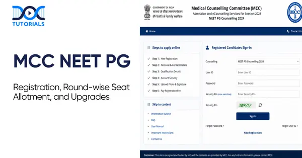
MCC NEET PG 2026: Registration Portal Guide, Round Schedule and Upgrade Options Explained

MCC NEET PG

MCC, or the Medical Counselling Committee, manages the seat allotment procedure in India for medical and dental courses. It is a government organisation that handles admissions to PG, UG, and super-speciality courses through a centralised online process. It allocates 15% of seats in government colleges, 50% of seats for PG courses and the rest in deemed universities. 

The All India Quota seat allocation is done as per the MCC NEET PG scores of the students, their subject preferences, and eligibility for reservation. The counselling is done through online registration, followed by choice filling and seat allotment, completely based on merit.

Here’s more on it!

Ace Your NEET PG Prep With DocTutorials NEET PG Online Live Power Pack

What is the MCC NEET PG Counselling Registration Procedure?

Candidates need to log in to the MCC website and open the NEET PG counselling for new registration. Create your login ID and password by entering your roll number, application number, date of birth, mobile number and email address. Next, you need to pay the counselling fee, opt for choice filling and wait for seat allocation.

Here are the steps to follow for the MCC NEET PG online registration counselling procedure:

Step 1: Visit the MCC Website

First, candidates need to visit the MCC website and select the NEET PG counselling option.

Step 2: Online Registration

Click on the link for “New Registration” and enter the following information to create your login ID and password:

  • Application number
  • Roll number
  • Date of birth
  • Mobile number and email address

Step 3: Pay the Fee for Counselling

Candidates need to pay a non-refundable registration fee and a refundable security deposit using their debit card, credit card, or net banking. After successful payment of the fees, you can participate in the counselling procedure.

Step 4: Choice Filling

After completing the MCC NEET PG counselling registration procedure, you can select the medical courses such as MD, MS, or Diploma and the medical college that you want to attend. You can select multiple options as per your preferences.

Step 5: Choice Finalisation

After making the final choices, the candidates need to lock them in before the deadline. Even if they miss manual locking, the preferences will be automatically locked in the system.

Step 6: Seat Allocation

The counselling body will announce the result of seat allocation as per the NEET PG rank of the candidates, their preferences, seat availability, etc.

Step 7: Reporting to College

After seat allocation, candidates need to download the allotment letter and get a printout of the same before reporting to the institution for document verification and the admission procedure.

What is the Seat Allotment Procedure for MCC NEET PG 2026?

The seat allotment procedure for MCC NEET PG includes six stages. It includes the registration, choice filling and locking for the preferred course and college, seat allocation based on the candidate’s rank, final seat allotment results, and reporting to the college allotted to the candidate.

The MCC allocates NEET PG seats to candidates based on several factors, including the candidate’s rank, preferences, reservation category, and seat availability. As already mentioned, the candidates need to fill out the choices for seats in their preferred colleges and courses. 

Here are the different stages of the seat allotment procedure for NEET PG:

  1. Registration for NEET PG counselling
  2. Choice filling for preferred seats
  3. Choice locking
  4. Seat allocation as per rank
  5. Results for seat allotment
  6. Reporting to the allotted college

What are the Round-wise Counselling Schedule for MCC NEET PG?

There are a total of three rounds for the MCC NEET PG counselling schedule. The first round involves seat allotment to the candidates as per their preference and merit. In the second round, the candidates can upgrade their seats, and the third round involves the allocation of the remaining seats.

The counselling procedure for MCC NEET PG 2026 involves multiple rounds. These rounds involve the following procedures:

  • Round 1

This is the seat allotment round. In this round, candidates are allotted seats in their preferred course and colleges as per their merit.

  • Round 2

This round includes the second phase of seat allotment. Seats not allotted in the first round will be allocated in this phase. Moreover, candidates who want to upgrade their seats are allowed to take part in this round.

  • Mop-Up Round

In the third round, the seats remaining after round 2 are allotted to the candidates.

  • Stray Vacancy Round (if applicable)

If any other seats remain vacant after the mop-up round, the authorities conduct a stray vacancy round. It is an optional round based on the seat allocation in the previous three rounds.

All candidates should stay updated with the counselling schedule for seat allocation and actively take part in all the rounds so that the chances of securing seats for them are increased.

What are the Upgrade Options in MCC NEET PG Counselling?

Vacant seats from the previous round are filled in Round 2 of NEET PG Counselling. Candidates who get a seat allotted in Round 1 can participate for upgrading of their allotted seat Candidates who were not allotted a seat in the previous round can also participate for the available vacancies. The process comprises registration, filling choices, seat allotment results and reporting to the allotted college within the given counselling schedule.

There are two purposes for the counselling in round 2. First, allocating seats to the candidates who have not secured any seat in the first round and allowing upgrades to better options for those who have already been allocated seats in round 1.

Here are the candidates who can participate in round 2:

  • Candidates who were not allocated seats in round 1
  • Candidates who want to upgrade their seats even after getting allocation in round 1
  • Fresh candidates who have missed the registration procedure of round 1, and if MCC allows them to reappear

Here are the considerations for round 1 strategy:

  • For Non-Allottees

This is the second chance for the non-allottees. The round 2 seats are usually available from candidates who have not reported to their allotted colleges in round 1, from round 1 allottees who want to upgrade their seats, and in case of any technical glitches.

  • For Round 1 Allotees

Seat upgradation is often risky as candidates need to surrender their allotted seat before the allotment of Round 2. In case the candidates did not find any better option even in round 2, they will lose their allotted seat and wait for the Mop-up round.

Here is the expected timeline for round 2 seat allotment for MCC NEET PG counselling:

EventsExpected Dates
Opening of RegistrationDate yet to be announced.
Deadline for Choice FillingDate yet to be announced.
Result for Seat AllotmentDate yet to be announced.
Reporting to Allotted CollegesDate yet to be announced.

FAQs about MCC NEET PG Registration

  1. Can I participate in Round 3 using my Round 2 seat as a safety?

No, you cannot attempt to qualify for Round 3 while keeping your Round 2 seat as a backup. According to MCC regulations, your Round 2 seat may be cancelled if you decide to upgrade or participate in Round 3 in the permitted manner.

  1. What happens if I’m given a Round 3 seat but choose not to participate?

If you do not participate in the round 3 seat allotment process, you may not be allowed to participate in future MCC rounds. Moreover,  your security deposit might be forfeited.

  1. Can I re-enter Round 3 if I quit Round 2?

Yes, you can re-register for Round 3 if you adhere to MCC’s resignation process and deadline, but in many situations, you may have to lose your security deposit. Resignation window notices (MCC), for instance, permitted resignations with college-generated resignation letters within certain dates. So, you can try again in Round 3, but it will cost you money.

  1. In 2026, how many rounds of NEET PG counselling would there be?

There are usually three major rounds, followed by mop-up and stray vacancy rounds for the remaining seats.

  1. What will happen if I miss the counselling registration deadline?

A candidate will not be able to register for that round of NEET PG counselling if they miss the counselling registration deadline. Instead, they would have to wait until the following round of NEET PG counselling, if there is one.

Conclusion

The MCC NEET PG counselling involves several steps and rounds. Candidates must stay updated and follow the strict guidelines to secure their preferred seat. However, merit plays a crucial role in allocating your preferred seat.

So, candidates should focus on securing a good score in the NEET PG exam. In this regard, DocTutorials is a reliable online coaching that guides candidates professionally with Quick Revision Program, video lectures, study materials, etc. For more information, join our NEET PG course today!

NEET PG Online Live Power Pack
DocTutorials App Download Now

Latest Blogs

Hirsutism: Causes, Symptoms, Diagnosis, and Treatment

Hirsutism

Hirsutism is characterised by the growth of coarse, terminal hair in females in a distribution pattern typically seen in males. It affects about 5-10% of women and is a frequent presenting complaint for cosmetic reasons in dermatology outpatient departments. Its importance lies in its physical presentation and the need for a systematic approach to its management.

For clinicians and NEET PG aspirants, a clear grasp of the endocrine basis, evaluation framework, and management options for hirsutism is essential. Keep reading to know more about the key mechanisms, diagnostic approach, and evidence-based hirsutism treatment.

What is Hirsutism?

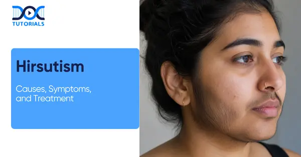
Hirsutism is when women develop thick, dark hair in areas like the face, lower abdomen, or inner thighs, where hair is usually minimal. It usually happens due to higher levels of male hormones, especially testosterone.

Hirsutism refers to the presence of thick, dark, coarse hair in women in areas where hair growth is typically minimal or absent. This includes the face, lower stomach and the inner thighs. Extra hair growth is often caused by excess male hormones called androgens. The main androgen involved is testosterone.

Hirsutism is to be distinguished from hypertrichosis, which is a separate condition of generalised excess hair growth that is not androgen-dependent and not limited to male-pattern distribution. Hirsutism, in contrast, is distributed in certain androgen-sensitive areas and has diagnostic implications for hormonal excess.

Although many conditions can lead to hirsutism, polycystic ovary syndrome and idiopathic hyperandrogenism account for more than 85% of cases. This distribution is directly relevant to clinical practice and exam preparation, as it informs both the diagnostic approach and the likelihood of finding a treatable underlying cause.

What are The Types of Hirsutism?

Hirsutism is classified according to its underlying origin. Identifying the correct type is an essential first step in directing both the diagnostic workup and the hirsutism treatment plan. Broadly, the condition falls into 2 principal categories:

  • Androgen-dependent
  • Androgen-independent

The following are the types of Hirsutism:

  1. Idiopathic Hirsutism: Occurs in women with regular menses, normal ovarian morphology, and normal androgen levels. It is a diagnosis of exclusion, thought to arise from increased follicular sensitivity or elevated 5-alpha-reductase activity within the skin.
  2. PCOS-Related Hirsutism: The most prevalent type, accounting for over 70% of cases. It is driven by chronic hyperandrogenism, anovulation, and insulin resistance working in combination.
  3. Adrenal Hirsutism: Arises from adrenal disorders such as congenital adrenal hyperplasia or Cushing syndrome, where excess adrenal androgen production stimulates terminal hair growth.
  4. Tumour-Induced Hirsutism: The rarest but most serious type, caused by androgen-secreting ovarian or adrenal tumours. It is characterised by a rapid onset and frequent signs of virilisation.
  5. Drug-Induced Hirsutism: Results from exogenous androgenic compounds and is typically reversible upon withdrawal of the offending medication.

What are the Causes of Hirsutism?

Hirsutism can have different causes, mainly related to hormones or sometimes other factors. The most common type is linked to conditions like polycystic ovary syndrome, while others may be due to adrenal issues, medications, or rarely, tumours. Knowing these causes helps doctors choose the right treatment.

Hirsutism develops when there is excess androgen present in the body, or when hair follicles are sensitive to normal androgens. Both ovaries and adrenal glands produce androgens in females. 

When production exceeds normal physiological levels or when the skin’s androgen receptors respond with disproportionate sensitivity, terminal hair growth is stimulated in male-patterned areas. The most common underlying hirsutism causes are:

  • Polycystic Ovary Syndrome (PCOS): The most common underlying cause is Polycystic Ovary Syndrome (PCOS), which accounts for the majority of cases.

In PCOS, hormonal imbalance leads to increased levels of free testosterone along with reduced sex hormone-binding globulin (SHBG), making more active androgen available in the body.

  • Idiopathic Hirsutism: Idiopathic hirsutism occurs despite normal androgen levels, due to increased peripheral 5-alpha-reductase activity or increased follicular androgen sensitivity.
  • Congenital Adrenal Hyperplasia (CAH): Less commonly, hirsutism may result from this condition. It is an inherited disorder affecting adrenal hormone production.
  • Cushing Syndrome: In this condition, excess cortisol is often accompanied by elevated androgen levels. This causes hirsutism.
  • Androgen-Secreting Tumours: In rare instances, androgen-secreting tumours of the ovaries or adrenal glands can directly increase androgen levels in the bloodstream, leading to hirsutism symptoms.
  • Medications: Certain medications can also contribute to hirsutism. Drugs such as danazol, minoxidil, anabolic steroids, and external testosterone are known to influence androgen activity and may trigger excessive hair growth.
  • Other Endocrine Disorders: Hyperprolactinaemia, hypothyroidism, hyperthyroidism, and acromegaly are uncommon but documented causes of isolated hirsutism.

What are the Risk Factors of Hirsutism?

Certain factors can increase the chance of developing hirsutism, such as a family history of hormonal conditions, being overweight, or having insulin resistance. It is also more commonly seen in women of certain ethnic backgrounds and can sometimes be triggered by certain medications.

Listed below are a few patient-specific factors that increase the likelihood of developing hirsutism:

  • Family History: Conditions such as PCOS and CAH have a heritable component, and a positive family history significantly raises clinical suspicion.
  • Ethnicity: Women of Mediterranean, Middle Eastern, and South Asian descent are more likely than other women to have extra body hair for no clear cause. This variation reflects differences in androgen sensitivity rather than androgen levels per se.
  • Obesity: Excess adipose tissue contributes to elevated androgen production and reduced sex hormone-binding globulin (SHBG) levels, resulting in higher concentrations of biologically active free testosterone in the circulation.
  • Insulin Resistance: Hyperinsulinaemia affects more than one-half of women with PCOS, triggering an increase in gonadotropin-releasing hormone pulse frequency, which subsequently increases production of ovarian and adrenal androgens whilst reducing hepatic SHBG production.
  • Prolonged Use of Certain Medications: Androgenic compounds, anabolic steroids, and drugs such as danazol and minoxidil represent a direct pharmacological cause that must be considered during clinical evaluation.

What are the Symptoms of Hirsutism?

Hirsutism causes slow, excessive growth of thick, dark hair in women on areas like the face, chest, and abdomen. In some cases, it may be accompanied by acne, irregular periods, or more severe signs like voice deepening.

Hirsutism refers to the abnormal growth of coarse hair in women on areas of the body where men normally develop terminal hair. The condition usually develops slowly, and it varies from one woman to another, depending first on how much androgen there is and second on how sensitive her hair follicles are to it.

The chin, upper lip, sideburns, chest, back and abdomen are common sites. Clinically observed key hirsutism symptoms are:

  • Facial Hair Growth: Coarse, dark hair appearing on the upper lip, chin, cheeks, and sideburns, often the earliest and most distressing feature for patients.
  • Body Hair Growth: Excess hair on the chest, abdomen, lower back, inner thighs, and buttocks in a distinctly male-pattern distribution.
  • Signs of Virilisation: In more advanced cases, hirsutism may be accompanied by voice deepening, male-patterned scalp hair loss, acne, reduced breast size, increased muscle mass, and clitoral enlargement.
  • Menstrual Irregularities: Particularly in PCOS-related hirsutism, irregular or absent menstrual cycles frequently accompany excess hair growth.
  • Rapid Onset: Women with a rapid onset of hirsutism over a few months or signs of virilisation are at high risk of having an androgen-secreting tumour and require urgent evaluation.

The psychological effects of hirsutism are usually downplayed. Many women experience emotional distress caused by the condition, resulting in reduced self-esteem, public participation, and overall quality of life.

Because of this, the patient’s emotional experience also needs to be cared for with as much care and thoroughness as the physical. That’s why hirsutism self-care and emotional well-being support are really necessary for these patients.

How is Hirsutism Diagnosed?

Hirsutism is diagnosed by learning your symptoms, checking your medical history, and doing a physical exam. Doctors may also use blood tests and scans to find the underlying cause, such as a hormonal imbalance.

The diagnosis of hirsutism requires a systematic and structured approach. The process starts with an extensive clinical examination and is followed by specific lab and imaging tests.

The most important diagnostic goal is to identify or exclude whether there is a significant underlying disorder, most notably androgen-secreting tumours, PCOS, CAH and Cushing syndrome.

The following are the diagnosis processes:

  • Clinical History: The first step is a thorough clinical history, ideally covering the timing and rate of hair growth, menstruation (regular/irregular), reproductive health, medication use, and family history of androgen-related disorders.
  • Ferriman-Gallwey Scoring: Ferriman-Gallwey scoring is a common diagnostic method for hirsutism within 9 body areas considered sensitive to androgens. Scores of 15 and higher indicate mild hirsutism, and scores greater than 25 indicate severe hirsutism.
  • Physical Examination: A thorough abdominal and pelvic examination is performed to exclude palpable masses. Acanthosis nigricans, a recognised marker of insulin resistance, should also be noted during examination.
  • Hormonal Blood Tests: Screening for levels of serum testosterone and 17-alpha-hydroxyprogesterone is sufficient in most cases. Women with irregular menses and hirsutism should be screened for thyroid dysfunction and prolactin disorders.
  • Specialised Endocrine Tests: According to the clinical findings, one may add a short dexamethasone suppression test for Cushing syndrome, an ACTH (Adrenocorticotropic Hormone Stimulation Test) stimulation test, or a carbohydrate tolerance test for insulin resistance syndrome.
  • Imaging Studies: If an androgen-secreting tumour is suspected, abdominal or pelvic ultrasound or a CT (computed tomography) scan may be employed. Pelvic ultrasound is also the primary imaging modality for confirming polycystic ovarian morphology.

What are the Treatment Options for Hirsutism?

Hirsutism is treated using a combination of medications and cosmetic methods. Medicines help reduce hormone levels, while options like laser hair removal or creams help manage visible hair. Lifestyle changes, especially weight management in some cases, can also improve symptoms over time.

Hirsutism treatment is generally classified as pharmacological and non-pharmacological, and both are often used in combination. Below are the treatment options for hirsutism:

1. Pharmacological Treatment

  • Combined Oral Contraceptives: Treatment of non-tumoral hirsutism includes oral contraception. It works by inhibiting the hypothalamic-pituitary axis through its progestogen component.

This reduces ovarian androgen production. The oestrogen component increases sex hormone-binding globulin. This dual action decreases both the production and the biological activity of circulating androgens.

  • Anti-Androgens: Anti-androgen therapy is often used when oral contraceptives alone are not enough. The most common anti-androgen for treating hirsutism is spironolactone.

The results are modest and can take at least six months to appear. Side effects may include irregular periods, and the hirsutism medication can also lead to birth defects. Therefore, they are prescribed along with contraception for women of reproductive age.

Flutamide is another hirsutism treatment option, but it is expensive and can potentially cause liver damage. Cyproterone acetate has strong progestogenic and anti-androgen effects. It decreases circulating testosterone and androstenedione levels by lowering luteinising hormone levels.

  • 5-Alpha Reductase Inhibitors: Finasteride is used to treat hirsutism and is preferred because it lacks oestrogenic or progestogenic activity. However, it is not recommended for women of childbearing age as there is a risk of ambiguous genitalia in the fetus.
  • Insulin-Sensitising Agents: In women with PCOS-related hirsutism and concurrent insulin resistance, metformin therapy and weight loss are typically recommended, as they can decrease serum testosterone levels and improve ovulation rates.
  • Topical Agents: Fine hair growth can be inhibited with twice-daily topical eflornithine. This agent works by inhibiting an enzyme required for hair follicle cell proliferation and is particularly useful for facial hirsutism.
  • Low-Dose Corticosteroids: Adrenal hyperandrogenism is slowed by low-dose corticosteroids, possibly in association with the treatments mentioned above. This approach is specifically relevant in cases of CAH-related hirsutism, where suppressing excess adrenal androgen production is the therapeutic goal.

2. Non-Pharmacological Treatment

The following cosmetic and procedural approaches are an important adjunct to pharmacological management, particularly given the delay in visible response to drug therapy:

  • Laser Hair Removal (Photoepilation): Photoepilation destroys pigmented terminal hair follicles via thermal damage from one of four laser types, depending on the patient’s hair colour and skin pigmentation.

Trials have shown hair reduction of 40% to 80%, depending on the type of laser used and the number of treatments provided. Despite its limitations, photoepilation is the preferred treatment for permanent hair removal, as it is up to 60 times faster than electrolysis.

  • Electrolysis: Electrolysis is less used now, except for coarse white hairs and also in cases of polycystic ovaries without insulin resistance. It remains, however, a reliable option for individuals in whom laser therapy is unsuitable due to hair or skin pigmentation.
  • Weight Management: In women with obesity-related hyperandrogenaemia, structured weight loss is an effective and evidence-based intervention.

Reduction in adipose tissue improves insulin sensitivity, lowers androgen levels, and increases SHBG, thereby reducing the degree of hirsutism independently of pharmacological treatment.

FAQs about Hirsutism

  1. Who does hirsutism affect?

This condition mostly affects women and people who were born female. While it can show up at any age, it’s most common during the years when people can have children. Especially around puberty, when hormone imbalances are more likely.

  1. How common is hirsutism? 

About 5 to 10% of women are affected by hirsutism. It’s actually a pretty common reason women visit a skin doctor: they’re concerned about how it looks. It’s also one of the most frequent hormone problems that doctors come across, no matter if they specialise in skin, women’s health, or hormones.

  1. Does hirsutism always mean PCOS?

No, not always. Even though PCOS is the reason for over 70% of cases, you can also get hirsutism from other things, like a certain type of adrenal problem that you’re born with, Cushing syndrome, tumours that produce male hormones, specific medications, or sometimes doctors just don’t know the exact cause.

  1. How can one reduce the risk of developing hirsutism?

Maintaining a healthy weight, managing insulin resistance, avoiding unnecessary androgenic medications, and seeking early medical review for menstrual irregularities are the most practical measures for reducing the risk of developing or worsening hirsutism.

  1. Do side effects of medication cause hirsutism?

Yes, they can. Some medications are known to cause extra hair growth. These include things like danazol, minoxidil, anabolic steroids, testosterone that’s given externally, and DHEA (dehydroepiandrosterone).

In most situations, if you stop taking the medicine that’s causing the extra hair, the hair growth usually reverses with a doctor’s guidance.

  1. Which tests does a patient need for hirsutism?

Initial tests typically include serum testosterone and 17-alpha-hydroxyprogesterone levels. Depending on clinical findings, additional tests may include thyroid function, prolactin, cortisol, a dexamethasone suppression test, pelvic ultrasound, or CT imaging to exclude tumours and other endocrine disorders.

  1. Does laser hair removal work for hirsutism?

Photoepilation has shown hair reduction of 40-80%, depending on the type of laser used and the number of treatments. It is the preferred method for long-term hair removal, though it works best on dark, coarse hair and requires multiple sessions.

  1. Can hirsutism go away on its own?

Hirsutism rarely resolves without intervention. When caused by a reversible factor such as medication or a temporary hormonal fluctuation, it may improve spontaneously.

In most cases, however, sustained management through pharmacological treatment, hair removal, or addressing the underlying cause is required for meaningful improvement.

Conclusion

Hirsutism is a potential marker of underlying endocrine pathology, including PCOS, congenital adrenal hyperplasia, Cushing syndrome and androgen-secreting tumours and warrants a thorough, structured approach to diagnosis rather than being relegated to a cosmetic issue.

Hirsutism treatment, whether pharmacological or procedural, should be individualised based on the hormonal profile, reproductive goals, and underlying aetiology. Hirsutism is a high-yield concept of Endocrinology, Gynaecology and Dermatology for NEET PG aspirants.

For additional guidance, DocTutorials can be your study companion. We offer crisp videos, clinical Qbank, exam-focused notes, flashcards, and mind maps. This helps ensure aspirants gain complete clarity over complex medical topics.

Join DocTutorials today and explore our NEET PG course to excel in your medical career!

NEET PG Online Live Power Pack
DocTutorials App Download Now

Latest Blogs

Actinomycosis: Causes, Symptoms, Diagnosis, and Treatment

Actinomycosis Symptoms

Actinomycosis is a condition in which the body develops a complex, coordinated immune response to a persistent infection or unexplained swelling. This condition demands a nuanced clinical understanding. It is a rare, chronic bacterial infection that often goes unrecognised in routine practice.

For those in the healthcare field or aspiring medical professionals, gaining a clear understanding of this disease is essential for delivering effective patient care and performing well on exams. Keep reading to know the causes, symptoms, diagnosis, and treatments of this condition.

What is Actinomycosis?

Actinomycosis is a bacterial infection that leads to painful abscesses and sometimes draining sinuses with yellowish discharge. It occurs when normally harmless bacteria enter deeper tissues following injury, leading to a persistent infection that can gradually spread if left untreated.

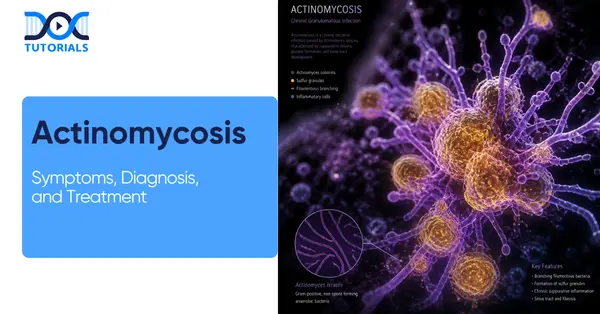
Actinomycosis is a subacute-to-chronic bacterial infection. It is mostly characterised by the formation of painful abscesses, tissue fibrosis, and draining sinus tracts that often discharge yellow “sulfur granules.”

It is a granulomatous disease and is caused by anaerobic or microaerophilic bacteria that normally reside harmlessly in the human mouth, digestive tract, and female reproductive system. When these bacteria breach the mucosal barrier, often due to trauma or surgery, they can trigger an invasive inflammatory process that spreads regardless of tissue planes.

Actinomycosis represents a failure to eliminate the bacterial threat to the human body. This leads to a long-term, destructive response that can last months or even years. Because the infection creates dense, “woody” masses of fibrous tissue, it often ignores anatomical boundaries, burrowing through muscle and bone to reach the skin’s surface.

What are the Causes of Actinomycosis?

Actinomycosis develops when Actinomyces bacteria enter deeper tissues through injury, dental work, or surgery. They grow in low-oxygen areas and can slowly cause an infection.

Actinomycosis is primarily caused by Actinomyces species, such as Actinomyces israelii, which transition from harmless commensals to pathogens when they invade deep tissues through mucosal breaks. The condition is further complicated by its polymicrobial nature.

Other organisms commonly associated with the infection include Aggregatibacter actinomycetemcomitans, Prevotella, Streptococcus, Enterobacteriaceae, Peptostreptococcus, and Staphylococcus.

This infection is fundamentally endogenous, occurring when the protective mucosal barriers of the mouth, respiratory system, or gastrointestinal tract are compromised. Actinomycosis involves the body’s own flora invading internal spaces.

These causes can be classified into several primary triggers:

  • Physical Trauma and Surgery: Common dental procedures, tooth extractions, jaw fractures, or abdominal surgeries provide a direct pathway for bacteria to bypass the mucosal lining.
  • Anaerobic Environments: Because Actinomyces are anaerobic, they thrive in low-oxygen niches often created by tissue necrosis or localised cell death caused by reduced blood supply.
  • Microbial Synergy: The presence of other co-infecting pathogenic microorganisms can facilitate the disease by consuming local oxygen. This creates the perfect opportunistic environment for Actinomyces to take root, forming dense fibrous masses that ignore standard anatomical boundaries.

What are the Risk Factors of Actinomycosis?

Actinomycosis is more likely in people with poor oral hygiene, those who have undergone dental surgery, or those who have sustained surgery or injury that breaks the body’s natural barriers. A weak immune system, long-term IUD use, smoking, or alcohol misuse can also increase the risk.

The typical risk factors for actinomycosis include poor oral hygiene, dental surgery, immunosuppression, and the long-term use of intrauterine devices (IUDs), which can facilitate bacterial invasion.

Actinomycosis is an opportunistic infection that exploits weaknesses in the body’s natural barriers. It is clinically characterised by its ability to mimic other diseases like malignancy or tuberculosis.

Below are some common risk factors associated with actinomycosis:

  • Poor Oral Hygiene and Dental Trauma: The most common form, cervicofacial actinomycosis, often follows tooth extractions, jaw fractures, or gingivitis.

An affected individual often has recently had dental work, poor oral hygiene, periodontal disease, radiation therapy, or trauma such as a broken jaw, causing local tissue damage.

  • Gastrointestinal Procedures: Appendicitis, perforated ulcers, or abdominal surgeries can release bacteria into the peritoneal cavity, leading to abdominal actinomycosis.
  • Intrauterine Devices (IUDs): Long-term use of IUDs is a recognised risk factor for pelvic actinomycosis, as the device can cause minor mucosal erosion where the bacteria can colonise.
  • Immunocompromised State: Those with a compromised immune system are at higher risk, as they are less able to fight off infections. Risk factors include HIV/AIDS (Human Immunodeficiency Virus/Acquired Immune Deficiency Syndrome), diabetes mellitus, malnutrition, and systemic immunosuppressive therapy.
  • Smoking and Alcohol Misuse: Reduction of alcohol abuse and improvement of dental hygiene may limit the occurrence of pulmonary, cervicofacial, and central nervous system actinomycosis.
  • Pulmonary Aspirations: Individuals with poor cough reflexes, often due to neurological conditions or alcohol use, may aspirate oral bacteria into their lungs, leading to thoracic actinomycosis.

What are the Symptoms of Actinomycosis?

Symptoms of actinomycosis depend on the area affected, but commonly include firm swelling, abscesses, and sometimes draining sinuses. People may also have pain, fever, cough, or abdominal or pelvic discomfort as the infection gradually spreads.

Actinomycosis presents in several distinct clinical forms, each with its own set of symptoms based on the organ system involved. The symptoms are rarely acute; instead, they are chronic 

and progressive.

The diseases and their symptoms are categorised based on their location:

  • Cervicofacial Actinomycosis (“Lumpy Jaw”): This accounts for about 50% of all cases. Symptoms include A slow-growing, non-tender, hard swelling that typically appears at the angle of the mandible (jawline).

Over time, the area becomes discoloured (red or purplish) and develops draining sinuses. While initially painless, it can become tender if secondary bacterial infections occur. Patients may have difficulty opening their mouths if the infection involves the masticatory muscles.

  • Thoracic Actinomycosis: This form results from inhaling bacteria from the mouth into the lungs. It often presents as a lung tumour or tuberculosis.

Patients report a productive cough, chest pain, and sometimes hemoptysis (coughing up blood). The infection can spread to the pleura, the chest wall, or even the ribs, creating visible abscesses on the chest wall.

  • Abdominal Actinomycosis: This usually follows a rupture in the intestinal tract. Patients often present with a palpable mass in the lower right quadrant of the abdomen (near the ileocecal region).

The pain is usually dull and constant, often accompanied by low-grade fever, weight loss, and changes in bowel habits. It is frequently mistaken for Crohn’s disease or a malignant tumour.

  • Pelvic Actinomycosis: Associated almost exclusively with IUD use. Vaginal discharge, pelvic pain, and abnormal uterine bleeding. If it spreads, it can form large masses in the pelvis that mimic ovarian cancer.

What is the Diagnosis of Actinomycosis?

Actinomycosis can be hard to diagnose because it looks like other diseases. Doctors usually confirm it with tests, scans, and examinations of samples from the affected area.

Diagnosis of actinomycosis is notoriously challenging. The diagnosis is frequently delayed because the clinical presentation can mimic other pathologies. They include malignancies, active Mycobacterium tuberculosis infection, nocardiosis, fungal infections, infarctions, or other granulomatous diseases.

Below are a few diagnosis methods for actinomycosis:

  • Clinical History: A thorough clinical history is the essential first step. The clinician should enquire about recent dental procedures, IUD use, surgical history, smoking, alcohol intake, and any history of immunosuppression.
  • Laboratory Investigations: Blood tests may reveal elevated white blood cell counts or other signs of infection. Cultures of pus or tissue samples can help identify the specific bacteria. However, cultural results must be interpreted with caution.

Gram staining of infected tissue or purulent material is more useful and sensitive than culture, as culture may be negative in 50% of cases. The negative rate is high due to previous antibiotic therapy, polymicrobial infection, failure to maintain an anaerobic environment during transportation and culture, or short-term incubation.

  • Histopathology: It is considered one of the most reliable diagnostic tools. The characteristic microscopic finding is necrosis of yellowish sulfur granules alongside filamentous gram-positive bacteria.
  • Imaging Studies: CT (computed tomography) or ultrasound often reveals mass lesions, and these are not infrequently mistaken for neoplasms. Hopkins Guides X-rays, CT scans, and MRI (magnetic resonance imaging) are all employed to assess the extent of disease, particularly in thoracic and abdominal forms.
  • Biopsy: Biopsy and aspiration are frequently required to obtain definitive material for examination. Culture performed through needle aspiration with incubation of a minimum of 14 days is recommended. Multiple biopsy specimens from different locations increase the likelihood of diagnosis.

What are the Treatment  Options for Actinomycosis?

Treatment of actinomycosis mainly involves long-term antibiotics, usually penicillin or similar drugs, taken for several months to clear the infection. In some cases, surgery may be needed to drain abscesses or remove infected tissue.

The cornerstone of actinomycosis treatment is high-dose, long-term antibiotic therapy, sometimes supplemented by surgical debridement. Because the infection is surrounded by dense, avascular fibrous tissue, antibiotics have difficulty reaching the bacteria.

Therefore, the treatment must be aggressive and sustained:

  • Antibiotic Therapy

Patients with actinomycosis require prolonged high-dose penicillin G or amoxicillin, typically over 6-12 months, though the duration of antimicrobial therapy may be reduced to 3 months in patients in whom optimal surgical resection of infected tissues has been performed.

The standard treatment protocol involves an initial phase of intravenous therapy followed by an oral step-down. High-dose intravenous penicillin G is usually effective for initial treatment, with oral penicillin or amoxicillin substituted after approximately 2-6 weeks.

For patients with documented penicillin allergy, viable alternatives exist. In cases of penicillin allergy, doxycycline is used. Sulfonamides such as sulfamethoxazole may also be used as an alternative regimen. Alternative antibiotics, including tetracycline, doxycycline, minocycline, clindamycin, erythromycin, ceftriaxone, and linezolid, have also been successful.

  • Surgical Intervention

Antibiotics alone are not always sufficient. Abscesses are drained, fistulas are surgically removed, and with prompt diagnosis and appropriate treatment, most people recover fully. In complex pelvic disease, extensive pelvic involvement may require drainage of intra-abdominal abscesses, sometimes with hysterectomy and salpingo-oophorectomy.

In cases of IUD-associated pelvic actinomycosis, device removal is an integral part of management. Treatment of IUD-associated pelvic actinomycosis involves removal of the IUD.

  • Adjunctive Therapies

Hyperbaric oxygen therapy may be used as an adjunct to conventional therapy when the disease process is refractory to antibiotics and surgical treatment. This modality is thought to inhibit the anaerobic organisms and potentiate antibiotic penetration into poorly vascularised fibrous tissue.

It is paramount that patients are counselled on the need to complete the full treatment course. Treatment can take a long time and requires patience, and frequent follow-up with the healthcare provider ensures the patient is on the correct antibiotics.

FAQs about Actinomycosis

  1. Is actinomycosis contagious?

No, actinomycosis is not contagious and cannot be transmitted from one person to another. The causative bacteria already exist as normal flora within the human body.

Infection arises only when mucosal barriers are breached by injury or surgical trauma. Since these bacteria cannot survive outside the human body, there is no risk of person-to-person transmission.

  1. What are the complications of actinomycosis?

If left untreated, actinomycosis can cause osteomyelitis, persistent sinus tract and fistula formation, bowel obstruction, and involvement of adjacent organs.

In severe cases, haematogenous spread may result in brain abscess and organ failure. Delayed diagnosis is the principal driver of serious complications across all anatomical forms of the disease.

  1. How soon after treatment will a patient feel better?

Clinical improvements, such as reduced swelling and diminished sinus tract discharge, may begin within several weeks of starting antibiotics. However, complete resolution takes months. Patients must complete the full six to twelve-month course, as premature cessation due to early symptomatic relief is a common and significant cause of relapse.

  1. Can actinomycosis be fatal?

Actinomycosis is rarely fatal when diagnosed and treated promptly, with most patients achieving full recovery.

However, delayed treatment, central nervous system involvement, or disseminated disease in immunocompromised individuals carries a considerably more serious prognosis. The timeliness of diagnosis remains the single most important determinant of patient outcome.

  1. Can actinomycosis be prevented?

Yes, it can be prevented. Preventive measures include maintaining good oral hygiene, attending regular dental reviews, avoiding tobacco and excessive alcohol, and replacing intrauterine devices within the recommended timeframe. Prompt medical attention following oral or abdominal trauma is also recommended.

  1. When should a patient see a healthcare provider for actinomycosis? 

A patient should seek prompt evaluation if they notice a slowly enlarging firm swelling around the jaw or neck, unexplained abdominal pain, persistent pelvic discomfort, unusual vaginal discharge, or any wound draining yellowish purulent material. Given that actinomycosis closely mimics malignancy, early specialist review is strongly recommended.

  1. What are the early signs of actinomycosis?

Early signs of actinomycosis are slowly developing, painless, firm swelling, most commonly around the jaw or neck, accompanied by mild localised discomfort. Skin overlying the swelling may appear reddish or bluish. In abdominal forms, early signs include dull abdominal pain and low-grade fever.

  1. Are there any dietary recommendations for those with actinomycosis?

There are no specific dietary guidelines for actinomycosis. However, a balanced, nutrient-rich diet supporting immune function is generally advisable during prolonged antibiotic therapy.

Patients should avoid alcohol entirely, as it impairs immune defences and may interact with certain antibiotics. Adequate hydration and maintenance of good oral hygiene during eating are also recommended.

  1. Can actinomycosis recur after treatment?

Yes, recurrence is possible, particularly when antibiotic therapy is discontinued prematurely. The bacteria can persist within dense fibrous tissue, surviving an inadequate treatment course.

Completing the full prescribed duration, typically six to twelve months, significantly reduces the risk of recurrence. Patients who have undergone surgical debridement alongside antibiotic therapy generally carry a lower risk of relapse.

  1. What are the causes of actinomycosis?

Actinomycosis usually happens when the body’s natural barrier is broken, like after dental work, injury, or surgery, letting bacteria enter deeper tissues. These bacteria grow well in low-oxygen areas and can slowly spread with the help of other microbes.

Conclusion

Actinomycosis is a rare but clinically significant chronic bacterial infection that demands a high index of suspicion from clinicians. Its ability to masquerade as malignancy, tuberculosis, or other granulomatous diseases makes early diagnosis critical.

For additional guidance, DocTutorials can be your study companion. We offer crisp videos, clinical Qbank, exam-focused notes, flashcards, and mind maps. This helps ensure aspirants gain complete clarity over complex medical topics.

Join DocTutorials today and explore our NEET PG course to excel in your medical career!

NEET PG Online Live Power Pack
DocTutorials App Download Now

Latest Blogs

Entropion: Causes, Symptoms, Diagnosis, and Treatment

Entropion Eye

Entropion is an eye condition characterised by the abnormal inward turning of the eyelids, leading to friction with the skin. It is common among elderly patients with weak eyelid muscles and can also be caused by scarring, inflammatory reactions, eyelid muscle spasm, or congenital factors.

Failure to treat entropion may lead to corneal damage, infections, and poor vision. To avoid the above complication, it is necessary to recognise entropion early and provide adequate treatment. It is important to understand the symptoms and treatment options of entropion eye, which is crucial for NEET PG preparation. Keep reading to know more!

What is Entropion?

Entropion is the inversion of the lower lid margin, during which the eyelashes start to come into contact with the ocular surface, leading to irritation, watering, and pain. While most of the time, only the bottom eyelid is affected, the condition can appear in both eyes simultaneously.

Entropion is a condition in which the eyelid folds inwards towards the eyeball. This results in friction between the eyelid skin and eyelashes and the eyeball’s surface, causing pain and irritation of the cornea (the front part of the eyeball). It is different from ectropion, wherein the eyelid folds inwards.

Entropion typically affects the lower eyelid and can be unilateral (one-sided) or bilateral (both sides). Patients suffer from redness, tearing, grittiness, and irritation. Even though the symptoms might be mild at first, they will continue to progress until the patient gets treatment.

What are the Types of Entropion?

There are 4 types of entropion, each with distinct causes, symptoms, and possible complications. These include cicatricial, spastic, involutional, and congenital entropion. Identifying the correct type of disorder allows specialists to choose the appropriate treatment method to achieve positive outcomes for a patient.

Depending on the causes of entropion eye, there are 4 major types:

1. Congenital Entropion

Congenital entropion is a relatively uncommon condition that affects infants and young children. It is caused by the failure of the eyelid muscles to develop properly, resulting in entropion.

It commonly affects the lower eyelid. The symptoms may include watering, redness, and constant eye rubbing. In some cases, it becomes apparent only when the affected person squints. Sometimes the issue resolves itself, but when the child is suffering from progressive entropion, the surgical procedure is needed.

2. Involutional Entropion

Involutional entropion is the most frequent case of this disease. This type of entropion occurs when the muscles of the eyelids weaken as people age. As a result, the eyelid rolls inwards.

Patients experience irritation, tearing, redness, and a gritty sensation in the eye. During the early stages, patients experience periodic entropion that can be alleviated by the use of lubricating drops, but eventually needs to be corrected surgically.

3. Cicatricial Entropion

It is the type of entropion where eyelids are rolled inward due to the formation of scar tissue in the conjunctiva area. In some cases, it affects the upper or lower eyelids and can be induced by trauma, infections, inflammation, or surgery.

Treatment options for cicatricial entropion include surgical removal of scars or eyelid reconstruction, treatment of underlying causes, and prevention.

4. Spastic Entropion

Spastic entropion occurs due to muscle spasm around the eye caused by irritation, dryness, or inflammation of the eye area. Spastic entropion is prevalent among older patients because of weaker muscles supporting the eyelids.

The severity of the symptoms varies depending on the severity of the spasm. Usually, the removal of irritation helps patients to overcome this issue, but sometimes it requires more invasive treatment.

What are the Causes of Entropion?

Entropion occurs when the normal anatomy of the eyelids is altered, affecting how the eyelids function. Most often, this condition results from age-related muscle weakness; however, there are many other causes, including scarring, infections, inflammation, or congenital abnormalities.

Entropion may result from any of the following causes:

  • Weak Eye Muscles: Ageing weakens the muscles around the eyes while stretching the tendons. This is the most frequent cause of entropion.
  • Scar Tissue: Scarred tissues on the eyelid due to chemical burns, injury, or surgery cause irregularity on the eyelid surface.
  • Eye Infection: Trachoma, an eye infection, is prevalent in developing countries across Africa, Asia, Latin America, the Middle East, and the Pacific Islands and is another identified cause of entropion. It also causes eyelid scarring, entropion, and blindness.
  • Inflammatory Reaction: Eye irritation from dryness and inflammation may cause the urge to rub or squeeze the eyelids together for relief. Such action can cause eyelid muscle spasms and inward-turning of the eyelid margins against the cornea.
  • Genetic Problem: Congenital entropion, which has been present since birth, occurs due to an additional eyelid fold causing inward eyelashes.

What are the Risk Factors of Entropion?

Multiple risk factors might contribute to the development of entropion. Ageing is the most common cause, while environmental conditions that expose people to excessive dust and smoke may also cause eye discomfort.

There are certain conditions that can make one prone to entropion, especially when they affect the structure of the eyelids, their muscle strength, or the eyes. Here are the common risk factors of entropion eye:

  • Age: Age is one of the conditions that could make one suffer from entropion because, as one gets older, there would be weakness in the muscles surrounding the eyelid.
  • Gender: Women are said to be at risk of suffering from entropion more than men due to their anatomy.
  • Exposure to Environmental Conditions: When one lives in environments that contain dust, wind, and pollutants, their eyes could become irritated.
  • Health Condition: Some medical conditions include Bell’s palsy, infection, or eye surgery, which can increase one’s risk of suffering from entropion.

What are the Symptoms of Entropion?

Common symptoms of entropion include irritation of the affected eye(s), redness, tearing, sensitivity to light, and a gritty sensation in the eye, usually accompanied by tearing. If left untreated, symptoms may become severe and include eye pain, vision changes, swelling, and discharge.

Signs and symptoms of Entropion may be mild or severe initially and include:

  • Irritation: Sensation of irritation in your eyes all the time in your eyes.
  • Redness: Your eye may be red and swollen due to irritation caused by lashes rubbing on your cornea.
  • Tears: Excess tearing due to irritation.
  • Sensitivity to Light:  Photophobia, or sensitivity to light, may occur.
  • Visual Effects: Visual problems may arise when irritation is severe.

Some symptoms are more serious due to a prolonged condition and require prompt medical care. These are:

  • Pain: Intense eye pain despite taking pain medication.
  • Vision Problems: Sudden vision problems or loss of vision.
  • Swelling: Rapidly increasing redness and swelling.
  • Discharge: Unusual secretion or discharge from the eyes.

What is the Diagnosis of Entropion?

The diagnosis of entropion is made by a thorough eye examination by a specialist. Although it is easy to identify entropion during an eye exam in clear cases, intermittent cases may require additional tests. The most commonly used examinations include the snap-back test, eyelid distraction test, orbicularis override test, and eyelid digital eversion test.

Diagnosis of entropion involves careful examination of the eye. Abnormal eyelid margin positioning can be seen in some cases, whereas intermittent entropion requires more careful inspection, as it does not occur at rest.

Tests used to examine and establish the diagnosis include the following:

  • Snap Back Test: This test assesses the eyelid’s ability to return to its original position when pulled downward.
  • Eyelid Distraction Test: This tests the looseness of the eyelid by pulling it out from its usual position.
  • Orbicularis Override Test: This test assesses eyelid position during a forceful blink, revealing muscle override of the eyelid.
  • Digital Eversion Test: This test assesses eyelid stability and identifies different types of entropion, such as cicatricial or involutional.

The table below shows the findings of the severity type of entropion, guiding appropriate treatment decisions:

TestNegative (Normal)MildModerateSevere / Positive
Snap-back TestEyelid returns quickly (<2 sec) to normal position.Returns slowly (>2 sec) before blink.Returns only after blink.Does not return even after blinking.
Eyelid Distraction TestThe eyelid can be pulled only 2–3 mm from the globe.Eyelid can be pulled ≥7 mm (laxity).
Orbicularis Override TestNormal alignment, even with forced blink.Lid margin turns inward on forced blink.
Digital Eversion TestNormal alignment at rest and on pulling.Entropion at rest, normal on gentle pull (involutional).Entropion persists when pulled (cicatricial).

What is the Treatment for Entropion?

The treatment of entropion is intended to relieve irritation and correct the abnormal placement of the eyelid. The type of approach to the problem should depend on the severity of the condition and the reasons for its development.

Treatment approaches for entropion focus on minimising irritation and fixing the inverted eyelid. This depends on factors such as the severity of the condition, the underlying cause, and whether it is temporary or permanent.

Non-surgical methods include temporary options or mild cases:

  • Eye Lubrication with Drops and Ointments: This keeps the eyes moisturised and minimises irritation from friction between the eyelids and the eyes. However, these cannot fix eyelid inversion.
  • Contact Lenses: Special contact lenses can serve as an external barrier to protect the cornea from irritation caused by lashes.
  • Use of Skin Adhesive: The eyelid is held in place by adhesive skin tape to ensure it moves outwards. This method is temporary and mostly used as a stopgap measure before definitive treatment.
  • Temporary Suturing: The eyelid is surgically stitched into a more favourable position. The stitches can also lead to scarring that maintains the position of the eyelid outside the eyeball for a few months.
  • Botox Treatment: This involves injecting small amounts of Botox into the muscles pulling the eyelid inward. This temporarily reduces the effect of these muscles.
  • Eyelash Removal (Epilation): This option entails the removal of misdirected eyelashes that irritate the cornea. They tend to regrow in their former positions.

Although non-surgical options can alleviate the symptoms, surgery is necessary for permanent correction. Surgical methods are performed under local anaesthesia with sedation, and patients can return home on the same day.

  • Tarsal Strip (Lateral Tarsal Strip Surgery): In this procedure, the eyelid is shortened and tightened.
  • Reinsertion of Retractors: It aims to strengthen the muscles involved in eyelid movement.
  • Debulking of Orbicularis Oculi: This weakens the muscle pulling the eyelid inward.
  • Eyelid Reconstruction: This is recommended in cases of traumatic damage or disease. Techniques like electrocautery or cryosurgery are used to permanently change lash direction.

What are the Complications of Entropion?

Untreated cases of entropion may result in serious consequences, such as worsening of symptoms, eye problems that are difficult to cure, or loss of vision. In this case, frequent friction of the eyelashes leads to corneal scratches and subsequent infection and corneal scarring, thus causing further difficulties and complications.

Entropion can cause serious eye damage over time due to persistent friction between the eyelashes and the cornea. Several complications can arise:

  • Corneal Scratches: The constant rubbing against the cornea causes scratch marks on the eye that can cause eye pain, light sensitivity, and loss of vision.
  • Eye Infections: The continual irritation makes the eyes vulnerable to infections by bacteria that can damage the eye.
  • Scarring: Persistent irritation can scar the cornea or eyelids, which can affect vision and complicate treatment.

What are the Prevention Strategies for Entropion?

Although not all cases of entropion are preventable, implementing eye care techniques can help reduce risk and preserve overall eye health. Thus, protecting your eyes from irritants, maintaining proper eyelid hygiene, following a balanced diet, and attending your eye appointments regularly will be crucial to avoiding complications.

Even though entropion is not fully preventable, some precautions will help in minimising its occurrence and maintaining eye wellness:

  • Eye Protection: Use sunglasses or goggles in dusty, windy, and polluted surroundings to avoid irritating and damaging your eyes.
  • Proper Eye Care: Cleanliness of the eyelids and maintenance of a clean surrounding of the eyes will prevent eye infections and inflammations.
  • Nutritious Diet: Eating a nutritious diet that contains vitamins and antioxidants ensures optimal eye wellness and healthy tissue.
  • Routine Eye Checkups: Periodic eye checks will ensure the early diagnosis of eyelid alterations, thus preventing complications.

Practising these simple measures will help you prevent entropion.

FAQs about Entropion

1. What is entropion of the eye?

Entropion is an ocular condition in which the eyelid folds itself inside, resulting in the eyelashes and skin rubbing on the surface of the eye. This leads to irritation, redness, watering, and pain. Over time, entropion can damage the cornea and affect a person’s vision.

2. What causes entropion?

Entropion occurs as a result of ageing and associated weakening of the eyelid muscles. It may also develop because of scars, infections, inflammation, muscle spasm, and congenital problems in rare cases.

3. Is entropion a serious condition?

Yes, this ocular condition can become quite severe if it is not treated appropriately. Friction may lead to the formation of abrasions, make the patient more prone to infections, and result in vision loss in certain cases.

4. What are the first symptoms of entropion?

First symptoms that might occur in the case of entropion are redness, watering, irritation, a feeling of grittiness or a foreign body presence, light sensitivity, and blurry vision, should the latter happen due to damage to the cornea.

5. Will the health condition of entropion go away without treatment?

Yes, entropion may go away spontaneously in certain cases, including mild ones associated with irritation or occurring in children. However, it is usually better to treat this problem with surgery since other solutions may bring only temporary relief.

6. How is entropion diagnosed?

The diagnosis of this condition requires performing a complete eye examination during which eyelid movement and eyelid laxity will be assessed, and a slit lamp test will be conducted.

7. What is the best treatment option for entropion?

Surgery remains the best treatment option since it is able to solve the problem on a fundamental level, unlike taping, injection of a chemical into the eyelid, or the application of eye drops.

8. Are eye drops capable of curing this entropion condition?

No, this ocular condition cannot be cured by using eye drops. In fact, these medications only help to relieve irritating symptoms by lubricating the eye.

9. Is entropion surgery dangerous?

No, this procedure is rather safe and common. The doctor uses local anaesthesia, which does not take too much time, and the patient recovers rapidly after it.

10. How to avoid the entropion eye condition?

To prevent the development of entropion, one should protect their eyes from dust and windy weather, maintain proper hygiene of the eyelid area, avoid infections, and have their eyes regularly checked.

Conclusion

The entropion eye may seem progressive, but with proper diagnosis and management, it can be treated effectively. The first step is to identify the symptoms and determine the underlying cause. Once the causative factor is identified, selecting the right treatment, whether non-surgical or surgical, can effectively prevent future complications.

For NEET-PG aspirants and students who wish to learn about entropion in depth for their board exams, DocTutorials can be your study partner in this journey.

We offer crisp videos, clinical Qbank, exam-focused notes, flashcards, and mind maps to ensure aspirants are well prepared and gain complete clarity on complex NEET PG concepts. Join DocTutorials today and explore our NEET PG course to excel in your medical career!

NEET PG Online Live Power Pack
DocTutorials App Download Now

Latest Blogs

Dyshidrosis: Symptoms, Treatment, and Prevention

Dyshidrosis Causes, Symptoms, and Treatment

Dyshidrosis is a recurrent skin condition that can cause uncomfortable flare-ups affecting the hands and feet, often interfering with daily activities. This condition is generally caused by factors such as stress, environmental triggers, and individual susceptibility.

Among individuals with hand eczema, 5% to 20% have dyshidrotic eczema, underscoring its clinical importance. For medical students, particularly in Dermatology and Venereology, understanding the causes, symptoms, and management of dyshidrosis is crucial for both exams and clinical practice. Keep reading to know more.

What is Dyshidrosis?

Dyshidrotic eczema (pompholyx) is a skin condition that causes intensely itchy, small, fluid-filled blisters on the fingers, palms, and feet. When these blisters dry, the skin often becomes flaky, cracked, and uncomfortable.

Dyshidrotic eczema, also known as dyshidrosis or pompholyx, is a condition in which the affected person develops itchy blisters and scaly skin. The condition typically develops on the webbed parts of the fingers, palms, and feet.

Blisters caused by this skin condition appear to be tiny beads filled with fluid. They range from 1 to 2 millimetres across, approximately the diameter of a pinhead. In some cases, the blisters may merge together to create one big blister. Once the blisters dry up, the affected skin becomes flaky and cracked.

What are the Clinical Features of Dyshidrotic Eczema?

Dyshidrotic eczema presents with itchy, fluid-filled blisters on the sides of the palms and soles, appearing symmetrically and with minimal redness. They typically heal over 2–3 weeks with peeling and may occur alongside other types of hand dermatitis.

Dyshidrotic eczema (pompholyx) is a long-term skin condition that is characterised by small, fluid-filled blisters (vesicles) located on the edges of the palms and soles of the feet. These vesicles are small, round in shape, and are generally symmetrical.

They do not have associated redness, and do not exist on any other parts of the body. The following are the clinical features of dyshidrosis:

  • The vesicles are usually very itchy.
  • Vesicles usually heal within two to three weeks following a peeling stage.
  • Vesicles can also occur with other forms of hand dermatitis.

What are the Causes of Dyshidrosis?

The main cause of dyshidrosis is unknown, but it is likely due to a mix of genetic and environmental factors. Common triggers include allergens, fungal infections, sweating or humidity, seasonal changes, and stress.

The exact cause of dyshidrotic eczema is not yet known. Although scientific evidences propose that genetics and the environment are likely to be the common cause of this skin condition.

It is not infectious, which means you cannot catch dyshidrotic eczema from another person, nor can it be transmitted by touch. These are some of the potential dyshidrotic eczema triggers, including but not limited to:

  • Exposure to Allergens/Irritants: Substances in your surroundings, such as nickel, personal care products, and certain foods, can be allergens or irritants.
  • Fungal Infections: One common example of a fungal infection that can trigger the condition is athlete’s foot, which can cause dyshidrosis of the feet.
  • Humidity: The excessive presence of moisture in the skin caused by constant washing of hands or prolonged use of gloves at work, sweaty feet and hands, and highly humid conditions can cause dyshidrosis flare-ups.
  • Changes in the Seasons: Warmer temperatures and seasonal allergies can worsen dyshidrotic eczema symptoms, while colder temperatures can alleviate them.
  • Stress: Physical and psychological stress are known to worsen symptoms of dyshidrotic eczema significantly.

What are the Risk Factors of Dyshidrosis?

Dyshidrosis can affect anyone, but it is more common in people with sensitive skin, high stress, or existing eczema. People with frequent exposure to metals such as nickel or cobalt also increase their risk.

Dyshidrotic eczema can affect anyone, although it is most common in the following:

  • People with Sensitive Skin

If you have sensitive skin, you are at increased risk of getting dyshidrotic eczema. More specifically, if you get a rash on your skin, the risk of that rash developing into dyshidrotic eczema is high.

  • People who Experience High Stress

When you experience any kind of physical or emotional stress, the probability of developing dyshidrotic eczema increases. If you tend to get stressed easily, this means that you’re vulnerable to dyshidrotic eczema.

  • Exposure to Metal

The chances of developing dyshidrotic eczema become high if you work in an industrial environment, where exposure to metals such as nickel or cobalt is common.

  • People with Atopic Dermatitis

Atopic dermatitis (also known as eczema) is an itchy, inflamed skin condition. This means that if you’re already suffering from atopic dermatitis, you have an increased risk of developing dyshidrotic eczema.

What are the Signs and Symptoms of Dyshidrosis?

Symptoms of dyshidrosis include small itchy blisters on the hands, along with pain, peeling, dryness, and cracking as they heal. Sometimes, larger blisters can form, but the condition usually remains limited to the hands.

Having dyshidrotic eczema means that your symptoms appear and disappear over time, but can last for a long period, like several weeks. The typical dyshidrosis symptoms are usually produced on the skin:

  • Small, firm blisters on hands
  • Pain or soreness
  • Itching
  • Peeling or scaling
  • Changes in color
  • Extra sweating
  • Dryness and cracking (after blisters go away)

Some of the rare but most serious symptoms associated with dyshidrotic eczema are large blisters that may develop on the back of the fingers and hands. However, there is no risk of having any blisters develop on any other part of the body.

What is the Diagnosis of Dyshidrosis?

Diagnosis of dyshidrosis involves a doctor examining the skin and reviewing your medical history to find out possible triggers. Tests such as allergy tests, fungal tests, or skin sampling may be performed to rule out other conditions and confirm the diagnosis.

In order for this disease to be diagnosed properly, a dermatologist consultation is crucial, as its symptoms may appear similar to other skin conditions like fungal infection, contact dermatitis, and psoriasis.

Diagnosing dyshidrosis involves the following methods:

  • Physical Exam: The doctor examines the skin on the affected like hands and feet, to check the density and appearance of the blisters, including their size.
  • Medical History: To find out the triggers of these blisters, the doctor may ask about the patient’s medical history to gather information about their past allergies, lifestyle choices, stress levels, and their environment.

The following tests for diagnosing dyshidrosis help determine the actual cause of the symptoms and develop an accurate treatment plan:

  • Allergy Test: This test detects sensitivity to allergens such as nickel, cobalt, fragrances, preservatives, and detergents. These findings help the patients to adjust themselves according to their surroundings to prevent flare-ups of dyshidrosis in the future.
  • Fungal Culture: In this test, a small skin sample is obtained and examined in the laboratory to assess the possibility of fungal infections, such as hand fungus or athlete’s foot, that may resemble dyshidrotic eczema.
  • Patch Test: In a patch test, a specific chemical is applied to small skin samples to detect possible triggers of eczema. Based on the patch test, a doctor can prescribe an appropriate treatment to avoid future flare-ups.
  • Skin Scraping/Biopsy: In some cases, the doctor performs this procedure to determine whether the condition is dyshidrosis or some other skin infections like tinea, psoriasis, or autoimmune disorders.

What are the Treatment Options for Dyshidrosis?

Treatment of dyshidrosis depends on severity and may include creams, medicines, or therapies to reduce itching and heal blisters. In some cases, phototherapy, antihistamines, or injections may be used to improve control.

After considering the symptoms and the seriousness of dyshidrotic eczema, your doctor will provide the right treatment option for you. There are various treatments available for dyshidrotic eczema, and they include:

  • Corticosteroids

The application of corticosteroid cream and ointment may be effective in reducing the visibility of blisters. After applying the corticosteroid cream, the affected area should be covered with plastic wrap or a clean, compressed cloth to speed up the healing process. Oral corticosteroids may be prescribed in cases of severe dyshidrotic eczema.

  • Immune Suppressant Ointment

When patients prefer a reduction in steroid use, immunosuppressants can be recommended by the doctor. The medication contains pimecrolimus and tacrolimus, which help in the healing of blisters.

  • Phototherapy

As an alternative, phototherapy can be recommended if other methods do not respond to treatment. This technique involves combining medication with ultraviolet (UV) rays.

  • Botulinum Toxin Injections

The treatment of severe cases of dyshidrotic eczema involves botulinum toxin injections.

  • Antihistamines

In mild cases of dyshidrotic eczema, antihistamines may also be prescribed by your doctor.

What are the Complications of Dyshidrosis?

Untreated dyshidrosis can lead to skin infections, thickened or cracked skin, and ongoing pain or discomfort. It can affect daily activities and cause nail changes and emotional stress due to its appearance and symptoms.

When the condition is left untreated or becomes a frequent cause of flare-ups, several complications may arise, affecting both the physical and psychological well-being of an individual. Frequent flare-ups of the condition typically impair the skin’s natural barrier, making one prone to irritation, infection, and sensitivity.

Additionally, the condition may impair the performance of everyday tasks such as typing, bathing, or walking, depending on whether it affects hands or feet. Possible complications of dyshidrosis include:

  • Development of secondary infections (most likely Staphylococcus aureus), thickening, and fissure formation due to chronic inflammation, as well as psychological discomfort from the appearance of lesions and pain associated with the condition.
  • Thickening of the skin and/or cracks due to frequent drying and blistering, followed by healing of the skin.
  • Chronic pain or discomfort, especially during flare-ups and periods of open lesions.
  • Nail abnormalities such as ridges, pitting, or thickening are due to the extension of eczema into the area beneath the nails.
  • Psychological discomfort and anxiety as a result of the appearance of blisters or itching.

What are the Prevention Strategies for Dyshidrosis?

Managing dyshidrosis focuses on habits like gentle skin care, regular moisturising, and avoiding over-washing. Staying away from triggers, protecting your hands, and keeping stress under control can help prevent flare-ups.

Despite having various methods of treating dyshidrotic eczema, there is no definitive treatment for the disease. The following lifestyle changes and practices can assist you in controlling your symptoms and stopping flare-ups:

  • Keep your Skin Clean and Moisturised

Cleanse and dry your hands and feet carefully, and apply moisturiser (emollient) regularly and each time after washing your hands.

  • Wash Your Hands Properly

Before washing your hands, take off your rings and other pieces of jewellery that may deposit soap and water over your skin, which will lead to irritation.

Wash your hands in warm rather than hot water without soap, to prevent dryness of the skin. Avoid washing your hands too often because excessive hand washing can irritate the skin as well.

  • Find Out Your Triggers and Avoid Them

Coming into contact with irritants and allergens directly leads to flare-ups. Avoid touching such substances and use gloves to prevent their contact.

  • Protect Your Hands by Wearing Gloves

Wear cotton gloves under waterproof gloves while doing laundry and protective gloves when cooking, gardening, cleaning, or when it is cold outdoors.

  • Control Your Stress Levels

Emotional stress is one of the most common factors that cause flare-ups. Engage yourself in activities like exercise, meditation, and breathing exercises, and opt for therapy consultations if required.

FAQs about Dyshidrosis

1. What is dyshidrosis?

It is a recurring skin disorder characterised by small blisters filled with fluid that affect the palm surface and the sides of the finger pads. They are usually very itchy and develop further into peeling or cracking of the affected areas.

2. What causes dyshidrosis?

Although no particular cause is evident, the triggers of dyshidrosis are assumed to be multifactorial. Emotional stress, allergic reactions (specifically to metals such as nickel and cobalt), hyperhidrosis, and irritants such as soap, detergents, and chemicals are among the common causes.

3. Who is susceptible to dyshidrosis?

People who have atopic dermatitis, allergies, or hypersensitive skin have a higher chance of having this disease. The presence of hyperhidrosis, exposure to frequent moisture, and a stressful environment can also contribute to developing this condition.

4. Is dyshidrosis contagious?

No, dyshidrosis is not an infectious skin ailment that transmits from person to person through physical contact, sharing of personal things, and exposure to an infected area.

5. What are the usual signs of dyshidrosis?

The typical symptom of dyshidrosis is a severe itching or burning sensation of the palms, which is later followed by vesicles. The development of small blisters may eventually form scales or peel off the skin. There could be painful fissures of the skin in some cases.

6. How do doctors diagnose dyshidrosis?

Diagnosing dyshidrosis is primarily based on visual identification and the history of patients’ experiences. When uncertainty occurs, patch testing for possible allergens or biopsy examination for ruling out fungal infection is performed.

7. How should dyshidrosis be treated?

Topical corticosteroids are commonly applied to the affected area to prevent inflammation and swelling. Emollients are used for restoring the integrity of the skin.

Antihistamines relieve itching. Other treatments may involve systemic drugs, phototherapy, and immunosuppressant medications.

8. Can dyshidrosis disappear spontaneously?

Yes, mild cases can naturally disappear in a few weeks. However, this skin condition is a recurring problem, so if left untreated, it can easily relapse.

9. How can dyshidrosis be prevented?

Avoiding triggers and irritants, keeping the affected area moist while maintaining the skin clean and dry, wearing gloves, and eliminating stressful situations are some preventive measures that can prevent dyshidrosis from occurring.

10. What are the complications of dyshidrosis?

Untreated dyshidrosis can cause a secondary infection caused by bacteria because of skin breakdown. Lichenification or thickening of the skin can occur, and chronic irritation or discomfort may develop.

Conclusion

Dyshidrotic eczema is a chronic but manageable skin condition that can be effectively controlled when diagnosed at an early stage, together with an appropriate treatment option. NEET-PG requires candidates to know everything related to dyshidrosis. For added guidance, DocTutorials can be your study partner.

We offer crisp videos, clinical Qbank, exam-focused notes, flashcards, and mindmaps to ensure aspirants are well-prepared and gain complete clarity over complex NEET PG concepts.

Join DocTutorials today and explore our NEET PG course to excel in your medical career!

NEET PG Online Live Power Pack
DocTutorials App Download Now

Latest Blogs

Layers of the Epidermis: Structure, Functions, and Disorders

layers of the Epidermis

Meningitis is the swelling of the membranes (meninges) that surround the brain and the spinal cord. It is a severe disorder which may develop quickly and has to be treated immediately. It can be triggered by different organisms (bacteria, viruses, fungi, parasites) or noninfectious factors (drugs, autoimmune processes).

Acute bacterial meningitis is life-threatening, but viral meningitis is typically mild. Treatment involves a rapid diagnosis (physical examination and CSF culture) and appropriate therapy (antibiotics, antivirals, steroids). This guide addresses the aetiology and pathophysiology of meningitis relevant to NEET-PG aspirants. Keep reading to know more.

What is the Pathophysiology of Meningitis?

Meningitis is characterised by the inflammation of the subarachnoid space. Bacterial infection causes a neutrophil-rich exudate, leading to brain oedema, raised intracranial pressure, and tissue damage. Viral meningitis presents a mild lymphocytic inflammation. Cases that are not treated can cause infarction, herniation or hydrocephalus.

In bacterial meningitis, the pathogens penetrate the meninges via the bloodstream or by direct spread. They multiply in the CSF, leading to a neutrophilic reaction with purulent exudates and oedema. This causes increased intracranial pressure and disorders such as infarction, herniation and subsequent hydrocephalus.

Viral meningitis is less severe and is characterised by lymphocytic inflammation. It leads to cytokine release and mild neuronal irritation, and minimal or no pus. The course is typically subacute, with good recovery, but some viruses, such as Herpes Simplex Virus (HSV), can lead to meningoencephalitis and require specific treatment.

Fungal meningitis and tuberculosis progress gradually. Cryptococcus is prevalent in immunocompromised individuals, and tuberculosis leads to basal exudates, vasculitis, and other complications such as hydrocephalus. CSF is usually characterised by elevated leukocytes, elevated protein, and low glucose.

What are the Causes of Meningitis?

There are various causes of meningitis. Types of infections are bacterial, viral, fungal, parasitic, and tubercular. Noninfectious causes include drugs, autoimmune disorders, and cancer. Identifying the cause is key to proper treatment and prevention.

Meningitis is caused by a broad spectrum of infectious and non-infectious agents, which may vary in severity, progression, and management. The cause of meningitis varies according to region and age.

Major causes include bacterial, viral, fungal, tubercular, parasitic, and non-infectious causes, each with its own clinical and pathological characteristics. Here are the causes of meningitis:

  • Bacterial Meningitis (Acute and Life-threatening): Streptococcus pneumoniae is the most prevalent, although Neisseria meningitidis is a source of outbreaks in the youth. Vaccination has reduced Hib cases.

Listeria affects all newborns, the older generation, and those with frail immune systems, whereas Group Bacillus, Streptococcus, and Escherichia coli are prevalent among newborns. Other bacteria (such as Staphylococcus aureus and Pseudomonas) can occur after injury or surgery.

  • Viral (Aseptic) Meningitis: Mainly caused by enteroviruses. HSV (particularly HSV-2) may lead to recurrent disease. Other causes include varicella-zoster, mumps, measles, HIV, and arboviruses. Usually milder with a lymphocytic response.
  • Fungal Meningitis: Uncommon in healthy people. Cryptococcus neoformans thrives most among immunodeficient patients. Others (Histoplasma, Coccidioides, Candida) cause chronic or subacute meningitis with high protein and lymphocytes.
  • Tubercular Meningitis: As a result of Mycobacterium tuberculosis. Slow onset with basal exudates, granulomas, and infarcts. CSF shows lymphocytes, very low glucose, and high protein. Common in India; needs specific therapy.
  • Parasitic and Other Causes: Parasites (e.g. Angiostrongylus, Gnathostoma) cause eosinophilic meningitis. Naegleria fowleri causes an acute, fatal infection. Others (cysticercosis, malaria) may involve the meninges. The non- infectious causes are malignancy, autoimmune disease, drugs and sarcoidosis.

Risk Factors of Meningitis

Poor housing hygiene increases the risk of meningococcal meningitis (a bacterial infection transmitted through respiratory droplets). Risk is increased by a lack of vaccination (pneumococcal, Hib, meningococcal). Immunosuppression (HIV, chemotherapy, and splenectomy) predisposes to many forms.

Ear/sinus infections, skull fractures or neurosurgery can directly introduce bacteria into the meninges. Infants inherit risk through maternal infection (GBS, E. coli). Pregnancy predisposes to Listeria.

What are the Symptoms and Signs of Meningitis?

Meningitis is usually characterised by fever, neck stiffness and headache. Other aspects are the altered mental status, photophobia, nausea/vomiting and rash. Infants might exhibit non-specific symptoms (irritability, bulging fontanelle). 

Meningitis results in a mixture of systemic and neurological manifestations. Symptoms can build up very quickly in acute cases, but can be mild or slow in some populations, such as infants or those with chronic infections.

Typical signs and red flags must be identified early to enable prompt diagnosis and treatment:

  • Patients typically experience rapid-onset fever and severe headache.
  • Neck stiffness (nuchal rigidity) is the typical feature: patients have difficulty flexing the neck.
  • Photophobia (sensitivity to light) is frequent.
  • Meningeal irritation also causes positive Kernig’s sign (pain on extending the knee when the hip is flexed) and Brudzinski’s sign (hip/knee flexion when the neck is flexed).
  • In addition to meningitis, neurological symptoms present can be confusion, drowsiness, or coma when encephalitis is present. In the severe cases, there may be seizures.
  • A typical rash (petechial or purpuric) indicates Neisseria meningitidis.
  • Headache and photophobia are prominent in adults.
  • In children and infants, presentation is more variable: children may reject feeds, become fussy or sleepy, develop a bulging fontanelle, or cry inconsolably.
  • Fever may even be absent in neonates. Among the red flags are a sudden increase in fever, an intense headache, neck soreness, and a change in consciousness. (“Classic triad: fever, headache, neck stiffness)

Systemic signs include:

  • Tachycardia
  • Hypotension
  • Septic meningitis may be accompanied by shock.

Rapid progression, rash, and Disseminated Intravascular Coagulation (DIC) are observed in meningococcemia. Chronic or tubercular meningitis can have a subacute onset with progressive confusion, cranial nerve palsies (Most IV nerve inducing vision in both eyes), and hydrocephalus (nausea/vomiting due to increased ICP).

What is the Diagnosis of Meningitis?

Diagnosis is based on clinical features and lab tests. Suspected cases should be urgently tested by history, exam, and lumbar puncture (CSF analysis), the gold standard. Treatment should not be withheld in possible cases of bacterial meningitis.

Suspected meningitis presents with fever and signs of meningitis (stiff neck, photophobia, altered mentation). Imaging and lumbar puncture are a part of the evaluation.

In cases of focal deficits, seizures, or changes in consciousness, a CT/MRI (computed tomography/magnetic resonance imaging) scan must be performed initially to exclude increased intracranial pressure or mass lesions.

  • Lumbar Puncture (LP)

Take CSF of cell count, chemistry (protein, glucose), Gram stain, and culture. Other tests can include PCR of viruses (enterovirus, HSV), Tuberculosis Polymerase Chain Reaction (TB PCR) or acid-fast stain (in the event of suspicion of TB), and fungal antigen (cryptococcal) in immunocompromised patients.

Laboratories will then perform specific tests with CSF or blood to identify the organism. An overview of typical CSF results is as follows:

CSF ParameterBacterial MeningitisViral MeningitisTubercular Meningitis
WBC (cells/µL)100–10,000 (mostly neutrophils)<100 (mostly lymphocytes)<100 (lymphocytes 50–1000)
Neutrophils (%)Predominant (>50%)It may seem (particularly early)Predominant early (50–1000 total)
Protein>1.0 g/L0.4–1.0 g/L1.0–5.0 g/L
Glucose (ratio CSF: serum)<0.4Normal (>0.6)<0.3
Lactate (mmol/L)>2.0 (often >4.0)<2.0>2.0
Gram stain/CultureFrequently positive for bacteria.Negative (PCR for viruses)May be negative (AFB stain, culture can take weeks)

Additional tests include:

  • Blood cultures must be taken. 
  • Brain imaging (CT/MRI) can demonstrate meningeal enhancement or exclude an abscess.

Other supportive tests:

  • Complete blood count (typically presents with leukocytosis), 
  • Inflammatory markers (CRP, procalcitonin),
  • Serum chemistry (to measure dehydration, organ functionality).

Polymerase chain reaction (PCR) tests (e.g. BioFire panel) can quickly detect bacterial and viral DNA (deoxyribonucleic acid) in CSF. Nonetheless, empirical therapy should not be delayed due to PCR results.

What is the Treatment for Meningitis?

The course of treatment is based on the cause. Bacterial meningitis is a medical emergency: administer high-dose IV antibiotics and usually IV corticosteroids. Viral meningitis is largely supportive; antivirals are administered in cases of suspected herpes. Fungal meningitis is treated using suitable antifungals. In every case, supportive care is essential.

Meningitis management necessitates cause-specific urgent treatment and supportive care to minimise mortality and complications. Since delays—especially in bacterial meningitis—can be life-threatening, therapy is often started empirically based on clinical suspicion and patient factors such as age and immune status.

After the underlying cause is diagnosed, treatment is customised accordingly, with efforts to reduce inflammation, prevent complications, and support essential functions. The treatment depends on the aetiology, but in any case, the main focus is to prevent complications and minimise mortality.

  • Bacterial Meningitis: Needs broad-spectrum IV antibiotics immediately, regardless of test results, depending on age and risk factors.

The therapy is then streamlined based on culture and sensitivity reports. Close contacts of Meningococcal cases require chemoprophylaxis.

  • Role of Steroids: Corticosteroids are also administered as an adjunct to minimise inflammation and neurological destruction, especially in bacterial or tubercular meningitis. They are best initiated at an early age.
  • Viral Meningitis: Usually mild and self-limiting. The treatment is primarily supportive, and antivirals are only used in certain infections,s such as herpes viruses.
  • Tubercular Meningitis: Managed with prolonged multi-drug anti-tubercular therapy along with steroids. It is vital to initiate early treatment to avoid long-term complications.
  • Fungal Meningitis: Needs long-term antifungal treatment, usually in stages (induction and maintenance). Immunocompromised people have a more intensive management.
  • Supportive and Adjunctive Care: Involves fluid and oxygen therapies, and surveillance for seizures and complications, such as elevated intracranial pressure or hydrocephalus. In some cases, surgical operations might be necessary.

Here is a table for key treatment modalities:

Type of MeningitisImmediate ManagementDefinitive / Targeted Therapy
BacterialUrgent empiric IV antibiotics (ceftriaxone/cefotaxime + vancomycin +/- ampicillin), administer dexamethasone, fluids, and observe closely.
Adjust antibiotics in accordance with culture susceptibility (e.g. penicillin, ceftriaxone, ampicillin).Time depends on the organism (7-21 days). The close contact chemoprophylaxis in meningococcal cases.
ViralSupportive care: hydration, antipyretics, observation; hospitalise for severe and uncertain diagnosis.Antivirals only if indicated (e.g. acyclovir for HSV/VZV). The majority of cases do not require special treatment.
TubercularEarly anti-TB therapy initiation using corticosteroids; treatment of elevated ICP and complications.
Long-term ATT regimen (HRZE → HR for 12 months). Steroids were maintained during the first weeks to minimise complications.
FungalBegin antifungal treatment immediately; control intracranial pressure (e.g. repeated LPs when necessary).Amphotericin B + flucytosine as induction and fluconazole as maintenance. Long-term treatment according to immune status.
Parasitic / OtherSupportive care and stabilisation; prompt diagnosis of the underlying cause.Antiparasitic drugs, when available (e.g. miltefosine), steroids in inflammatory forms, and underlying disease (autoimmune, malignancy, etc.).

What are the Major Complications of Meningitis?

Meningitis can lead to serious, sometimes permanent complications such as hearing loss, seizures, memory and concentration problems, limb amputations (after severe sepsis), and even death. Bacterial meningitis carries the highest risk, with up to 1 in 5 survivors experiencing long-term effects.

Here are some of the key complications of meningitis based on neurological, physical and systemic classification:

Neurological Complications

  • Hearing Loss (Partial or Total): One of the most common long-term effects.
  • Vision Loss: Damage to the optic nerves or brain pathways.
  • Seizures (Epilepsy): Recurrent episodes due to brain irritation.
  • Cognitive Problems: Memory loss, poor concentration, and learning difficulties.
  • Movement and Balance Issues: Coordination problems, muscle weakness.
  • Behavioural Changes: Personality shifts, emotional instability, or developmental delays in children.

Physical Complications

  • Limb Loss/Amputation: Especially in meningococcal meningitis, where sepsis damages tissues.
  • Bone and Joint Problems: Arthritis or chronic pain due to infection spreading.
  • Kidney Damage: From septicemia or complications of treatment.

Systemic Complications

  • Sepsis: Widespread infection in the bloodstream, often fatal if untreated.
  • Increased Intracranial Pressure: Swelling of the brain leading to coma or death.
  • Hydrocephalus: Buildup of cerebrospinal fluid in the brain.
  • Blood Vessel Damage: Stroke or reduced blood supply to parts of the brain.

What is the Prevention for Meningitis?

Prevention is crucial, especially when there are complications of meningitis. The most important measures are vaccination (Hib, pneumococcal, meningococcal), prophylactic antibiotics among close contacts, and good hygiene. Avoid sharing drinks, cover coughs, and ensure proper sanitation. To minimise the risk of listeria, pregnant women must avoid unpasteurised foods.

The prevention of meningitis aims to minimise exposure to infectious agents and enhance immunity, particularly against vaccine-preventable pathogens.

The combination of immunisation, timely prophylaxis, hygiene measures, and lifestyle measures is important in reducing the risk of infection and managing outbreaks.

  • Vaccination: Hib and Streptococcus pneumoniae conjugate vaccines are commonly administered to children. At-risk groups are vaccinated against Neisseria meningitidis. These greatly reduce the incidence of bacterial meningitis.
  • Antibiotic Prophylaxis: Antibiotics (rifampin, ciprofloxacin, or ceftriaxone) should be administered to close contacts of meningococcal cases. Unvaccinated people may need prophylaxis with Hib exposure; this is not standard with pneumococcus.
  • General Hygiene: Handwashing, using separate utensils/drinks, covering coughs, and maintaining space will reduce the transmission. Masks or avoiding crowds may be advised during outbreaks.
  • Safe Food Practices: To prevent listeria, avoid unpasteurised dairy and cook meats properly, particularly in pregnant or immunocompromised people.
  • Vector Control: Prevention of some viral meningitides (arboviruses) may occur through avoidance of mosquito/tick bites, i.e., the use of insect repellent and protective clothing.

FAQs about Meningitis

  1. How quickly can meningitis become dangerous?

Bacterial meningitis may deteriorate in a matter of hours and cause serious complications or death. It is important to have early medical attention.

  1. Can meningitis spread from person to person?

Some types, particularly meningococcal meningitis, are transmitted via respiratory droplets (coughing, sneezing, close contact).

  1. Who is at the highest risk of meningitis?

People at risk are infants, elderly people, immunocompromised patients, and those who live in crowded environments.

  1. Can meningitis occur without fever?

Yes, particularly in neonates, elderly patients or immunocompromised patients where the symptoms can be atypical.

  1. Why is a lumbar puncture important in meningitis?

Lumbar puncture in meningitis assists in the diagnosis and determining the exact cause through analysis of cerebrospinal fluid.

  1. Can meningitis cause long-term disability?

Yes, survivors can have complications such as hearing loss, learning disabilities or neurological impairments.

  1. Is meningitis always contagious?

No, not every form is contagious, fungal, tubercular, or non-infectious; they do not normally pass on between individuals.

  1. What is the role of imaging in meningitis?

In some cases, CT or MRI is performed before lumbar puncture to eliminate elevated intracranial pressure or brain lesions.

  1. Can meningitis recur?

Yes, meningitis can also possibly have recurrent episodes, particularly with some viral infections or immune defects.

  1. Why is early treatment started before confirmation?

Delays in treatment, particularly in bacterial meningitis, may lead to high mortality and morbidity.

Conclusion

Meningitis is a severe disorder that entails inflammation of the meninges and has diverse etiologies. Bacterial forms result in neutrophilic exudates, and viral forms present lymphocytic inflammation. It is important to identify the symptoms early and analyse the CSF as soon as possible. Early intervention and therapy are life-saving.

For NEET-PG students, a clear understanding of the pathology and management of meningitis is crucial not only for exam preparation but also for future clinical practice. For additional guidance, DocTutorials can be your study companion. We offer crisp videos, clinical Qbank, exam-focused notes, flashcards, and mind maps. This helps ensure aspirants gain complete clarity over complex medical topics.

Join DocTutorials today and explore our NEET PG course to excel in your medical career!

NEET PG Online Live Power Pack
DocTutorials App Download Now

Latest Blogs

NEET PG Online Live Power Pack
DocTutorials App Download Now