neet-pg Archives - Page 17 of 20 -

Cerebellum: Structure, Pathways, and Functions
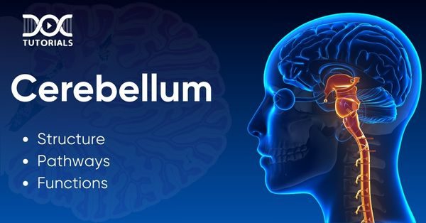
Cerebellum

The term ‘cerebellum’ is derived from the Latin word for ‘little brain’. Its primary function is to facilitate motor control and enable well-timed and smooth bodily responses. However, the cerebellum also handles many other cortical functions, including the experience of pleasure and fear, and even speech control.

In this guide, you can get a comprehensive idea about the anatomy of the cerebellum. Moreover, we will walk you through its functions and development before finally arriving at the relevance of different cerebellar disorders. 

Keep reading!

What is the Cerebellum?

The cerebellum is a section of the body’s central nervous system (CNS) that helps you to control your movements. 

During embryonic development, the brain and its associated components are formed from the anterior part of the neural tube. From here, the hindbrain gets divided into the myelencephalon (inferior) and metencephalon (superior) parts. Consequently, we witness the formation of the cerebellum in the latter division. 

What is the Difference Between the Cerebellum and Cerebrum?

Located at the bottom of your brain, the cerebellum is a small section placed near the back of your head. Conversely, the cerebrum constitutes the largest component of the human brain, surrounding the front side and top of the cerebellum. 

Anatomical Structure and Divisions of the Cerebellum

As far as location is concerned, the cerebellum is a part of the hindbrain situated inside the posterior cranial fossa within the skull cavity. Parts of it are located dorsal to the brainstem, caudal to the cerebrum, and within the tentorium cerebelli. 

The middle, inferior, and superior cerebellar peduncles link the cerebellum to other corners of the brain, such as the midbrain, pons, and medulla. 

Anatomical Features of the Cerebellum

This part of the brain is divided by a midline vermis into right and left hemispheres. Some of its tissues act as landmarks to mark the cerebellum into different lobes. Here’s how it takes place:

  • The primary fissure is responsible for dividing the cerebellum into larger posterior and smaller anterior lobes.
  • Then, the horizontal fissure distinguishes the superior and inferior sections (although functionally, this section may not be that prominent).
  • After this, the posterolateral fissure divides the flocculonodular lobe. 

Proceeding towards the cerebellum’s internal structure, the arrangement may not seem that dissimilar when compared to the cerebrum: 

  • Deep within the cerebellum’s white matter lie the cerebellar nuclei. They are clusters of grey matter that serve as crucial connection points to the cerebellar cortex. 
  • The cerebellar nuclei comprise interposed nuclei (combined emboliform and globose), fastigial, and dentate. 

In the table below, you can have a quick look at some of the vital cells and fibres connected with the cerebellum:

Cells/Fibres of CerebellumLocation
Parallel Fibres/Granule CellsSituated within the parallel fibres (molecular layer) and the granule cells
Purkinje CellsForms an integral part of the Purkinje layer, whereas the dendrites stretch into the molecular layer. Finally, the axons extend from the cerebellar cortex. 
Basket CellsPresent in the molecular layer
Stellate CellsPresent in the molecular layer
Golgi CellsLocated in the granular layer
Climbing FibresCan be found in cerebellar grey matter and white matter along with the brain stem
Mossy FibresSituated in the brain stem apart from cerebellar grey and white matter

Regarding its divisions, the cerebellum can be associated with three major functional segments. These sections collectively support the functional anatomy of the cerebellum. The list includes:

Cerebellar DivisionsPotential/ Identified Functional RegionsPedunclesParavermal cortices, vermis, and the anterior lobe’s cortex
VestibulocerebellumFlocculonodular lobe and vermisInferior cerebellar peduncleHelps in coordinating conjugate eye movements and establishing balance
SpinocerebellumParavermal cortices, vermis and anterior lobe’s cortexSuperior and inferior cerebellar pedunclesFacilitates posture correction, muscle coordination, and corrects motor activities
CerebrocerebellumCortex of the cerebellar hemispheresMiddle and superior cerebellar pedunclesInitiates movements and helps to control voluntary movements

What are the Pathways of the Cerebellum?

To stay connected with other segments of the nervous system, the cerebellum is dependent on three major pathways:

  1. Superior Cerebellar Peduncle 

Also referred to as the Brachium Conjunctivum, this peduncle is primarily an efferent pathway, meaning it carries signals to the muscles away from the nervous system. These fibres originate within the deep cerebellar nuclei and later on project to the red nucleus via the thalamus. 

The superior cerebellar peduncle aids in motor coordination and enables the adoption of habits by transmitting processed behaviours from the cerebellum to the higher motor centres.

  1. Inferior Cerebellar Peduncle 

The inferior cerebellar peduncle comprises both efferent and afferent fibres. As a result, these connections collect signal inputs from the spinal cord, brainstem tegmentum, and vestibular nuclei. Additionally, this segment is responsible for transmitting outputs to both the reticular formation and vestibular nuclei.

The cerebellar inferior peduncle is overall vital for correcting posture, balance, and ocular reflexes by coordinating spinal inputs.  

  1. Middle Cerebellar Peduncle

The middle peduncle, or Brachium Pontis, is an afferent pathway that collects inputs from the pontine nuclei. Next, these fibres of the cerebellum expand to the cerebellum after extending past the midline. 

In terms of motor planning and learning, the middle cerebellar peduncle is important for feeding cortical information into cerebellar functions. 

What are the Functions of the Cerebellum?

The cerebellum is known for fulfilling different functions that are related to muscle coordination and movements. These include:

  • Maintaining Balance: The cerebellum is composed of distinctive sensors that can detect shifts in movements and balance accordingly. Therefore, it distributes signals for the body to adapt to various situations and move about.
  • Controlling Movements: Almost all the movements need to combine different muscle groups. In this respect, the cerebellum plays a huge part as it times muscle actions and facilitates smooth movements.
  • Motor Learning: The cerebellum guides our body to practice and master movements that require precision and fine-tuning. For instance, when it comes to practising how to play a musical instrument or learning how to swim, the cerebellum plays a major role. 

Additionally, the cerebellum aids vision by coordinating eye movements. Also, researchers strongly speak for its involvement in developing thoughts, a particular mood, or even processing language. However, these assumptions still need to gather full-range exploration.

What are the Symptoms of Cerebellar Disorders?

Some signs of a cerebellar disorder are as follows:

  • Facing challenges with walking and mobility
  • Lack of muscle coordination
  • Difficulty in speaking or slurred speech
  • Headaches
  • Abnormal eye movements

Besides these symptoms, over the period, medical researchers have found many disorders associated with the cerebellum. These include:

  • Brain bleeds
  • Toxins
  • Stroke
  • Genetic anomalies
  • Infection
  • Cancer

However, to date, the fundamental symptom of cerebellar dysfunction is ataxia. It is the medical term used to describe a notable loss of muscle control and coordination. Beyond the primary symptom of ataxia, which is muscle control loss, those diagnosed can even encounter the following challenges:

  • Tiredness
  • Difficulty in swallowing
  • Blurry vision
  • Challenges faced when it comes to precise muscle control
  • Changes in thoughts and mood

Several factors may cause ataxia, including: 

  • Genetics 
  • Head injury 
  • Stroke 
  • Cerebral palsy and more 

How to Minimise the Chances of Damage to the Cerebellum?

Several actions can be taken to prevent damage to your cerebellum, such as:

  • Prioritise Having a Balanced Diet

Vitamin B12 is very essential for your brain. So, you need to ensure sufficient intake of vitamin B12 in your diet to maintain a healthy cerebellum.

  • Do not Overlook Infections

Consider promptly addressing infections, particularly eye infections, to prevent them from spreading to the brain and causing serious medical repercussions.

  • Stick to Prescribed Medications

Several drugs can put stress on your brain, especially your cerebellum. Thus, you should consult a doctor before taking any medicine to avoid these issues. 

  • Wear Safety Gear as Needed

You must protect your head and neck from injuries by consistently using appropriate safety gear during work, bike riding, and recreational activities.

FAQs About Cerebellum

  1. What is the main job of the cerebellum?

Primarily, the cerebellum is meant for controlling the body’s balance while walking, standing, or performing other complicated motor functions. 

  1. At what age does the cerebellum develop?

Cerebellar growth starts in the first trimester, roughly 30 days post-conception, and completes around two years after birth.

  1. Is the cerebellum in two parts?

Yes, the cerebellum is formed of two hemispheres that are connected together by the vermis. Located in the hindbrain, the cerebellum collects inputs and transmits output signals through a limited number of cells. 

  1. How to cure cerebellum damage?

To date, no permanent cure for cerebellar damage has been found. However, supportive medical treatments can help minimise the effects of symptoms. 

  1. How to improve your cerebellum?

You can bring many lifestyle changes to improve the overall health of your cerebellum. To lower the risk of a cerebellar injury or infection, consider protecting your head while riding a bike, exercising regularly, quitting smoking, limiting alcohol, and promptly taking care of eye infections.

Conclusion

Overall, the cerebellum is one of the most critical components of the brain. With time, advancements in medical technologies have helped doctors diagnose the various conditions that can affect the cerebellum early and thereby assisted them in finding effective and timely solutions.

On the DocTutorials website, you can explore various medical subjects while delving into the intricacies of diseases and treatments with detailed explanations and visuals. With our strategic approach to teaching, NEET aspirants can gain crystal-clear knowledge and be well on their way to achieving a top score.

Also, they can find top-quality video lectures, question banks, expert guidance, mock tests, and more. Kickstart your NEET PG 2025 preparation today!

NEET PG Online Live Power Pack
DocTutorials App Download Now

Latest Blogs

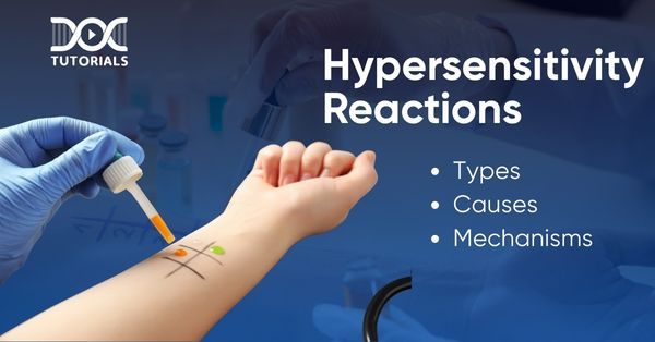
Hypersensitivity Reactions: Types, Causes, and Mechanisms

Hypersensitivity

The human immune system creates a strong shield of defence against pathogens. However, at times, it may ‘overreact’, resulting in undesirable outcomes. These effects on someone’s body are not generated by antigens but by several inflammatory mechanisms in which the immune cells are directly involved. 

These mechanisms cause hypersensitivity reactions, which can lead to different diseases. In many cases, pathologists find a genetic susceptibility to these reactions or a triggering event like an infection can be the root cause of the problem. 

In this guide, you will learn in detail about the four types of hypersensitivity reactions, popularly termed the Gell and Coombs classification. Additionally, we will explain the various causes of hypersensitivity, their mechanisms, and more. 

What are Hypersensitivity Reactions?

Hypersensitivity reactions sum up excessive or abnormal responses of a human body’s immune system to different foreign substances or allergens. 

When an already sensitised individual is re-exposed to a similar allergen, they are highly likely to experience allergic reactions. These hyperreactions can cause excessive tissue damage over time. 

What are the Different Types of Hypersensitivity Reactions?

To date, medical researchers have found four different types of hypersensitivity reactions. However, some studies suggest that a potential fifth type can exist. But, most recognised bodies prefer to include this fifth type as a subset of type 2 hypersensitivity reaction.  

Here are the major hypersensitivity types that you must be aware of:

  1. Type 1 Hypersensitivity Reaction

This type of hypersensitivity reaction triggers an immediate response. It can happen when a person is directly susceptible to an antigen. In these circumstances, the concerned individual’s body responds to the antigen by forming a definite type of antibody, termed Immunoglobulin E (IgE). 

The following elements can activate type 1 hypersensitivity responses:

  • Animal sources like rats, cats or bee stings
  • Selected food products like seafood, nuts, and soy
  • Environmental agents like latex, dust, mould, etc.
  • Allergic conditions like asthma, conjunctivitis, and allergic rhinitis

In this type of hypersensitivity, there is a sensitisation stage and an effect stage: 

  • During the first stage, the person does not experience any symptoms. 
  • However, the second stage is triggered by re-exposure, when the body finally recognises the antigen and generates a response as symptoms. 

Some common symptoms of a type 1 hypersensitivity reaction include:

  • Rash
  • Hives
  • Flushing
  • Itching
  • Wheezing
  • Oedema
  • Stomach cramps
  • Rhinitis

Treatment Options for Type 1 Hypersensitivity Reaction

To cure the above-mentioned effects, some people need immediate medical intervention. After the doctor suspects type 1 hypersensitivity, they first review the patient’s previous medical records before suggesting specific blood and allergy tests. 

Then, they provide treatment options like:

  • Systemic glucocorticoids
  • Adrenaline or epinephrine
  • Antihistamines

Also, the concerned individuals must avoid the recognised allergen moving forward. 

  1. Type 2 Hypersensitivity Reaction

Much similar to the type 1 hypersensitivity reaction,  type 2 reactions also result from antibodies. As a matter of fact: 

  • Both type 2 and type 3 hypersensitivity are generated from the same category of antibodies, IgE. 
  • The fundamental difference between type 2 and type 3 hypersensitivities lies in the type of antigen that triggers an immune response.

From a type 2 hypersensitivity, cytotoxic reactions take place where healthy cells are damaged as they encounter the antigens. This can lead to long-term tissue damage, causing conditions like:

  • Autoimmune hemolytic anaemia occurs when the red blood cells (RBC) burst happens spontaneously 
  • Thrombocytopenia or an acute blood disorder that occurs when there isn’t enough platelets
  • Autoimmune conditions like Grave’s disease
  • Autoimmune neutropenia, when the human body automatically destroys neutrophils responsible for healing infections

Treatment Options for Type 2 Hypersensitivity Reaction

Cephalosporins, penicillin, thiazides, and methyldopa are among the drugs that are known to induce type 2 hypersensitivity reactions. To prevent the unusual action of these antibodies, doctors can prescribe some treatment options, such as:

  • Intravenous immunoglobulin infusions
  • Systemic glucocorticoids
  • Cyclophosphamide and cyclosporine agents
  • Plasmapheresis
  1. Type 3 Hypersensitivity Reaction

In this type of hypersensitivity, antibodies and antigens combine to produce complexes in the blood vessels, skin, kidney tissues, and joints. A series of bodily reactions then follow, causing a great deal of tissue damage. 

Some causes of a type 3 hypersensitivity response can be:

  • Intake of drugs that include proteins from various organisms, like antivenins
  • A drug infliximab, which is often recommended for controlling autoimmune conditions
  • Animal stimulations like tick bites or insect stings

The various side-effects of type 3 hypersensitivity can feature:

  • Small-vessel vasculitis
  • Rheumatoid arthritis
  • Serum sickness
  • Lupus
  • Henoch-Schönlein purpura

Treatment Options for Type 3 Hypersensitivity Reaction

While treating type 3 hypersensitivity, doctors can do the following:

  • Conduct different assessments that include blood and urine tests along with imaging scans. 
  • Recommend different forms of immunosuppression with systemic glucocorticoids as well as some disease-modifying drugs based on the severity of the hypersensitivity response. 
  1. Type 4 Hypersensitivity Reaction

Very different from the other forms of hypersensitivity, all type 4 hypersensitivity examples are cell-mediated. In contrast to antibody-caused reactions, type 4 hypersensitivity responses result from T cells. Medical experts further distinguish these reactions into 4a, 4b, 4c and 4d. 

Additionally, when compared to the other types of hypersensitivity, this type causes a delayed response. The three major subsets of type 4 hypersensitivity are:

  • Tuberculin-type hypersensitivity
  • Contact dermatitis
  • Granulomatous-type hypersensitivity

The most common hypersensitivity reactions in this category are triggered by: 

  • Exposure to certain metals 
  • Poison ivy 
  • Drugs like anticonvulsants or antibiotics 

Doctors can diagnose contact dermatitis by suggesting skin patch tests or a skin biopsy. However, experts are more likely to use a chest X-ray when treating tuberculin-type hypersensitivity.

However, it is tricky to diagnose granulomatous-type hypersensitivity, and healthcare providers often stick to the following assessments before starting their treatment:

  • X-ray
  • Lymph node biopsy
  • Enzyme analysis
  • Salivary gland test

Treatment Options for Type 4 Hypersensitivity Reaction

Doctors usually suggest the following treatment options for type 4 hypersensitivity reactions:

  • If the patient is found to have contact dermatitis, then the healthcare provider can recommend availing topical steroids. 
  • Similarly, the treatment specifics vary from one case to another. 
  • For tuberculin-type hypersensitivity, pathologists typically resort to the regular method for diagnosing tuberculosis. 

Some typical treatments for tuberculin-type hypersensitivity are:

  • Isoniazid
  • Rifampin
  • Ethambutol
  • Pyrazinamide

Finally, granulomatous hypersensitivity treatment is approached as condition-specific, although in most cases, the doctors apply a steroid therapy.

Non Allergic Hypersensitivity Reaction

Some people can experience a non-immune anaphylactic reaction or non-allergic hypersensitivity to specific foods or drugs: 

  • These reactions are a bit complex to identify separately, but they are often noticed as they result in direct symptoms. 
  • Unlike allergic hypersensitivity reactions, they do not involve the release of T cells or antibodies. 

What is the Mechanism of Hypersensitivity?

As already discussed, hypersensitivity diseases are primarily segregated depending on the type of immune reaction and the underlying mechanism causing cell or tissue injuries. The mechanisms highlight some scenarios that are mainly reliant on antibodies while others rely on T cells. 

Depending on the way normal cellular functions are disrupted, these are the main hypersensitivity mechanisms observed to date:

  1. Immediate Hypersensitivity (Type I)

This is an IgE-mediated allergic reaction triggered by allergens, leading to rapid mast cell degranulation and release of inflammatory mediators. It manifests as conditions like asthma, hay fever, and anaphylaxis.

  1. Antibody-mediated Hypersensitivity (Type II)

This involves Immunoglobulin G (IgG) or Immunoglobulin M (IgM) antibodies binding to cells or tissues, causing damage through complement activation or antibody-dependent cellular cytotoxicity. 

Some instances include transfusion reactions and autoimmune hemolytic anaemia.

  1. Immune Complex-mediated Hypersensitivity (Type III)

This occurs when antigen-antibody complexes deposit in tissues, activating complement and attracting neutrophils, resulting in inflammation and tissue damage.

  1. T Cell-Mediated Hypersensitivity (Type IV)

Type 4 hypersensitivity is a delayed-type hypersensitivity mediated by T cells, which release cytokines and cause inflammation upon encountering an antigen.

FAQs About Hypersensitivity Reactions

  1. What are the main differences between type I and type II hypersensitivity reactions?

Type I hypersensitivity is characterised by allergic reactions like anaphylaxis and hives. They are caused by IgE-mediated mechanisms, while type II hypersensitivity is a cytotoxic response caused by antibodies that result in long-term cell damage or tissue destruction. The latter symptoms are commonly noticed in autoimmune disorders. 

  1. What are some common triggers for type IV hypersensitivity reactions?

A type four hypersensitivity reaction, a cell-mediated response to allergens like those in contact dermatitis or tuberculin skin tests, involves the activation of T cells that release inflammatory cytokines, causing a localised reaction.

  1. What are the long-term effects of untreated hypersensitivity reactions?

Apart from the medically established symptoms, chronic allergies or untreated hypersensitivity can deteriorate the overall quality of life. Some possible symptoms are frequent respiratory issues, skin allergies and conditions, poor digestive health, and so on. 

  1. What is a severe life-threatening hypersensitivity reaction?

Anaphylaxis is a life-threatening fatal hypersensitivity response. Its symptoms can arise within seconds or minutes after the sensitised person is exposed to the allergen. 

  1. Does hypersensitivity go away?

It is possible for hypersensitivity to disappear, particularly when the root cause is discovered and avoided. However, recovery time and severity of the disease vary.

Conclusion

Hypersensitivity reactions can take a time frame of a few minutes to hours or even days to generate harmful symptoms. Luckily, most of them can be detected by timely diagnosis followed by proper medication and treatment. Thus, it is highly suggested to visit a trusted immunologist upon facing the above-mentioned symptoms. 

While the study of hypersensitivity can seem vast, you can start learning about it from the basics through DocTutorials. Also, if you are preparing for an upcoming competitive medical examination, you can rely on our high-quality video lectures, study materials, mock tests, and more. So, consider starting your NEET PG 2025 preparation today and realise your dream of securing the dream medical seat!

NEET PG Online Live Power Pack
DocTutorials App Download Now

Latest Blogs

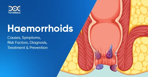
Haemorrhoids: Causes, Symptoms, Risk Factors, Diagnosis, Treatment, and Prevention

Haemorrhoids

According to a recent study, almost 11% of the Indian population suffers from haemorrhoids or piles. This condition can arise from a number of factors like age, chronic constipation, poor dietary choices, and more. Although uncomfortable, haemorrhoids or piles do get better on their own if one adopts the proper remedies. 

Thus, it is crucial to understand the types, symptoms, diagnosis, and treatment options for haemorrhoids to effectively manage this medical condition. Also, if you are a medical aspirant or a healthcare professional, learning in detail about piles is crucial for expanding your knowledge base and providing care to patients.   

Keep reading!

What are Haemorrhoids?

Haemorrhoids are swollen veins which are found both inside and on the outside of the anus and rectum. These swollen veins may cause itching, pain, and bleeding. Also known as piles, haemorrhoids are categorised as either internal, which lie inside the rectum, or external, which form under the skin around the anus. 

What are the Different Types of Haemorrhoids?

The classification of haemorrhoids depends on the site where the engorged vein arises. Check them out below:  

  1. External Haemorrhoids

These are swollen veins that appear under the skin around the anus, which is the end part of the large intestine and facilitates the excretion of stool. External haemorrhoids cause itching and soreness, and they can often cause bleeding. Although this problem is not threatening, it is capable of creating severe pain and inflammation.

  1. Internal Haemorrhoids

This category consists of engorged veins that occur inside the rectum, which is the portion of the gastrointestinal tract between the colon (large intestine) and the anus. Internal haemorrhoids can cause bleeding but are usually not painful.

  1. Prolapsed Haemorrhoids

Prolapsed haemorrhoids can cause severe pain or bleeding. Both internal and external haemorrhoids can become prolapsed or extend and protrude out of the anus. 

  1. Thrombosed Haemorrhoids

When a blood clot has formed, an external haemorrhoid may be blue or purple in colour. This condition is known as thrombosis or thrombosed haemorrhoids.

What are the Causes and Risk Factors of Haemorrhoids?

Although medical practitioners have not fully discovered why haemorrhoids appear, they might occur because of the following risk factors:

  • Ageing: Haemorrhoids are largely prevalent in people between 45 and 65, yet they may affect younger adults as well as children.
  • Chronic Constipation: People suffering from chronic constipation habitually put pressure on the blood vessel walls of the rectum and anus for bowel movement. This often leads to haemorrhoids.
  • Pregnancy: The increased pressure of the developing foetus and the uterus during pregnancy compresses the pelvic region and results in haemorrhoids in around 35% of pregnant individuals.
  • Diarrhoea: Frequent diarrhoea has the potential to cause the growth of haemorrhoids.
  • Diet: A diet that lacks adequate fibre could be responsible for the growth of haemorrhoids.
  • Prolonged Sitting: Sitting for long periods, especially on the toilet, is a possible risk factor that can cause haemorrhoids.
  • Heavy Exercises: Heavy exercises and weight lifting on a repeated basis may be contributing factors leading to haemorrhoids.
  • Weight: Too much obesity can cause haemorrhoids, possibly because of increased intra-abdominal pressure.

What are the Symptoms of Haemorrhoids? 

Internal haemorrhoids usually do not show any symptoms. You might notice blood in your stool, on the toilet tissue after you have wiped, or in the bowl.

Symptoms of prolapsed haemorrhoids can include:

  • Pain, especially with bowel movements
  • Hard lumps near the anus

External haemorrhoids can be symptomatic and appear with:

  • Pain while sitting 
  • Rectal bleeding
  • Itching in the anus
  • Swelling

Lastly, the symptoms of thrombosed haemorrhoids can include:

  • Severe pain
  • Itching
  • Bleeding

How is the Diagnosis of Haemorrhoids Performed? 

A healthcare provider diagnoses haemorrhoids by evaluating the symptoms and performing a physical examination. Other procedures may involve:

  • Digital Rectal Examination: The doctor places a lubricated, gloved finger into the rectum to feel for dilated veins.
  • Anoscopy: The medical practitioner uses an anoscope, a lighted tube, to examine the lining of the anus and rectum.
  • Sigmoidoscopy: The doctor uses a sigmoidoscope, a lighted tube that contains a camera, to see the inside of the lower portion of the colon (sigmoid) and the rectum. Sigmoidoscopy is classified into two types: flexible sigmoidoscopy and rigid sigmoidoscopy (proctoscopy). 

What are the Treatment Options for Haemorrhoids? 

Although haemorrhoids may usually heal with lifestyle changes, in certain cases, additional medical care is required. Some common treatments for haemorrhoids vary by severity and kind and can be grouped into the following:

Home Therapies

  • Hydration: Sufficient fluid intake softens the stools and reduces pressure, thus lessening the symptoms of haemorrhoids.
  • Topical Creams: External haemorrhoid pain may be relieved by over-the-counter (OTC) creams that reduce swelling, itching, and irritation.
  • Fibre Supplements: Supplements containing fibre like methylcellulose and psyllium can help relieve constipation, thus reducing the pressure on the anus and decreasing the chances of haemorrhoids.
  • Cold Compress: Ice or a cold compress applied to the area can possibly alleviate pain and swelling.
  • Warm Water Bath: Taking a warm water bath in a tub while seated can bring relief from pain and inflammation.

Medications

  • Zinc Oxide: Zinc oxide creams assist in minimising irritation and itching.
  • Witch Hazel: Its anti-inflammatory and astringent properties provide relief from haemorrhoid pain, itching, and burning sensations.
  • Lidocaine: Lidocaine creams or suppositories give relief from haemorrhoid pain.
  • Painkillers: OTC painkillers such as ibuprofen or acetaminophen may assist in alleviating pain from haemorrhoids.

Non-surgical Treatments

  • Rubber Band Ligation: A process that involves the use of an elastic band to encircle the haemorrhoid and slow down the blood supply, making it shrink.
  • Sclerotherapy: Injection of a solution into the haemorrhoid to form scar tissue that shuts off the blood supply, making it shrink.
  • Electrocoagulation: This technique uses electric current to prevent the blood flow to a haemorrhoid.
  • Infrared Coagulation: In this method, a small probe is inserted into the rectum to transmit heat and reduce the pain of the haemorrhoid.

Surgical Treatment

  • Haemorrhoidectomy: This is a surgical technique that helps eliminate large or prolapsed haemorrhoids.
  • Stapling: In this method, a stapling device is utilised to eliminate an internal haemorrhoid or pull a prolapsed internal haemorrhoid inside the anus and hold it in position.

What are the Prevention Tips for Haemorrhoids? 

Lifestyle modifications can lower the risk of haemorrhoids. Here are some common prevention tips: 

  • Have A Balanced Diet: Eat plenty of fibre-containing foods such as fruits, vegetables, and whole grains to soften stools. Adequate water intake and taking over-the-counter fibre supplements can also prevent constipation, thus reducing the chances of haemorrhoids.
  • Visit The Bathroom When Required: Constantly holding back stool may lead to constipation and worsen the risk of having haemorrhoids. Hence, go to the bathroom whenever required.
  • Maintain Activity Levels: Exercise daily to regularise bowel movements and keep the possibility of haemorrhoids at bay.
  • Maintain Normal Weight: Maintain a standard body weight, as obesity can increase the risk of haemorrhoids.

FAQs About Haemorrhoids

  1. How long will it take for a haemorrhoid to heal?

Although most haemorrhoids improve in a few days to weeks with self-care and home remedies, some may last longer or become chronic. If haemorrhoids have lasted over a week or two, it is best to see a doctor.

  1. Are piles dangerous?

Piles do not normally cause serious problems, but in some cases, they can lead to complications. External piles can become inflamed and swollen, resulting in the formation of ulcers. Additionally, skin tags can occur when the inside of a haemorrhoid contracts, but the skin remains.

  1. How are piles and haemorrhoids different?

Piles and haemorrhoids are actually the same medical condition, signifying enlarged veins in the lower rectum and anus. In normal cases, these veins act as cushions to the anus and regulate stool. When they get inflamed or swollen, they may produce some symptoms like pain, bleeding, etc. 

  1. What is the ideal treatment for piles?

Several treatments are available for piles, including creams, ointments, suppositories, tablets, and fibre supplements. Home treatments such as increasing fibre intake and modifying lifestyle are also beneficial.

  1. When do you need to see a doctor for haemorrhoids?

You need to go see a doctor if you notice any of the following:

  • Ongoing or heavy bleeding, with or without pain
  • Pus from haemorrhoids
  • High fever
  • Extreme pain

Conclusion

Overall, haemorrhoids and piles are the same condition in which the veins in the anus or rectum become swollen. Understanding the causes, symptoms, risk factors, and treatment of piles can help you deal with this common condition effectively. 

For aspiring medical students studying for the NEET PG exam, learning about haemorrhoids in detail is essential. In this regard, DocTutorials can serve as an ideal solution. With professional video courses, Quick Revision Programs (QRPs), and interactive tools, DocTutorials ensures you are all set to crack your exams and ready to face any medical challenges with confidence. Check out our NEET PG study materials today!

NEET PG Online Live Power Pack
DocTutorials App Download Now

Latest Blogs

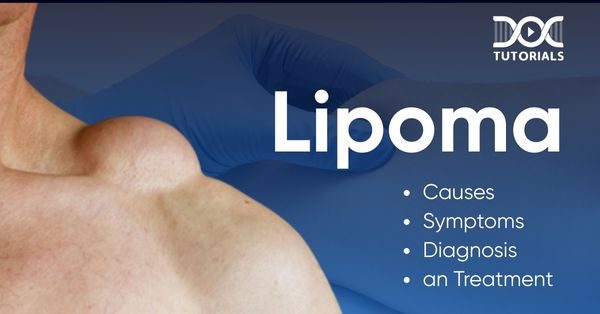
Lipoma: Causes, Symptoms, Diagnosis, and Treatment

Lipoma

Occurring in about 1% of the population in India, lipomas are lumps of fatty tissue that occur just under the skin, most commonly among people who are in their 50s or 60s. These are painless lumps of fatty tissue that develop just under the skin. 

While they are usually painless and do not pose health risks, there are instances where they may require medical intervention. It is imperative to understand the causes, symptoms, and treatment of lipomas, particularly for those who will need to be able to differentiate them from dangerous conditions. 

Keep reading to learn more about this condition!

What is Lipoma?

A lipoma is a soft, rounded, or oval fatty tissue lump that develops just under the skin. It is usually movable on palpation and is generally painless. Lipomas may be of any size, ranging from as small as a pea to several inches in diameter. They are most commonly located on the upper body, arms, or thighs but can be found anywhere on the body.

These soft tissue tumours are benign and develop slowly, and they do not present serious health threats. Although they are generally harmless and need not be treated, people should consider having them removed by physicians if the lipoma is irritating, painful, or indicates growth.

Where do Lipomas form?

Lipomas can grow anywhere in the human body. Although they do not develop on the muscles, brain, or other internal organs, most of them form under the skin and mainly in these places in the body:

  • Arms
  • Legs
  • Neck
  • Shoulders
  • Trunk (chest and torso)
  • Back
  • Forehead

Causes of Lipoma

Healthcare professionals are unsure of the main causes of lipomas. However, there are several conditions that can result in the growth of lipomas in the body:

  • Dercum’s Disease: It is an unusual disorder involving the formation of painful lipomas, most often found on the trunk, legs, and arms. It has also been known as adiposis dolorosa or Anders’ syndrome.
  • Gardner Syndrome: It is a form of familial adenomatous polyposis (FAP) that results in the development of lipomas and other health issues.
  • Madelung’s Disease: It is a condition which often occurs in men who consume excessive alcohol. It also goes by the name multiple symmetric lipomatosis, which leads to lipomas growing on the neck and shoulders.
  • Hereditary Multiple Lipomatosis: Also referred to as familial multiple lipomatosis, it is a hereditary disorder that is transmitted through family branches.

Risk Factors of Lipoma

There are a few risk factors that may increase your chances of developing a lipoma. These include:

  • Age: Lipomas are more common among people 40 to 60. Although the growths occur at any age, they are most common in this age group.
  • Genetics: Genetic influences seem to have a relevant impact on the occurrence of lipomas since they usually appear in families.
  • Obesity: Anyone with obesity tends to develop lipoma sooner or later.

Symptoms of Lipoma

Mentioned below are the common symptoms that may manifest if someone has lipoma:

  • A Soft, Movable Lump: Lipomas are most often soft to the touch and can be easily shifted beneath the skin. They can range in size from a tiny bump to a large, easily observable mass.
  • Painless or Mild Pain: Lipomas are usually not painful. However, if they compress nearby nerves, muscles, or other tissues, they can cause pain, discomfort, or stiffness.
  • Feels Doughy and Slippery: When you press the lipoma, you will find it soft and doughy so that it can move freely with the pressure of the fingers.
  • Stays in One Place: It remains limited and stays in a particular area because it is covered by a thin fibrous sheath that prevents it from infiltrating surrounding tissues.
  • Other Symptoms: Anyone having a lipoma on or near the bowels may experience vomiting, nausea, and constipation.

Diagnosis of Lipoma

Large lipomas can be seen as bulging, appearing clearly as rounded masses on the skin surface. To diagnose a lipoma, your doctor can perform the following tests:

  • A physical examination
  • Biopsy to sample tissue to be analysed in the lab
  • Imaging procedures, such as an X-ray, MRI, or CT scan, if the lipoma is large, has unusual features, or appears to be deeper than the fatty tissue.

Treatment Options for Lipoma

There are several treatment methods available for the management of lipomas, some of which are:

  • Surgery

The most widespread way of eliminating lipomas is surgery. This is not a complicated process and is done under local anaesthesia. The surgeon removes the lump, removing the fatty tissue in the process. This method is often successful, and the chances of the recurrence of the lipoma are negligible.

  • Liposuction

While treating larger lipomas, liposuction can be an option. Liposuction uses a thin tube inserted through the skin into the lipoma to remove the fat cells by suction. Liposuction is less invasive than open surgery, but there is a higher risk of remaining behind lipoma tissue, which might result in recurrence.

  • Steroid Injections

In some instances, steroid injections may be used to shrink a lipoma. While this is not a permanent solution, it can reduce the size of the lump, especially when surgery is not feasible or is not desired.

Best Foods to consume for reducing Lipomas

Certain foods are believed to have a positive effect on hormonal regulation and metabolism, which can also be helpful for individuals with lipomas:

  • Vegetables

Vegetables contain a high amount of nutrients, fibre, and antioxidants. They promote overall health and are believed to reduce inflammation that may exacerbate lipomas. Some ideal options include broccoli, kale, spinach, and other green vegetables.

  • Turmeric

Turmeric contains a substance named curcumin, which is an anti-inflammatory and antioxidant. It can reduce body inflammation, which is also said to be associated with the development of lipomas.

  • Fruits

Fruits are rich in fibre, vitamins, and antioxidants. Blueberries, raspberries, and pears are especially beneficial for people with lipomas.

  • Omega-3 Fatty Acids

Omega-3 fatty acids may reduce inflammation and restrict the growth of fat tissue beneath the skin. Salmon, mackerel, herring, and cod liver oil are some good sources. Being rich in Omega-3 fatty acids, these items are great to help reduce lipomas. 

  • Olive Oil

Olive oil is high in monounsaturated fats, which are good for the heart. It also possesses antioxidants that may reduce inflammation and help lessen lipomas. 

  • Green Tea

Green tea is a healthy beverage for individuals with lipomas. It is rich in antioxidants and can increase fat oxidation and metabolism, possibly aiding in decelerating the growth of lipoma.

FAQs About Lipomas

  1. Are lipomas and liposarcomas same?

No, lipomas are different from liposarcomas. While liposarcomas are malignant fat cell tumours, lipomas are not. Even their appearances tend to be nearly indistinguishable from each other.

  1. What are the types of lipoma?

Lipomas are grouped into many different subtypes, such as angiomyolipomas, angiolipomas, myelolipomas, ossifying lipomas, chondroid lipomas, spindle cell lipomas, fibrolipomas, hibernomas, pleomorphic lipomas, and neural fibrolipomas.

  1. How much time do lipomas take to grow?

In general, lipomas develop gradually for months or years, usually averaging between 1 to 10 centimetres in size.

  1. What foods must be avoided for lipoma?

It is better to avoid foods and drinks that contain a lot of sugar, such as sweets, candies, and pastries, because they can raise blood glucose levels and result in fat accumulation. One should also limit the consumption of alcohol since it can disrupt metabolic processes and trigger fat deposition, thus escalating the risk of lipoma.

  1. What are the complications of lipoma?

Lipomas are generally harmless and do not lead to any complications, but at times, they can be troublesome when they become large or put pressure on nerves or other tissues.

Conclusion 

Lipomas are normally benign tumours that form from fatty tissues under the skin. Knowing the causes, symptoms, diagnosis, and treatments can help manage lipomas effectively. For students aspiring to a career in medicine, a thorough knowledge of lipomas and other medical conditions is important to excel in exams like NEET PG. DocTutorials offers cutting-edge learning techniques and NEET PG study materials, which enable students to acquire the expertise required to understand and remember complex medical concepts.

NEET PG Online Live Power Pack
DocTutorials App Download Now

Latest Blogs

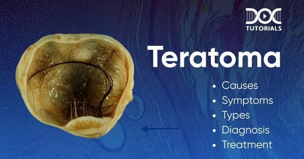
Teratoma: Causes, Symptoms, Types, Diagnosis, and Treatment

Teratoma

The incidence rate of teratomas is about 1.2 to 14.2 cases per 100,000 population per year, with the majority being ovarian teratomas. Teratomas are more common in women of reproductive age. A type of the same, mature cystic teratomas, comprise 10% to 20% of all ovarian neoplasms, and they are one of the most common germ cell tumours.

In this article, we will explore all the information you require about teratomas, their types, causes, risk factors, and treatment options. 

What is a Teratoma?

A teratoma is a germ-cell tumour that arises from germinal layers in the body. It stands out from conventional tumours as it has a distinct composition and can contain multiple types of tissues, such as hair, teeth, bone, etc. 

Most teratomas are benign, while some can become malignant and metastasise to a different location in the body. They usually occur in the ovary, testis, and tailbone region. 

Causes  of Teratoma

To date, the exact cause of teratomas is unknown, and it has an idiopathic cause. However, several factors are known to accelerate the process of tumour formation. The suspected causes of teratoma include:

  • Genetic conditions affecting the central nervous system, genitourinary tract, and the spine
  • Changes in embryonic stem cells during development

While a foetus is in its developmental stage, some of the cells which do not differentiate, retain the ability to turn into other types of cells. These types of cells are then known as pluripotent cells.

Risk Factors of Teratoma

Some of the risk factors are known, which help in turning ovarian teratomas into malignant tumours. These include:

  • Older age
  • Larger tumour size
  • Elevated levels of tumour marker CA-125
  • Post-menopausal women

There are also a number of risk factors which do not cause the tumour directly but help in accelerating the tumour formation. These include:

  • Low birth weight 
  • Cryptorchidism
  • Hypospadias
  • Maternal bleeding 
  • Increased maternal age
  • Retained placenta
  • Jaundice

What are the Various Types of Teratomas?

Teratomas are classified based on various criteria, like nature, location, etc.

  • Based on the Nature of a Teratoma 
    • Mature Teratoma: These teratomas are usually benign in nature and are non-cancerous.
    •  Immature Teratoma: These are the malignant and cancerous types of teratomas. They are more commonly seen in children.
  • Based on the Location of a Teratoma
    • Ovarian Teratoma: The most common type seen in females. These rarely turn cancerous.
    • Testicular Teratoma: The most common type seen in males. In young aged males, these are non-cancerous. However, in older males, they have a 50% chance of becoming malignant.
    • Sacrococcygeal Teratoma: They are commonly seen in children. However, these have a very rare chance of incidence, about 1 in 40000 live births. The location is near the tailbone, and the female gender is predominantly affected.

Apart from these, a rare type of teratoma is the Fetiform teratoma, with an incidence of 1 in 50000 people. It is a type of dermoid cyst that closely resembles a malformed fetus. However, due to the absence of a placenta and an amniotic sac, this fetus can never develop to full term. Usually, it is diagnosed within the first 18 months of life.

What are the Various Symptoms of a Teratoma?

It is difficult to track the presence of a teratoma in the body until it becomes a grave concern. An individual tends to be completely healthy and asymptomatic in the early stages. Some of the generalised symptoms of teratoma include:

  • Pain
  • Bleeding
  • Swelling
  • Sudden unexplained weight loss
  • Elevated levels of beta-hCG (beta-human Chorionic Gonadotropin)
  • Elevated levels of alpha-fetoprotein ( a tumour marker)

A few symptoms which are specific for a particular type of teratoma include:

  • Ovarian Teratoma
    • Abdominal pain
    • Pelvic pain
    • NMDA Encephalitis (Rare)
  • Testicular Teratoma
    • Lump in testicle
    • Swelling of one or both testicles
  • Sacrococcygeal Teratoma
    • Coccydynia (Pain in the tailbone)
    • Visible mass in the tailbone area
    • Abdominal Pain
    • Constipation
    • Dysuria (Pain during urination)
    • Pubic swelling
    • Weakness in lower limbs

How to Diagnose a Teratoma?

If a teratoma is suspected in the body, a physician would do a general physical examination of your body. If there is no visible anomaly, they would proceed to the advanced tests, which include:

  • Imaging tests:
    • X-rays
    • Computed Tomography Scan (CT Scan)
    • MRI
    • Ultrasonography
  • Blood tests: These are performed to ensure that the tumour markers and other indices are within the normal range for each age and gender.
  • Biopsy: One of the most accurate tests to confirm the presence of a teratoma. It can also help us to identify whether the teratoma is benign or malignant.

What are the Treatment Options Available?

The most common option is to undergo surgery. However, if the surgery is in a vital area of the body, such that it poses a threat to the life of the person, then the doctor may choose other options like:

  • Chemotherapy
  • Radiotherapy
  • Chemoradiation (a mix of the two above)

Currently, no preventive measures are available. However, regular checkups and a healthy lifestyle may help reduce the risk to some extent.

FAQs about Teratomas

  1. What are the odds of having a teratoma develop on your face?

Though it is rare for a teratoma to develop on a face, the odds are not nil. Teratomas may sometimes develop on the face and neck, with a probability of 6 in 100 cases.

  1. What makes the teratoma develop teeth and hair?

Teratomas are a type of germ cell tumour (GCT). These germ cells are responsible for the development of all the structures of the human body, from the skin to the tiniest blood vessels. Hence, teratomas can also develop different types of tissues like hair, bone, teeth, muscles, and nerves.

  1. Which teratoma is the most common?

The mature cystic teratoma, which is benign in nature, has the highest rate of incidence among all cases of teratoma.

  1. Can a teratoma have a heartbeat?

Yes. Since the teratoma is a germ cell tumour, it can also produce cardiac muscles, which can theoretically have a heartbeat. However, practically speaking, the chances are very scarce.

  1. What is the average size of a teratoma?

Studies reveal that the mean size of a teratoma with torsion is 6.5 ± 2.6 cm, and for teratomas that can rupture, the mean size is about 5.3 ± 2.6 cm.

Conclusion

Teratomas are a topic of major complications. However, with the advancement in technology, one can surely hope for betterment in the future. With DocTutorials, you can access a plethora of video lectures, notes, Qbanks, and other resources to help you ace your NEET PG exam. Log in and enjoy the demo videos for free.

NEET PG Online Live Power Pack
DocTutorials App Download Now

Latest Blogs

Dr Sandeep Sharma – Pediatrics

Dr Sandeep Sharma | Pediatrics

Dr. Sandeep Sharma, one of the best faculties in NEET PG Pediatrics, brings a decade of teaching experience. His engaging lectures, case discussions, and in-depth explanations simplify complex topics. Widely admired for his unique style, he has mentored countless NEET PG aspirants, enabling them to confidently approach Pediatrics and secure top ranks in medical exams.

About Subject

What is Pediatrics?

Pediatrics is the branch of medicine that deals with the medical care of infants, children, and adolescents. It addresses a wide range of health issues, from preventive health care to the diagnosis and treatment of acute and chronic diseases in young patients.

Pediatrics

20 topics

  • General Pediatrics
  • Nutrition
  • Infections
  • Metabolism
  • Adolescents
  • Genetics & Syndromes
  • Fluid Management
  • Neonatology

Concise High-Yield Videos

75 Videos

Exam-Focused QRP Videos

31 Videos

Crisp Qbank

36 Chapters 780 MCQs

Notes

Speciality of Pediatrics In DocTutorials

At DocTutorials, we are pioneers in simplifying Pediatrics for students like you who are preparing for their NEET PG exam. Our dedicated faculty makes sure to enhance your understanding of Pediatrics concepts with clear explanations using illustrations, Mind maps, and Flashcards. We prioritize effective learning through meticulously curated videos, integrated Edition 5 Notes, Qbank, QRP, Grand Tests, and Elite tests, tailored specifically for NEET PG Pediatrics and designed for your exam success.

High-Yield Videos

Top Quality Video lectures for conceptual understanding.

Qbank

Faculty-Curated Notes

Seamless integration with lectures for efficient learning.

Dr Vandana Puri – Pathology

Vandana Poori

Dr. Vandana Puri, one of the best faculties in NEET PG Pathology, has over 18 years of teaching experience, earned her MBBS from Coimbatore Medical College and holds an MD from LHMC and DM from AIIMS, New Delhi. Author of multiple textbooks and 70+ publications, she is known for her innovative approach and engaging presence on Instagram and Facebook, inspiring medical students.

About Subject

What is Pathology?

Pathology is the study of diseases, focusing on their causes, development, and effects on the body. It involves examining tissues, organs, and bodily fluids to understand disease processes, aiding in diagnosis, and guiding treatment plans.

Pathology

5 topics

  • General Pathology
  • Systemic Pathology
  • Haematology
  • Blood Banking
  • Techniques

Concise High-Yield Videos

150 Videos

Exam-Focused QRP Videos

66 Videos

Crisp Qbank

40 Chapters 1468 MCQs

Notes

Speciality of Pathology In DocTutorials

At DocTutorials, we are pioneers in simplifying Pathology for students like you who are preparing for their NEET PG exam. Our dedicated faculty makes sure to enhance your understanding of Pathology concepts with clear explanations using illustrations, Mind maps, and Flashcards. We prioritize effective learning through meticulously curated videos, integrated Edition 5 Notes, Qbank, QRP, Grand Tests, and Elite tests, tailored specifically for NEET PG Pathology and designed for your exam success.

High-Yield Videos

Top Quality Video lectures for conceptual understanding.

Qbank

Faculty-Curated Notes

Seamless integration with lectures for efficient learning.

Crack INI CET with Ease

Achieve INI CET Success with Our 360° Learning Approach!

INI CET Banner

Faculty For Successful INI CET Exam

We at DocTutorials believe that “combining top-quality education materials with focused conceptual training by specialists can make any student achieve their dreams”.

Rajesh Kaushal
Dr Rajesh Kaushal

Anatomy

Read Bio

Priyansh
Dr Priyansh Jain

Biochemistry

Read Bio

Dr Naveen Porwal | Physiology
Dr Naveen Porwal

Physiology

Read Bio

Dr Sonu Panwar | NEET PG
Dr Sonu Panwar

Microbiology

Read Bio

Dr Nileshraj | Pharmacology
Dr Nileshraj

Pharmacology

Read Bio

Dr Rajiv Dhawan | ENT
Dr Rajiv Dhawan

ENT

Read Bio

Dr Jayaprakash K | FMT
Dr Jayaprakash K

FMT

Read Bio

Dr Shivani Jain | Ophthalmology
Dr Shivani Jain

Ophthalmology

Read Bio

Dr Rajasi | PSM
Dr Rajasi

PSM

Read Bio

Dr Pallavi | Dermatology
Dr Pallavi

Dermatology

Read Bio

Dr Rahul Rajeev | Medicine
Dr Rahul Rajeev

Medicine

Read Bio

Dr Gayathri | ObGyn
Dr Gayathri

ObGyn

Read Bio

Dr Rohit Daniel | Orthopaedics
Dr Rohith Daniel

Orthopaedics

Read Bio

Dr Sandeep Sharma | Pediatrics
Dr Sandeep Sharma

Pediatrics

Read Bio

Dr Nadeem Zafar | Psychiatry
Dr Nadeem Zafar

Psychiatry

Read Bio

Dr Khaleel Ahmed | Radiology
Dr Khaleel Ahmed

Radiology

Read Bio

Dr Rajamahendran | Surgery
Dr Rajamahendran

Surgery

Read Bio

Dr Swati Singh | Anaesthesia
Dr Swati Singh

Anaesthesia

Read Bio

Vandana Poori
Dr Vandana Puri

Pathology

Read Bio

Why DocTutorials for INI CET Online Coaching

01

Learn With Video Lectures

Effortlessly master INI CET with crisp video lectures, expert explanations, case discussions, and comprehensive revision videos—covering the entire syllabus for effective and easy preparation.

  • Recorded video lectures
  • Live sessions
  • Integrated MCQs discussion videos [IMDVs]
  • Tests and discussion videos [T&Ds]
INI CET Video Lectures
INI CET Qbank

02

Faculty Reviewed Qbank

Our INI CET Qbank features faculty-reviewed MCQs, IBQs, and PYQs, making it an essential resource for effective exam preparation.

  • Faculty reviewed MCQs & IBQs
  • Custom module for personalized prep
  • Bookmark tough & important questions
  • Quizzes with the latest exam patterns

03

Quick Revision Program [QRP]

QRP provides detailed video explanations, sharpens skills, enhances medical knowledge, and boosts confidence to master the INI CET exam.

  • 240+ crisp and concise revision videos
  • Quick recall on every INI CET subject
  • Easy to catch up on important topics
  • Practice faculty-selected MCQs & PYQs
INI CET QRP Videos
INI CET Test Series

04

INI CET Test Series

Our test series helps identify strengths, weaknesses, and areas for improvement, streamlining focus and simplifying INI CET preparation efficiently.

  • Grand tests with subject analysis
  • Topic tests
  • Faculty curated Elite tests [T&D]
  • System-based integrated tests [SBITs]

05

Mentorship & Guidance at its Best

DocTutorials offers expert mentorship to simplify complex subjects, guiding students effectively for INI CET success with top medical faculty support.

  • Expert guidance
  • Mentorship by top faculty
  • Faculty with unique teaching styles
  • Deep exploration of subjects
INI CET Faculty

Your INI CET Exam Preparation Starts Here

Get the Best App for INI CET Online Coaching

Download the Best App for Online INI CET Preparation

Doc App | App Store
Doc App | PlayStore
DocTutorials App QR Code

Learn Anytime, Anywhere with DocTutorials App

DocTutorials Doctors Vector

Hear It From RANKERS

Choose your INI CET course and achieve your dream.

Study Smarter , Not Harder

NEET PG Hard Copy Notes

Unlock INI CET success with expert-crafted notes—designed by doctors, for doctors! Get high-quality digital notes on our app, your ultimate weapon for last-minute revision. Prefer hard copies? We’ve got you covered—order printed notes for in-depth study and ace your INI CET exam preparation!

Don’t just study; study smart!

NEET PG Registration 2027: An Essential Guide For Exam Prep

NEET PG registration 2026

The NEET PG registration, which is conducted online, is a crucial step in the exam process. Filling out the NEET PG application form determines your eligibility for the exam. Candidates must provide accurate details, including their MBBS qualification information, to complete the registration successfully.

In this comprehensive guide, we will detail some strategies to prepare for the exam and fill out the NEET PG application form 2027.

Crack NEET PG with DocTutorials NEET PG Online Live Power Pack. Highest Exam Strike Rate.

Why Is It Important To Prepare For NEET PG? 

NEET PG exam is your gateway to securing a seat in prestigious postgraduate medical courses like MD, MS, and PG Diploma programs. The NEET PG 2027 exam tests your knowledge, clinical skills, and aptitude, so thorough preparation is essential to achieve a higher rank. There is no NEET PG attempt limit, but you would waste a year if you were unprepared. A good score not only opens doors to top medical colleges but also shapes your future opportunities in the medical field.

Manage Your Time To Prepare For NEET PG

As the competition is intense and the syllabus is extensive, it helps to be prepared so you can tackle challenging questions effectively. Effective time management is an essential aspect when preparing for the NEET PG 2027 exam. Create a balanced preparation approach before filling out the NEET PG exam application form with these helpful tips and ensure progress without compromising your well-being.

Create A Study Schedule

  • Set clear goals with daily, weekly, and monthly targets. 
  • Prioritise subjects you find challenging while revising other topics regularly. Focus on high-yield topics.
  • Maintain a timetable to track your progress and stay organised.
  • Include slots in your study schedule for revising.
  • Don’t overload and be realistic in setting a schedule.

Take Regular Breaks

Taking short breaks during study sessions helps maintain focus and prevents mental fatigue.

Studies have shown that regular breaks improve memory retention and a refreshed mind can absorb information better. This will make your study sessions more effective.

Balance Study With Other Commitments

When you think of the NEET PG 2026 registration, you also need to consider other responsibilities you have. 

  • Identify and prioritise your commitments so you may allocate time for them accordingly.
  • Create a consistent daily schedule balancing study hours with other activities.
  • Stay organised with apps or to-do lists to manage your tasks.

NEET PG 2027: Exam Overview

The NEET PG exam is conducted annually in India by the National Board of Examinations in Medical Sciences (NBEMS) under the guidelines of the National Medical Commission (NMC). Here is additional information about NEET PG that you should consider before completing the application form for NEET PG 2026.

Feature Details 
Exam Name National Eligibility cum Entrance Test for Post Graduate Courses (NEET PG) 
Conducting Body National Board of Examinations in Medical Sciences (NBEMS) 
Mode of Examination Computer-Based Test (CBT) 
Exam Frequency Once a year 
Courses Offered MD, MS, PG Diploma, and Post-MBBS DNB Programs 
Exam Duration 3 hours 30 minutes (210 minutes) 
Total Questions 180 Multiple Choice Questions 
Total Marks 720 Marks 
Marking Scheme +4 marks for a correct answer   -1 mark for an incorrect answer   0 marks for an unattempted question  
Application Fee ₹3,500 (General, OBC, EWS)   ₹2,500 (SC, ST, PwBD)  

Important Dates for NEET PG 2027 

The tabular representation below showcases the expected timeline on which different events regarding the NEET PG 2027 exam are likely to take place: 

Event Expected Timeline (Tentative) 
Start Date for Online Application 1st Week of July 2027 
Last Date for Online Application 3rd Week of July 2027 
Issue of Admit Card 4th Week of August 2027 
Date of Examination Last Week of August 2027 
Declaration of Result Last Week of September 2027 

NEET PG 2027: Registration

  • Step 1: Visit the official NBEMS website and register by providing your name, contact information, and other required details. 
  • Step 2: Use your credentials to log in and complete the NEET PG 2027 application form. You need to enter your personal details and educational qualifications here. Furthermore, you need to select your preferred exam centre. 
  • Step 3: Upload scanned copies of your required documents, such as MBBS certificates, internship certificates, passport-size photographs, and your signature. 
  • Step 4: Complete the payment of the application fee online. Following a recent fee reduction implemented by the NBEMS, the NEET PG 2027 application fee for general, unreserved, OBC, and EWS candidates is exactly ₹3,500 and for SC, ST, and PwD candidates, it is ₹2,500. Payment must be made through online modes such as credit card, debit card, net banking, or UPI. 
  • Step 5: Submit the application form after reviewing all the details. Take a printout of this page for future use. 

Study Strategies for NEET PG

Efficient prep is needed for NEET PG, which includes a structured approach, effective study techniques, and the right resources. Here are some tips to boost your chances of success.

  • Make a timetable covering all subjects, giving more time to your weaker areas.
  • Use learning techniques such as concept mapping, summarisation, and self-quizzes.
  • Solve previous year’s papers and do mock tests.
  • Revise regularly to reinforce concepts.

Some standard textbooks are listed below by subject.

NEET PG SUBJECTSTUDY MATERIAL FOR NEET PG 
AnatomySelf-assessment and Review of Anatomy by Rajesh Kaushal
MedicineComplete Review of Medicine for NBE by Deepak Marwah or Mudit Khanna
PharmacologyReview of Pharmacology by Gobind Rai Garg, Sparsh Gupta, and Ranjan Patel
BiochemistrySelf-assessment and Review of Biochemistry by Rebecca James
PathologyReview of Pathology and Genetics by Sparsh Gupta
SurgerySurgery Essence by Pritesh Singh SRB

Tips To Retain Information

  • Use mnemonics and acronyms for easier information recall. 
  • Engaging in discussions with your study group to strengthen understanding and memory.
  • Flashcards are an effective tool to reinforce key concepts.

Your Guide to NEET PG Preparation in Your Final Year

Balancing your final-year studies with NEET PG exam preparation can be challenging, but a strategic approach can help you manage both effectively. Integrate NEET PG topics into your regular coursework, allocate dedicated time for revision, and focus on high-yield topics. 

To revise important topics, use active learning methods like flashcards, group discussions, and solving previous years’ question papers. 

Managing stress is equally important, so take regular breaks and ensure that you get sufficient sleep. Physical exercise and meditation can also help you focus. 

If you have a disciplined routine, you can balance your final-year studies and NEET PG preparation without burnout.

Taking Notes for NEET PG Preparation

Using notes actively can significantly improve your understanding of study topics and boost your NEET PG performance. Detailed notes should summarise complex topics in an easy format. Instead of writing long sentences, focus on key information to make your notes more exam-oriented.

Organise and review your notes using headings, bullet points, and colour coding. You should also regularly update your notes with new insights from mock tests and lectures.

Instead of reading passively, convert your notes into flashcards, make mind maps, or discuss them with peers. 

Common Subjects Covered in NEET PG

The NEET PG exam syllabus is based on the MBBS curriculum and covers pre-clinical, para-clinical, and clinical subjects. Here’s a full list:

Pre-Clinical Subjects

  • Anatomy
  • Physiology
  • Biochemistry

Para-Clinical Subjects

  • Pathology
  • Pharmacology
  • Microbiology
  • Forensic Medicine
  • Social and Preventive Medicine 

Clinical Subjects

  • General Medicine (Including Dermatology & Venereology, Psychiatry)
  • General Surgery (Including Orthopedics, Anesthesia, and Radiology)
  • Obstetrics and Gynecology
  • Paediatrics
  • Ophthalmology
  • Otorhinolaryngology (ENT)

Each subject has a different weightage in the NEET PG exam. Clinical subjects generally have more importance, but a well-structured plan that covers all areas is key to scoring well. 

For a detailed list of recommended resources for each subject, check expert recommendations and coaching institute guidelines to make sure you don’t miss out on important topics.

Common Mistakes to Avoid in NEET PG Preparation

Preparing for NEET PG requires discipline and a strategic approach. There is no NEET PG age limit, but you would rather crack the test on your first try. You might not be able to complete the syllabus if you procrastinate or lack consistency. Regular revision and daily practice are essential to staying on track.

Another thing to avoid is relying on last-minute cramming. While quick revisions are helpful, cramming lots of information in a short time does not help information retention and instead increases your stress levels. 

Many candidates also ignore self-care and mental health aspects, leading to burnout. Prioritise your health and proper sleep to enhance concentration and overall performance. 

Conclusion

Before your NEET PG registration, you need to successfully prepare a structured study plan, do consistent revision, and have smart study techniques. Avoid procrastination, last-minute cramming, and neglecting your well-being. Utilise the right study materials, organise notes effectively, and manage stress to enhance performance.

For aspiring NEET PG candidates, remaining dedicated, disciplined, and motivated is essential for success. With hard work and perseverance, you can reach your goal of securing a top rank in NEET PG and advancing your medical career.

Latest Blogs

NEET PG Exam 2025- Date, Pattern, Marking Scheme, Subject Wise Weightage, and Exam Mode

NEET PG 2025

NEET PG Exam 2025 is the ultimate gateway for medical graduates aspiring to pursue postgraduate courses in medicine, including MD, MS, and PG diploma programs. As one of the most competitive exams in the country, excelling in NEET PG exam 2025 requires thorough preparation, a solid strategy, and unwavering dedication.

This comprehensive guide provides insights into eligibility criteria, application process and fees, exam mode, and structure, admit card details, subject-wise weightage, marking scheme, and top preparation tips to help you navigate NEET PG 2025 effectively.

NEET PG 2025 Exam Date

NEET PG Exam 2025 is tentatively scheduled for June 15, 2025, as announced by the National Medical Commission (NMC). Candidates like you who are aiming to appear for the NEET PG exam 2025 must ensure the completion of their mandatory internship by July 31, 2025, as this is a crucial eligibility criterion. Mark these dates on your calendar and start planning your preparation accordingly.

For NEET PG 2025 Exam Date Official Notification Click Here

NEET PG Exam 2025 Eligibility Criteria

To be eligible for NEET PG Exam 2025, candidates like you must meet the following criteria:

  1. Educational Qualification:
    • You must have an MBBS degree recognized by the National Medical Commission (NMC) or the Medical Council of India (MCI).
    • Completion of a mandatory one-year internship on or before July 31, 2025.
  2. Provisional Registration:
    • Candidates with provisional registration must submit proof of the same during the application process.
  3. Foreign Nationals:
    • Foreign medical graduates must qualify for the FMGE (Foreign Medical Graduate Examination) and have MCI/NMC registration.

NEET PG Exam 2025 Application Process

The NEET PG Exam 2025 application process involves the following steps:

Registration

Visit the official NBE website and register using your email ID and phone number. A unique user ID and password will be generated.

Filling the Application Form

Enter your personal details, educational qualifications, and internship completion date. Make sure the details are accurate, as discrepancies in the provided information can lead to disqualification.

Uploading Documents

  1. Passport-sized photograph
  2. Scanned signature
  3. Internship completion certificate
  4. MBBS degree certificate

Fee Payment

NEET PG Exam 2025 application fee can be paid online via net banking, credit card, or debit card. The expected fee structure is:

  • General/OBC: ₹4,250
  • SC/ST/PwD: ₹3,250

Confirmation

Upon successful submission of the application & payment, download and save the confirmation page for future reference.

NEET PG Exam 2025 Admit Card

The admit card is a crucial document required to appear for the exam. It will be available for download on the NBE website approximately 10-15 days before the exam date. Follow these steps to access your admit card:

  1. Visit the official NBE website and log in with your credentials.
  2. Download the admit card and check all details, including your name, roll number, exam center, and timings.
  3. Print the admit card and ensure you carry a valid photo ID proof to the exam center.

Note: Any discrepancies in the admit card must be reported to the NBE immediately.

NEET PG 2025 Exam Mode and Structure

The NEET PG Exam 2025 will be conducted in online mode as a computer-based test across various centers in India. The exam will adhere to the following structure and format.

  • Total Questions: 200
  • Number of Sections: 5
  • Total Questions Per Section: 40
  • Duration: 3 hours 30 minutes
  • Time for Each Section: 42 minutes
  • Type of Questions: Multiple Choice Questions (MCQs)
  • Language: English only
  • Marking Scheme:
    • +4 marks for each correct answer
    • -1 mark for each incorrect answer
    • 0 marks for unattempted questions

NEET PG Exam 2025 – Subject Wise Mark Distribution/Weightage

The NEET PG Exam 2025 curriculum covers a wide range of subjects, making it essential to understand the subject-wise weightage for strategic preparation. Below is a breakdown of the question distribution across all subjects.

SubjectWeightage
Anatomy3-5
Physiology3-5
Biochemistry3-5
Pharmacology20-25
Pathology20-25
Microbiology15-20
Forensic Medicine10-15
Social and Preventive Medicine25-30
GeneralMedicine – Dermatology, Venereology, Psychiatry40-50
General Surgery – Orthopedics, Anesthesia, Radiodiagnosis50-55
Obstetrics and Gynaecology20-23
Pediatrics5-7
Ophthalmology10-13
ENT4-6

Note: This table is for reference purposes only. Do not rely solely on it, as it is intended for informational use. Understanding the weightage can help allocate preparation time more effectively.

NEET PG Exam 2025 Marking Scheme

NEET PG Exam 2025 marking scheme is designed to reward accuracy while penalizing incorrect answers. Here’s how it works:

  • Each correct answer: +4 marks
  • Each incorrect answer: -1 mark
  • Unanswered questions: 0 marks

This system emphasizes the importance of selective answering to maximize your score.

5 Best NEET PG 2025 Preparation Tips

  • Understand the Syllabus

Cover all 19 subjects systematically. Focus on high-yield topics like pathology, pharmacology, and general medicine.

  • Practice MCQs

Regular practice with MCQs helps improve speed and accuracy. Utilize Qbanks with explanations.

  • Revision Videos

Incorporate concise revision videos into your schedule for a quick recap of critical topics.

  • Mock Tests

Take full-length mock tests under timed conditions to simulate the actual exam.

  • Focus on Weak Areas

Identify weak subjects through mock tests and allocate extra time to strengthen them

Conclusion

NEET PG 2025 is more than just an exam; it’s the stepping stone to your dream medical career. With a structured preparation strategy, consistent practice, and the right resources, success is well within your reach. Stay updated with official announcements and prepare diligently.

Good luck with your NEET PG Exam 2025 preparation!

Latest Blogs

NEET PG Online Live Power Pack
DocTutorials App Download Now