neet pg online coaching Archives -

NEET PG Books for 2027: Your Essential Resources to Crack the Exam
NEET PG Books | DocTutorials

Preparing for NEET PG? The right NEET PG books can make all the difference between an average score and a top rank. NEET PG books play a crucial role in exam preparation. The quality of a candidate’s preparation largely depends on the study materials they use, including books and notes.

Selecting the best options from a wide range becomes a challenge for many students. Whether you are aiming to strengthen your basics or master high-yield topics, the right study material can streamline your preparation. In this guide, you will learn about the best NEET PG books for each subject, how to use them effectively, and key strategies to enhance your preparation.

Which Are the Best Books for NEET PG Preparation?

The following is a list of the best books to prepare for the NEET PG exam 2027:

SubjectsBooks nameAuthor Name
PharmacologyReview of PharmacologyGobind Rai Garg, Sparsh Gupta, Ranjan Patel
AnatomyENT for the entrance examRajesh Kaushal 
ENTSelf-assessment and review of basic anatomy and forensic medicineManisha Sinha and Sachin Budhiraja
Forensic MedicineSelf-assessment and review of BiochemistryFinal Edge Image-Based
BiochemistrySelf assessment and review of BiochemistryRebecca James
MicrobiologySelf-assessment and review of obstetrics & GynecologyRachna Chaurasia / Apurba Sastry / Ananthanarayan 
MedicineComplete review of medicine for NBEDeepak Marwah or Mudit Khanna
PathologyReview of Pathology and GeneticsSparsh Gupta & Devesh Mishra
PediatricsReview of Microbiology and ImmunologyTaruna Mehra or OP Ghai
PhysiologyReview of PhysiologyDr. Soumen Manna Dr. Krishna Kumar
PSMCommunity medicine (PSM)Vivek Jain
SurgerySurgery essencePritesh Singh SRB
OBS and GynaeFinal Edge Image-BasedSakshi Arora
RadiologyReview of RadiologyRajat Jain
OphthalmologyComprehensive OphthalmologyAK Khurana Ruchi Rai
OrthopedicsOrthopedics quick reviewApurv Mehra
PsychiatryReview of PsychiatryPraveen Tripathi
AnesthesiaPROAFS Anesthesia for NBEProfs by Vivek Jain
DermatologyReview of DermatologySaurabh Jindal
Image-BasedFinal Edge Image BasedDeepak Marwah

How to Use NEET PG Books Effectively for Exam Preparation?

To ensure complete coverage of the syllabus, you must use your NEET PG study materials wisely. Referring to the right resources minimises the chances of missing out on important topics. A well-structured study plan and the best books for NEET PG can help streamline your preparation for the NEET PG exam 2027.

  1. Choose the Right Books

Selecting books by reputed authors is crucial for comprehensive preparation. Look for study materials that cover a major portion of the NEET PG syllabus. Some of the recommended books include Review of Physiology,  Review of Radiology, and Review of Pathology and Genetics. Adopting a subject-wise approach is the most effective strategy. You must focus on one subject at a time, strengthen your understanding, and then move on to the next.

  1. Follow a Subject-Wise Plan

Organising your preparation subject-wise ensures that you give equal attention to all topics. Research each topic thoroughly and take notes, as they will be invaluable for revision. Having structured notes makes it easier to retain key concepts and revise efficiently.

  1. Practice MCQs Regularly

Once you are confident about your concepts, start solving multiple-choice questions. Practicing from a book for NEET PG MCQ will help you get familiar with the exam pattern and improve problem-solving skills. Additionally, solving questions from the PYQ NEET PG book will give you insight into frequently asked questions and help you assess your performance.

  1. Focus on Concept-Based Learning

Instead of rote memorisation, focus on understanding the core concepts. Superficial reading will not help you tackle complex questions. A strong conceptual foundation allows you to answer confidently, no matter how the questions are framed.

  1. Prioritise Thorough Revision

Revision is just as important as learning new topics. After completing each subject, review it multiple times before the exam. You must aim to revise every subject at least three to four times, ensuring that you retain crucial information and reinforce your understanding. A well-planned revision strategy can make a significant difference in your final performance.

What Are the Benefits of the NEET PG Books for 2027?

NEET PG books for 2027 play a crucial role in exam preparation by offering comprehensive knowledge, concept clarity, and ample practice opportunities. Here are the key benefits of using them:

  • Complete Syllabus Coverage: NEET PG books thoroughly cover the entire syllabus, ensuring you gain all the essential information required for the exam.
  • In-Depth Concept Explanations: These books provide well-structured explanations, often supplemented with diagrams, tables, and illustrations, making it easier to understand and retain information.
  • Extensive Practice Material: With multiple-choice questions (MCQs), as well as short and long-answer questions, these books offer plenty of practice exercises to evaluate your understanding and pinpoint weak areas.
  • Regular Updates: The content is frequently revised to align with the latest syllabus and exam pattern changes, ensuring you always study with up-to-date material.

Preparation Strategy for NEET PG 2027 Exam

The following are some key tips to help you prepare efficiently and stay on track for the exam:

  1. Effective Time Management

Start by analysing how you currently spend your time and then create a structured daily schedule. Use a calendar and set reminders for important tasks, allocating specific time slots for each activity. Staying consistent with your routine is key to maintaining a steady study pace.

  • Keep a journal and a calendar
  • Set priorities
  • Follow a proper schedule
  • Take care of your health
  • Establish a disciplined routine
  1. Practice with Sample Papers

Incorporating NEET PG sample papers into your study plan is crucial. Aim to solve at least one mock test or a previous year’s paper daily. Choosing the best book for NEET PG previous year question papers can give you an edge by helping you understand question patterns, assess your performance, and get familiar with the difficulty level of the exam.

  1. Understand the Exam Pattern

Your study strategy should align with the NEET PG exam format and marking scheme. While answering questions, stay calm and focused to avoid unnecessary mistakes that could affect your final score.

  1. Go Through the Syllabus Thoroughly

A thorough understanding of the NEET PG syllabus is the foundation of your preparation. Simply skimming through topic names is not enough—you need to grasp the key subjects and subtopics covered in the exam. Identify high-weightage topics and focus more on them while ensuring you do not neglect less emphasised sections.

NEET PG Exam Pattern 2027

Having a clear understanding of the NEET PG exam pattern is essential for effective preparation. Here is a detailed breakdown of the exam structure:

Exam ComponentDetails
Mode of ExaminationComputer-based (Online Mode)
Language of ExaminationEnglish only (No regional languages)
Exam Duration3.5 hours (9:00 AM – 12:30 PM)
Types of QuestionsObjective-type (Multiple Choice Questions)
Total Marks720
Total Number of Questions186
Marking Scheme+4 for each correct answer, -1 for each incorrect answer
Number of Questions Per SectionPart A: 50 questions
Part B: 100 questions
Part C: 150 questions

FAQs on NEET PG Books

  1. What is the top book for Pharmacology?

For Pharmacology, Review of Pharmacology by Gobind Rai Garg, Sparsh Gupta, and Ranjan Patel is widely regarded as the best choice.

  1. Which anatomy book is the best for NEET PG?

Among the numerous anatomy books for NEET PG, Self-Assessment and Review of Anatomy by Rajesh Kaushal stands out as the most recommended book for Anatomy.

  1. How many books are necessary for NEET PG preparation?

Instead of focusing on the number of books, you should prioritise the quality of study materials. As long as you have a thorough understanding of the concepts, the number of books they use becomes less important.

  1. What is the mode of NEET PG 2026 counselling?

The NEET PG 2026 counselling process will take place online. To participate, candidates must first register and then select their preferred colleges and specialties for admission to postgraduate courses.

  1. Do top NEET PG books contain practice questions?

The majority of NEET PG books provide sample test papers for practice.

  1. How many hours should I study daily for NEET PG?

For NEET PG, it is not necessary to study for long hours. Quality is preferred over quantity. One should ideally study for 6-8 hours a day. This will help you be consistent and stay on track with your studies. It is ideal to have 45-50 minutes of in-depth study followed by short breaks in between.

  1. What is the best way to revise for NEET PG?

It is necessary to revise for NEET PG regularly. Revisions should be done in a scheduled manner and strictly followed. This will help you cover all your subjects well before the exam.

  1. Is solving MCQs important for NEET PG preparation?

Yes, it is necessary to solve MCQs for NEET PG. This will help you get familiar with the exam and gain confidence to face it well.

  1. Can I buy all NEET PG books online in one place?

Yes, you can buy almost all NEET PG preparation books, including complete sets, subject-wise guides, and previous year question banks (PYQs) from a single source using trusted online platforms.

  1. Which subject book should I buy first for NEET PG preparation?

For NEET PG, it’s best to begin with high-yield, foundational subjects to build a strong base. Pathology (using Review of Pathology by Sparsh Gupta or Devesh Mishra) and Anatomy (using Review of Anatomy by Rajesh Kaushal or DAMS) are ideal starting points, as they are core subjects with high exam weightage.

Conclusion 

The NEET PG 2027 books serve as essential resources to strengthen your concepts, enhance problem-solving skills, and streamline your preparation. By selecting the right books, following a structured study plan, and consistently revising, you can improve your chances of securing a top rank. Understanding the exam pattern, practising MCQs, and managing time effectively will further boost your confidence. With the right strategy and dedication, you can stay ahead in your preparation and achieve success in NEET PG 2027.

With DocTutorials, you gain access to a comprehensive question bank, previous years’ questions, high-yield study materials, and expert-led practice sessions tailored to the latest exam trends. Their structured approach ensures you stay ahead in your preparation and maximise your performance.

Start your journey with DocTutorials today and take a step closer to achieving your NEET PG 2027 goals.


NEET PG Online Live Power Pack
DocTutorials App Download Now

Latest Blogs

NBEMS NEET PG 2026: What the NBEMS Portal Offers, Login Process, and Key Features Explained 

NBEMS NEET pg 2026

NEET PG 2026 is considered one of the most crucial medical entrance exams in India for students who hold an MBBS degree and want to enrol in post-graduation courses like MD, MS, PG Diploma, DNB, and DrNB. The exam is conducted by the National Board of Examinations in Medical Sciences (NBEMS), and is the sole entrance to post-graduate medical education in the country. With more than 2.42 lakh candidates appearing for the examination in recent years, the competition continues to grow each year. 

To simplify application, examination and result-related processes, NBEMS has a dedicated portal as a central hub for all candidates. Understanding how the NBEMS portal works, its login process, and its key features can help aspirants navigate the NEET PG journey more efficiently. Continue reading to learn about NBEMS NEET PG 2026 and its role in assisting candidates during the admission process.

About NEET PG 2026

The NEET PG 2026 is a mandatory national-level computer-based entrance examination conducted for MBBS graduates aspiring to secure admission into MD, MS, PG Diploma, and DNB courses across India. It serves as the single gateway for 50% of All India Quota seats, private medical colleges, and armed forces institutions, excluding premier Institutes of National Importance like AIIMS and JIPMER.

The National Eligibility cum Entrance Test for Postgraduate courses (NEET PG) is a national-level entrance examination conducted for admission to various MD, MS, PG Diploma, Post DNB, and NBEMS Diploma programmes across India.

Qualifying the NEET PG 2026 examination is mandatory for candidates seeking admission to MCI/NMC-recognised government and private medical colleges offering postgraduate medical courses, except for Institutes of National Importance (INIs). Admissions to INI institutions are conducted separately through the INI-CET examination. Besides, if foreign nationals want to take admission to a PG Diploma or MD/MS program in India, they will also have to pass NEET PG.

NEET PG 2026 serves as the gateway for admission to the following:

  • All India Quota seats are available at 50% in the States and UTs.
  • Private medical colleges and medical institutes across the country (deemed universities).
  • Medical institutions under the Armed Forces Medical Services.
  • Post-MBBS DNB courses with NBEMS Diploma programmes.

Admissions to the following institutions are not conducted through NEET PG:

  • AIIMS, New Delhi and other AIIMS institutions
  • PGIMER, Chandigarh
  • JIPMER, Puducherry
  • NIMHANS, Bengaluru
  • SCTIMST, Thiruvananthapuram

NEET PG 2026 Highlights

To prepare for the entrance exam for the postgraduate medical program, candidates must have an understanding of the important information and test format. The NEET PG 2026 application form can be submitted online through the official NBEMS portal.

The table below provides an overview of the examination:

ParticularsDetails
ExaminationNEET PG – National Eligibility cum Entrance Test for Postgraduate Courses
Conducting AuthorityNational Board of Examinations in Medical Sciences (NBEMS)
Exam LevelNational Level
FrequencyOnce Every Year
Mode of ExaminationComputer-Based Test (CBT)
LanguageEnglish
Duration3 Hours 30 Minutes
Number of Questions180
Maximum Marks720
Marking Scheme+4 marks for each correct answer, -1 mark for each incorrect answer, and 0 marks for unanswered questions
Negative Marking25% negative marking, i.e., -1 mark per incorrect response

Conducting Body of NEET PG

The National Board of Examinations in Medical Sciences (NBEMS) was set up in 1975 as the official body to ensure uniformity in postgraduate medical education and conduct the NEET PG exam. Beyond managing student results and merit lists, NBEMS oversees several other critical medical screening and certification tests nationwide, including NEET-MDS, NEET-SS, and FMGE.

The National Board of Examinations in Medical Sciences (NBEMS) was established by the Government of India in 1975 with the objective of enhancing the quality of medical education and maintaining uniform standards in postgraduate medical examinations throughout the country. The organisation also strives to maximise the use of the existing healthcare infrastructure to increase medical training and assessment.

NBEMS is the conducting body of the NEET PG exam, the declaration of the NEET PG result and providing the NEET PG merit list to the designated counselling authorities for seat allotment.

NBEMS organises several medical examinations at the national level apart from NEET PG, such as:

  • NEET-MDS
  • NEET-SS
  • DNB-PDCET (Post Diploma Centralised Entrance Test)
  • Fellowship Entrance Test (FET)
  • Foreign Medical Graduate Examination (FMGE)
  • Foreign Dental Screening Test (FDST)
  • MRE/DRE
  • Fellowship Exit Examination (FEE)
  • DNB/DrNB Final Examinations (Theory and Practical)

The examinations are used by NBEMS to set a national standard for the assessment of the knowledge, skills and competency of medical graduates and postgraduate candidates in different medical domains.

How to Complete the NEET PG 2026 Application Through the NBEMS Portal

To apply online, candidates must register on the official portal and provide all the personal, academic, and internship details and then generate the Applicant ID. To complete the submission successfully, applicants must select their preferred exam city on a first-come, first-served basis, upload properly formatted documents, and pay the requisite registration fee online.

The NEET PG 2026 application process is conducted entirely online through the NBEMS portal. An official notification and information bulletin by the National Board of Examinations in Medical Sciences (NBEMS) will be issued that will include important dates, criteria and detailed instructions for applying. The process for the application, based on the admission cycle in the last year, is expected to be as follows:

  1. Register on the NBEMS Portal

Candidates need to refer to the official NBEMS examination website and link the NEET PG 2026 application after it gets activated. Applicants will need to fill in basic details at the time of registration, including their name, date of birth, mobile number, email address and other personal information.

Once successful registration is completed, a unique Applicant ID and Applicant password are generated. These credentials are necessary for future logins, application updates, downloading of the admit card and other activities related to the result.

  1. Complete the NEET PG 2026 Application Form

After logging in, candidates need to fill out the online application form with accurate information. The typical form will request information on:

  • Personal information
  • MBBS qualification details
  • Medical college and university information
  • Internship commencement and completion dates
  • Medical registration details
  • Category information (General, OBC, SC, ST, EWS, etc.)
  • Preferred examination cities

Applicants should carefully verify all entries before submission. Information submitted in the application form is considered official and may be used throughout the admission and counselling process.

  1. Upload Required Documents

The NBEMS portal requires candidates to upload scanned copies of specific documents according to prescribed guidelines. These generally include:

  • Recent passport-size photograph with a plain background
  • Scanned signature
  • Thumb impression image
  • Additional supporting documents, if required

Depending on the notification, candidates may also need to provide documents such as:

  • MBBS degree or provisional certificate
  • Internship completion certificate
  • Category or reservation certificates

NBEMS specifies file size, dimensions, and format requirements in the information bulletin. Adhering to these guidelines will help avoid rejection of your application because of image problems.

  1. Choose Your Preferred Exam City

Candidates need to opt for their choice of city in which they wish to appear for the examination from the list of available test centres in India. City allotment is typically on a first-come, first-served basis.

Since exam city selection often follows a first-come, first-served approach, early registration increases the chances of securing a preferred location. When the number of applicants for a given city exceeds the capacity, they will be required to choose a different city.

  1. Pay the Examination Fee Online

Once the candidate has filled out the application form, they need to pay the examination fee using the online payment gateway by the candidate through any of the following methods:

  • Credit Card
  • Debit Card
  • Internet Banking
  • Other approved digital payment methods

Based on past years, the fee structure is anticipated to be as follows:

  • ₹3,500 for GEN/OBC/EWS candidates
  • ₹2,500 for SC, ST & PWD candidates

Additional taxes and transaction fees may apply. Before moving on, candidates must ensure that the payment is confirmed as successful.

  1. Download the Confirmation Page

Once completed the application form and payment process are completed, candidates need to download the application/confirmation page.

The confirmation page consists of the following information:

  • Application number
  • Personal details of the candidate
  • Exam city preferences
  • Payment confirmation

It is advised to maintain a copy of the application/confirmation page for future reference, especially during the counselling and verification of documents.

  1. Use the NBEMS Correction Window if Needed

The correction facility is usually provided by NBEMS after the application’s closing. During this period, candidates can modify certain editable details in their submitted applications.

A separate window for correcting images is also provided to candidates whose uploaded photo/signature/thumb impression does not meet the prescribed criteria.

  1. Download NEET PG 2026 Admit Card

The admit card can be downloaded by candidates whose applications get approved before taking the examination.

The admit card consists of the following details:

  • Date and timing of examination and shift
  • Time of reporting at the centre
  • Exam centre detail
  • Instructions to the candidates

It is the responsibility of the candidates to download, print and bring the admit card along with a valid photo ID proof to the exam centre.

  • Important Guidelines for Using the NBEMS Portal

The candidates are strictly advised to follow the official NBEMS application process and notifications while filling out the application form. Don’t click on unofficial websites, agents or third-party services that promise admissions or help with applications.

Always double-check website addresses and directions printed in the official information bulletin. Using the official NBEMS portal ensures accurate information, secure application submission, and timely updates regarding NEET PG 2026.

Key Features and Services Offered on the NBEMS Portal

The NBEMS portal is a single multi-user platform that acts as a digital single window solution offering login gateways for institutes, trainees and faculty directly involved in training and academic records management. It also provides separate dedicated portals for institutional accreditation, examination schedules, grievance redressal, and post-qualification degree or convocation services.

The NBEMS portal serves as an integrated platform for all medical graduates, trainees, faculty and institutions affiliated with NBEMS. It simplifies access to exams, accreditation services, training management and other vital academic and administrative processes.

The following are the main sections and services within the NBEMS portal:

  • Institute Login

The Institute Login section is meant to enable NBEMS-accredited institutions to carry out their official online activities. Institutions can update records, submit necessary documents and manage trainees’ information through this portal, and ensure regulatory compliance. It serves as a communication centre between NBEMS and institutions.

  • Trainees Login

The Trainees Login portal provides access to all important academic and training-related information for post-graduate trainees. Users have the ability to monitor progress in their training, submit required reports, update personal information and access notifications for their NBEMS training. It assists trainees in effectively navigating their learning process.

  • Faculty Login

The Faculty Login section is for the faculty members of institutions affiliated with NBEMS. It allows them to carry out teaching activities, check trainee activities, provide academic information and engage in various NBEMS-related activities. This helps to ensure transparency and proper academic supervision.

  • Accreditation Section

The Accreditation Section offers resources and services to institutions that are applying for accreditation or renewal of NBEMS. Institutions can submit applications, upload compliance documents, monitor accreditation status and view guidelines. This section ensures that educational facilities are fit for the training of postgraduate medical students.

  • Exams Conducted by NBEMS

This section gives information about all the medical entrance/certification exams conducted by NBEMS, such as NEET PG, DNB, DrNB and more. Notices, application form, admit card, exam schedule, result notification, etc. are made available on the website for the candidates. It gives the primary source of information for examinations.

  • Grievance Redressal Portal

The Grievance Redressal Portal is a platform where candidates, trainees, faculty and institutions can raise their concerns and ask for resolutions on their issues. Through the portal, users can submit complaints, track complaints, and get updates.

  • Training & Monitoring

The Training & Monitoring section supports NBEMS in monitoring and ensuring the quality of and progress in all postgraduate medical education programs. It provides an easy means of submitting and reviewing training records, assessments and compliance reports.

  • Degree & Convocation Services

This section contains information about the issuance of degrees, convocation ceremonies and certification of degrees provided by NBEMS. Qualified candidates can get updates on degree distribution, eligibility and important convocation announcements.

FAQs About NBEMS NEET PG 2026

  1. Who conducts NEET PG 2026 and NEET MDS 2026?

Both NEET PG 2026 and NEET MDS 2026 are being organised by the National Board of Examinations in Medical Sciences (NBEMS). The organisation is responsible for managing the examination process, releasing results, and coordinating with counselling authorities.

  1. What is the role of the NBEMS portal for NEET PG aspirants?

The NBEMS portal serves as the official online platform for NEET PG-related activities, including registration, application submission, admit card download, result access, and official notifications. It acts as a central hub throughout the admission process.

  1. Is qualifying NEET PG compulsory for PG admissions in India?

Yes, it is mandatory to clear NEET PG to enrol in MD, MS, PG Diploma, DNB and DrNB courses provided by various medical institutions in India.

  1. How can candidates register on the NBEMS portal?

The candidates can register on the official NBEMS website using their personal information, email address, and mobile number. Upon successful registration, an Applicant ID and password are generated for future logins.

  1. What documents are generally required while filling out the NEET PG application form?

Typically, candidates are required to submit a photo, their signature, a thumbprint and other required documents (as detailed in the information bulletin). All files must be of the required size and format.

  1. Can candidates edit their NEET PG application form after submission?

Yes, in general, NBEMS offers an application correction window where the applicant can change some editable information after the due date for application submission. Discrepancies related to the image can also be corrected separately.

  1. How are exam cities allotted for NEET PG 2026?

Exam city allocation typically depends on the availability of the exam city when the application is submitted. Candidates who have completed the application forms early will have better chances of getting their desired examination city.

  1. What services are available under the Trainees Login section of the NBEMS portal?

The Trainees Login section allows post-graduate trainees to keep track of training progress, upload academic records, update personal details and receive pertinent notifications regarding their NBEMS training programmes.

  1. How does the Grievance Redressal Portal help candidates?

The Grievance Redressal Portal allows candidates, trainees, faculty and institutions to file their complaints and queries. Users can also track the progress and resolution status of their grievances online.

  1. What is the function of the Training & Monitoring section?

The Training & Monitoring section supports NBEMS to assess the quality and progress of postgraduate medical training programmes. It facilitates institutions and trainees to submit training records, assessments and compliance reports.

Conclusion

The NBEMS portal plays a crucial role in simplifying the entire NEET PG 2026 journey, from registration and application submission to admit card download, result access, and post-examination services. It is the official digital platform of NBEMS where candidates, trainees, faculty members and institutions can have a central system to handle the important academic and administrative processes. The knowledge of the portal’s login procedure, application process, and essential features can guide aspirants to avoid common pitfalls and remain informed during the entire admission process.

NEET PG Online Live Power Pack
DocTutorials App Download Now

Latest Blogs

NEET PG Applicants 2026: How many candidates registered, appeared, and the actual competition ratio 

NEET PG Applicants

Lakhs of candidates appear for NEET PG annually to get admissions in MD/MS, PG diploma, and DNB courses. One such question often asked by candidates preparing for NEET PG exams is the following: How many candidates apply for the NEET PG exam, and what is the actual degree of competition among those who appear in the NEET PG?

Although the numbers of registrations and appearances were lower in previous years, the official data for the most recent cycle shows a clear picture of rising competition.

Discover how many candidates took NEET PG in the last 5 years, find out the total PG seats available in each state, and see which specialities are trending! Click below to grab the PDF now!

What Is NEET PG?

NEET PG is a national-level entrance examination organised by NBEMS for getting admission in postgraduate medical programmes. It acts as an entry test for MD/MS, PG Diploma, and DNB courses. 

This exam is computer-based, and according to the latest pattern, there are 180 multiple-choice questions with a total of 720 marks. 

How Many Students Registered for NEET PG?

Over the last few years, the number of candidates applying for NEET PG has been continuously above two lakh.

YearRegistered Candidates
20242,28,540
20252,42,493
20262,73,096

The record number of 2,73,096 applicants in 2026 reflects the increasing interest of medical graduates in opting for specializations in postgraduate courses. 

How Many Students Actually Appeared?

Not all applicants appear in the examination. But their participation ratio is always very high.

YearAppeared Candidates
20242,16,136
20252,30,114
20262,65,980

In NEET PG 2026, a massive 2,65,980 candidates appeared for the examination out of 2,73,096 registrations. This means that over 97 percent of registered candidates ultimately took the exam. 

NEET PG 2027: Expected Number of Candidates

As of now, NBEMS has not officially provided any statistics regarding the exact number of candidates for the upcoming NEET PG 2027. However, looking at the consistent upward trend, multiple reports estimate that the total number of competitors in the examination will easily exceed 2.8 lakh candidates next year.

The exact number will only become known when NBEMS shares the final examination statistics.

What Is the Actual Competition Ratio in NEET PG?

The competition in NEET PG becomes clearer when the number of candidates is compared with the available postgraduate seats.

According to the latest 2026 seat matrix data, there are around: 

  • 42,936 government PG medical seats 
  • 86,360 total PG medical seats in government medical colleges, private institutes, deemed universities, and DNB hospitals. 

Considering the overall numbers from the latest 2026 data: 

  • Number of candidates appeared: 2,65,980 
  • Total number of PG seats: 86,360 This makes up to one seat for approximately three candidates overall. 

Considering only the highly competitive government PG medical seats: 

  • Number of candidates appeared: 2,65,980 
  • Number of government PG seats: 42,936 It means that one government seat is shared among roughly six applicants. 

These numbers prove NEET PG to be one of the most competitive examinations for admission to PG colleges among all medical entrance tests held in India.

Why the Competition Is So High?

Two main reasons determine the level of competition:

1. Candidate Pool Size

Over two lakh MBBS graduates participate in NEET PG every year. Each year, the size of the candidate pool grows with the increasing number of medical colleges producing MBBS graduates.

2. Limited Number of PG Medical Seats

Even though the number of PG medical seats keeps growing every year, there are still fewer places than the number of candidates registering to appear in the NEET PG examination.

What This Means for Aspirants

Statistics clearly demonstrate that securing a postgraduate medical seat requires much more than simply passing the exam. You will be competing against top MBBS graduates nationwide for a highly limited number of clinical seats. 

Looking at the most recent NEET PG 2026 data: 

  • 2,73,096 registered candidates 
  • 2,65,980 appeared as candidates 
  • Over 2.8 lakh candidates are expected to apply for NEET PG 2027 
  • Approximately 86,360 total PG seats are available across India 

Therefore, it is incredibly important that future applicants prepare properly to secure their desired medical specialization and dream college. 

Frequently Asked Questions

1. How many students will register for NEET PG 2027?

While the official numbers for NEET PG 2027 will only be revealed closer to the exam date, current trends indicate that well over 2.8 lakh candidates are expected to register next year.

2. How many students appeared for NEET PG 2026?

According to the latest official exam statistics, exactly 2,65,980 students appeared for the NEET PG 2026 exam out of the 2,73,096 candidates who successfully registered.

3. How many candidates appeared for NEET PG 2025?

During the 2025 cycle, a total of 2,30,114 candidates appeared for the examination, while 2,42,493 had officially registered.

4. What is the actual competition ratio in NEET PG?

When looking at the latest 2026 figures, over 2.65 lakh candidates competed for approximately 86,360 total postgraduate medical seats. This brings the overall ratio to roughly one seat for every three candidates, though competition for top government clinical seats is much steeper at roughly one seat for every six applicants.

5. How many government PG medical seats are offered through NEET PG?

India currently offers approximately 42,936 government postgraduate medical seats according to the most recent 2026 seat matrix updates.

6. Is NEET PG one of the most competitive medical entrance exams in India?

Yes. With well over two lakh candidates taking the test every year for a limited pool of seats, NEET PG remains one of the toughest and most competitive medical entrance exams in the country.

7. Why is NEET PG so competitive?

The primary reason for such high competition is the sheer volume of graduating MBBS doctors who appear every year, combined with a strong preference for highly limited clinical branches like General Medicine or Radiodiagnosis.

8. Does registering for NEET PG guarantee a seat?

No, simply registering only confirms your eligibility to take the test. Actual admission depends entirely on your final score, your All India rank, seat availability, category reservations, and your choices during the counselling rounds.

9. How many questions are asked in the NEET PG exam?

Following the latest updated exam pattern, NEET PG now consists of 180 multiple choice questions. The exam carries a total of 720 marks, where every correct answer awards 4 marks and 1 mark is deducted for every wrong answer.

10. When will the candidate statistics for NEET PG 2027 be released officially?

The NBEMS typically publishes official registration and attendance numbers shortly after the exam is conducted. Statistics for the 2027 session will most likely be made public around August 2027.

Conclusion

Although the exact numbers for NEET PG 2027 are not yet available, recent exam trends show a massive surge in medical graduates pursuing higher education. During the recent 2026 session, a record-breaking 2,73,096 candidates registered, and 2,65,980 actually appeared for the exam. With the total postgraduate medical seat count sitting around 86,360, the competition remains incredibly high.

Only dedicated preparation, consistent practice, and a clear understanding of the new exam pattern will help you secure your desired clinical seat in the upcoming session. Unlock your potential with DocTutorials! Our comprehensive support in preparation, revision, and expert faculty makes conquering NEET PG not just achievable, but a stepping stone to your dreams. Join our meticulously crafted NEET PG Plans today and take the decisive step towards your success!

NEET PG Online Live Power Pack
DocTutorials App Download Now

Latest Blogs

NEET PG Rank-wise Seat Allotment PDF 2026: How to Download and Use it for Choice Filling

NEET PG Rank-wise Seat Allotment PDF

After every counselling, the Medical Counselling Committee publishes the NEET PG rank wise seat allotment PDF. It will be announced on the official MCC website, mcc.nic.in. You can get the download link once it is available on the website.  

After qualifying the NEET PG exam, candidates need to complete the registration procedure for counselling. On behalf of MCC, DGHS administers four rounds of NEET PG counselling at mcc.nic.in for 50% of the All India Quota and 100% of seats in deemed and central universities, ESIC, and AFMS. After each round, the authority releases the 2026 NEET PG seat allotment list PDF.

Here’s more on it!

What does NEET PG Rank-wise Seat Allotment Refers to?

MCC releases the NEET PG 2026 rank wise seat allotment result in PDF format after each round of counselling. The rank wise allotment is based on rank, choices, availability of seats and reservation. Candidates whose names appear in the list have to visit their respective colleges for verification of documents and payment of the full tuition fee to secure admission and preserve their seats.

MCC provides the NEET PG rank-wise seat allotment PDF format following the conclusion of each round of NEET PG counselling in 2026. The NEET PG rank, college and course selections, available seats, reservation requirements, and other variables are taken into consideration when allocating NEET PG seats.

To confirm NEET PG 2026 admission, candidates whose names appear in the NEET PG seat allotment result PDF must subsequently show up at the designated college for document verification. To guarantee a spot during the NEET PG rank-wise seat allocation, candidates must pay the entire tuition price.

How to Download the Rank-wise NEET PG Seat Allotment Result?

You can download the NEET PG rank wise seat allotment PDF in a few easy steps. Go to mcc.nic.in, click on NEET PG Medical Counselling, and then click on the link for 2026 counselling. Pick your round (Round 1/2, Mop-Up or Stray) and open the PDF result. Search with your roll number by Ctrl+F, download the rank-wise allotment PDF and log in to MCC to download your personal allotment letter for college reporting.

Here are the steps to follow while downloading the NEET PG rank wise seat allotment PDF:

Step 1: Visit the official website of mcc.nic.in and select NEET PG Medical Counselling

Step 2: Navigate the NEET PG 2026 counselling link from the menu on the homepage

Step 3: Choose your counselling round as per relevance: Round 1, Round 2, Mop-Up Round, or Stray Vacancy Round.

Step 4: The seat allotment result will be published in PDF form along with the candidate’s roll numbers, ranks, and allotted colleges.

Step 5: Check your details by pressing Ctrl + F and entering your roll number to find the seat allotted for you. 

Step 6: Download the NEET PG rank wise seat allotment PDF and save the printout for reporting in college or future references.

Step 7: Download Your Personal Allotment Letter. If you find your name in the list, sign in to the MCC portal with your login details to access your personal allotment letter, necessary for college reporting.

What are the Details Mentioned in the Result of the NEET PG Seat Allotment Result?

The details in the NEET PG seat allotment result include the candidate’s name, roll number, all-India rank, counselling rounds, category, allotted college, course or speciality, type of seat, joining date, etc.

The seat allotment result for NEET PG includes crucial information about the seats assigned to any candidate during each round. Candidates can download the result in PDF form or receive the allotment letter personally. Here are the details included in the NEET PG rank wise seat allotment PDF:

DetailDescription
Name of the CandidateFull name according to the NEET PG registration 
Roll Number of NEET PGUnique roll number assigned to a candidate during the exam 
All India Rank (AIR)Rank secured by the candidate in NEET PG 2026 
CategoryGeneral/SC/ST/OBC/EWS/PwD and quota (AIQ/State/Management/NRI) 
Rounds of CounsellingRound 1, Round 2, Mop-Up, or Stray Vacancy Round 
Allotted College NameThe medical college or institute’s name that has been allotted 
Allotted Course/SpecialtyAllotted MD, MS, PG Diploma branch or course
Seat TypeGovernment, Private, Deemed, Management or NRI seat type 
Reporting or Joining DatesLast date to report to the college 
Instructions for ReportingGuidelines for verification of documents and fee payment 

Candidates should verify each and every detail carefully. If any discrepancy is found in the candidate’s name, category, allotted course, or roll number, they should immediately report it to the counselling authority.

Candidates should decide whether they want to accept the seat or upgrade it in the second round after downloading the allotment result PDF.

What is the NEET PG Rank-wise Seat Allotment List of State Quota?

The state quota for NEET PG rank-wise seat allotment includes AFMS PG medical admission, AP PG medical admission, Assam PG medical admission, Bihar PG medical admission, BHU PG medical admission, BVP PG medical admission, Goa PG medical admission, etc.

Here are the names of the state quota seat allotment for NEET PG rank wise seat allotment:

  • AFMS PG medical admission
  • AP PG medical admission
  • Assam PG medical admission
  • Bihar PG medical admission
  • BHU PG medical admission
  • BVP PG medical admission
  • DU PG medical counselling
  • Goa PG medical admission
  • Himachal Pradesh PG medical admission
  • Haryana PG medical admission
  • Jharkhand PG medical admission
  • Karnataka PG medical counselling
  • Kerala PG medical admission
  • Maharashtra PG medical counselling
  • MP PG medical admission
  • Odisha PG medical admission
  • Punjab PG medical admission
  • Uttarakhand PG medical admission
  • UP PG medical admission
  • West Bengal PG medical admission
  • J&K PG medical admission
  • Gujarat PG medical admissions
  • Telangana PG medical admission

What are the Different Types of NEET PG Rank-wise Seat Allotment?

The NEET PG rank-wise seat allotment PDF is released in two types – MCC-conducted counselling (50% AIQ, 100% central/deemed universities, ESIC, AFMS) available at mcc.nic.in and State Authority-conducted counselling (100% private college seats, 50% state quota) available at the respective state counselling websites.

The NEET PG rank wise seat allotment PDF is published in two types. The table below explains them:

Serial NumberType of NEET PG CounsellingType of NEET PG Seat Allotment Result
1MCC Conducted CounsellingNEET PG rank-wise seat distribution is available at mcc.nic.in for 50% AIQ and 100% central and deemed universities, including ESIC and AFMS institutes.
2State Authority Conducted CounsellingThe NEET PG seat allocation results for 100% private medical college seats and 50% state quota seats are posted on the relevant state website.

FAQs about NEET PG Rank Wise Seat Allotment PDF

  1. Who is the conducting body for NEET PG rank wise seat allotment?

The seat allotment for All India Quota, central, as well as deemed universities, is conducted by the MCC. However, the allocation of state quota seats is conducted by the state government.

  1. Where can I find the NEET PG seat allotment result?

For the seats of All India Quota, the seat allotment result is published on the MCC website. However, results for state quota seats are published on the state counselling portals of the respective states.

  1. For how many rounds is the NEET PG seat allotment procedure conducted?

The NEET PG seat allotment procedure is performed in four total steps. They are named as round 1, round 2, Mop-up round, and Stray Vacancy round. 

  1. Is it possible to upgrade my seat if I don’t like the first allocation?

Yes, candidates who are allocated seats in the first round can appear in the second round to upgrade their seats if the first one is not suitable for them.

  1. Can I participate in the Mop-up Round to upgrade my seat?

Usually, candidates who have not received any allocation yet need to appear in the Mop-up Round for seat allocation. However, new guidelines are released every year, so keep an eye on that. 

Conclusion

The NEET PG rank wise seat allotment PDF is published every year after seat allocation for PG candidates after each round. The seats are allotted to the candidates based on their merit. So, if you want admission to your preferred college, you should score good marks in the NEET PG exam.

In this regard, DocTutorials can help you professionally guide. Our reputed and reliable faculty, video lectures, Quick Revision Programmes, quality study materials, etc., help in preparing the candidate efficiently. 

Join our NEET PG course today to strengthen your success!

NEET PG Online Live Power Pack
DocTutorials App Download Now

Latest Blogs

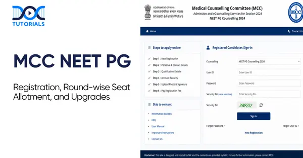
MCC NEET PG 2026: Registration Portal Guide, Round Schedule and Upgrade Options Explained

MCC NEET PG

MCC, or the Medical Counselling Committee, manages the seat allotment procedure in India for medical and dental courses. It is a government organisation that handles admissions to PG, UG, and super-speciality courses through a centralised online process. It allocates 15% of seats in government colleges, 50% of seats for PG courses and the rest in deemed universities. 

The All India Quota seat allocation is done as per the MCC NEET PG scores of the students, their subject preferences, and eligibility for reservation. The counselling is done through online registration, followed by choice filling and seat allotment, completely based on merit.

Here’s more on it!

Ace Your NEET PG Prep With DocTutorials NEET PG Online Live Power Pack

What is the MCC NEET PG Counselling Registration Procedure?

Candidates need to log in to the MCC website and open the NEET PG counselling for new registration. Create your login ID and password by entering your roll number, application number, date of birth, mobile number and email address. Next, you need to pay the counselling fee, opt for choice filling and wait for seat allocation.

Here are the steps to follow for the MCC NEET PG online registration counselling procedure:

Step 1: Visit the MCC Website

First, candidates need to visit the MCC website and select the NEET PG counselling option.

Step 2: Online Registration

Click on the link for “New Registration” and enter the following information to create your login ID and password:

  • Application number
  • Roll number
  • Date of birth
  • Mobile number and email address

Step 3: Pay the Fee for Counselling

Candidates need to pay a non-refundable registration fee and a refundable security deposit using their debit card, credit card, or net banking. After successful payment of the fees, you can participate in the counselling procedure.

Step 4: Choice Filling

After completing the MCC NEET PG counselling registration procedure, you can select the medical courses such as MD, MS, or Diploma and the medical college that you want to attend. You can select multiple options as per your preferences.

Step 5: Choice Finalisation

After making the final choices, the candidates need to lock them in before the deadline. Even if they miss manual locking, the preferences will be automatically locked in the system.

Step 6: Seat Allocation

The counselling body will announce the result of seat allocation as per the NEET PG rank of the candidates, their preferences, seat availability, etc.

Step 7: Reporting to College

After seat allocation, candidates need to download the allotment letter and get a printout of the same before reporting to the institution for document verification and the admission procedure.

What is the Seat Allotment Procedure for MCC NEET PG 2026?

The seat allotment procedure for MCC NEET PG includes six stages. It includes the registration, choice filling and locking for the preferred course and college, seat allocation based on the candidate’s rank, final seat allotment results, and reporting to the college allotted to the candidate.

The MCC allocates NEET PG seats to candidates based on several factors, including the candidate’s rank, preferences, reservation category, and seat availability. As already mentioned, the candidates need to fill out the choices for seats in their preferred colleges and courses. 

Here are the different stages of the seat allotment procedure for NEET PG:

  1. Registration for NEET PG counselling
  2. Choice filling for preferred seats
  3. Choice locking
  4. Seat allocation as per rank
  5. Results for seat allotment
  6. Reporting to the allotted college

What are the Round-wise Counselling Schedule for MCC NEET PG?

There are a total of three rounds for the MCC NEET PG counselling schedule. The first round involves seat allotment to the candidates as per their preference and merit. In the second round, the candidates can upgrade their seats, and the third round involves the allocation of the remaining seats.

The counselling procedure for MCC NEET PG 2026 involves multiple rounds. These rounds involve the following procedures:

  • Round 1

This is the seat allotment round. In this round, candidates are allotted seats in their preferred course and colleges as per their merit.

  • Round 2

This round includes the second phase of seat allotment. Seats not allotted in the first round will be allocated in this phase. Moreover, candidates who want to upgrade their seats are allowed to take part in this round.

  • Mop-Up Round

In the third round, the seats remaining after round 2 are allotted to the candidates.

  • Stray Vacancy Round (if applicable)

If any other seats remain vacant after the mop-up round, the authorities conduct a stray vacancy round. It is an optional round based on the seat allocation in the previous three rounds.

All candidates should stay updated with the counselling schedule for seat allocation and actively take part in all the rounds so that the chances of securing seats for them are increased.

What are the Upgrade Options in MCC NEET PG Counselling?

Vacant seats from the previous round are filled in Round 2 of NEET PG Counselling. Candidates who get a seat allotted in Round 1 can participate for upgrading of their allotted seat Candidates who were not allotted a seat in the previous round can also participate for the available vacancies. The process comprises registration, filling choices, seat allotment results and reporting to the allotted college within the given counselling schedule.

There are two purposes for the counselling in round 2. First, allocating seats to the candidates who have not secured any seat in the first round and allowing upgrades to better options for those who have already been allocated seats in round 1.

Here are the candidates who can participate in round 2:

  • Candidates who were not allocated seats in round 1
  • Candidates who want to upgrade their seats even after getting allocation in round 1
  • Fresh candidates who have missed the registration procedure of round 1, and if MCC allows them to reappear

Here are the considerations for round 1 strategy:

  • For Non-Allottees

This is the second chance for the non-allottees. The round 2 seats are usually available from candidates who have not reported to their allotted colleges in round 1, from round 1 allottees who want to upgrade their seats, and in case of any technical glitches.

  • For Round 1 Allotees

Seat upgradation is often risky as candidates need to surrender their allotted seat before the allotment of Round 2. In case the candidates did not find any better option even in round 2, they will lose their allotted seat and wait for the Mop-up round.

Here is the expected timeline for round 2 seat allotment for MCC NEET PG counselling:

EventsExpected Dates
Opening of RegistrationDate yet to be announced.
Deadline for Choice FillingDate yet to be announced.
Result for Seat AllotmentDate yet to be announced.
Reporting to Allotted CollegesDate yet to be announced.

FAQs about MCC NEET PG Registration

  1. Can I participate in Round 3 using my Round 2 seat as a safety?

No, you cannot attempt to qualify for Round 3 while keeping your Round 2 seat as a backup. According to MCC regulations, your Round 2 seat may be cancelled if you decide to upgrade or participate in Round 3 in the permitted manner.

  1. What happens if I’m given a Round 3 seat but choose not to participate?

If you do not participate in the round 3 seat allotment process, you may not be allowed to participate in future MCC rounds. Moreover,  your security deposit might be forfeited.

  1. Can I re-enter Round 3 if I quit Round 2?

Yes, you can re-register for Round 3 if you adhere to MCC’s resignation process and deadline, but in many situations, you may have to lose your security deposit. Resignation window notices (MCC), for instance, permitted resignations with college-generated resignation letters within certain dates. So, you can try again in Round 3, but it will cost you money.

  1. In 2026, how many rounds of NEET PG counselling would there be?

There are usually three major rounds, followed by mop-up and stray vacancy rounds for the remaining seats.

  1. What will happen if I miss the counselling registration deadline?

A candidate will not be able to register for that round of NEET PG counselling if they miss the counselling registration deadline. Instead, they would have to wait until the following round of NEET PG counselling, if there is one.

Conclusion

The MCC NEET PG counselling involves several steps and rounds. Candidates must stay updated and follow the strict guidelines to secure their preferred seat. However, merit plays a crucial role in allocating your preferred seat.

So, candidates should focus on securing a good score in the NEET PG exam. In this regard, DocTutorials is a reliable online coaching that guides candidates professionally with Quick Revision Program, video lectures, study materials, etc. For more information, join our NEET PG course today!

NEET PG Online Live Power Pack
DocTutorials App Download Now

Latest Blogs

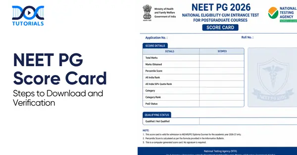
NEET PG Score Card 2026: How to Download, Verify Details and Use it for Counselling

Neet pg score card

Medical graduates who want to pursue the MD, MS, or PG Diploma course in India need to have a qualified NEET PG score card. The NBEMS (National Board of Medical Sciences) conduct the NEET PG exam, which determines the candidate’s eligibility for PG medical admission across government, private, and other reputed universities.

Several MBBS graduates appear for the NEET PG examination every year, and after the result is out, they need to download the NEET PG score board to proceed with counselling, document verification, admission, and further reference.

What are the Steps to Download NEET PG Score Card?

Candidates can download the NEET PG score card from the official website of NBEMS. They need to click on the “NEET PG” option and select the link of score card under the application login. Log in with your ID and password and click on the “Download Score Card” option.

The NEET PG score card is a crucial and mandatory document required for the physical verification and counselling procedure. It includes the detailed metrics of the candidate’s performance and rank as per their category. Here are the steps for the NEET PG score card download:

Step 1: Log in to the official website of NBEMS at nbe.edu.in.

Step 2: Click on the “NEET PG” option on the homepage

Step 3: Select the link for “NEET PG 2026 Score Card” under the section for application login.

Step 4: Fill in the user ID and password that were generated during the registration procedure.

Step 5: After logging in, select the “Download Score Card” option on the dashboard.

Step 6: After downloading the PDF, verify if your photograph and signature are clearly visible on it.

Step 7: Print a minimum of three copies of the score card for counselling, submission to the university, and personal records.

Note: In case of any technical issues while downloading the NEET PG score card, contact 011-45593000, NBEMS candidate helpline. Before calling, keep your admit card handy as you need to verify your roll number. 

What is a NEET PG Score Card?

Issued by the NBEMS, the NEET PG score card is an essential document for the candidates who qualified the exam, reflecting their detailed score and performance. It includes details of the candidates, their roll number, NEET PG score, category-wise percentage, All India rank, qualification status, etc.

The NEET PG score card is a crucial document for the candidates appearing for PG courses. It is issued by the NBEMS to reflect the performance of a candidate in the NEET PG exam. It includes information regarding the candidate’s roll number, score, All India rank, qualification status, and category-wise percentage.

It is mandatory to download this score card for every candidate willing to participate in AIQ (All India Quota) counselling, state counselling, as well as admission in any reputed private institute.

What are the Details Mentioned on the NEET PG Score Card?

Candidates should verify all the details available in their NEET PG score card to avoid any last-minute hassles. The card includes details such as the candidate’s name, roll number, application ID, category, All India Rank, NEET PG score of the candidate, percentage, examination session, and qualifying status.

After downloading the NEET PG score card, you should verify all the information mentioned on it carefully to avoid any further issues. Here are the details that are mentioned on the score card:

  • Name of the candidate
  • Roll number of the candidate
  • Category of the candidate (SC/ST/OBC/UR/EWS)
  • Application ID
  • All India Rank
  • Candidate’s NEET PG score out of 800
  • Score in percentage
  • Examination session
  • Qualifying status

Note: If you find any issues, you should report them immediately to NBEMS.

What are the Documents Required for Verification Post Result?

Candidates need to provide documents such as the NEET PG score card, MBBS degree certificate or provisional certificate, internship certificate, permanent or provisional registration, caste certificate, etc. to seek admission after qualifying the NEET PG 2026 exam.

If a candidate is allotted a seat for a PG course based on their rank in NEET PG 2026, they must provide the original documents as requested by the medical college. If you fail to provide any of the following documents, your admission may be immediately cancelled:   

  • NEET PG score board and admit card
  • Provisional certificate or MBBS degree certificate
  • Certificate of internship completion (it should be completed within the cutoff date, which is generally within June or July)
  • MBBS professional marks the cards of all the years
  • Provisional or permanent registration certificate as issued by the MCI, NMC, or State Medical Council
  •  Valid photo ID, which includes PAN card, Aadhaar card, or passport
  • Higher secondary certificate to prove date of birth
  • Caste certificate for SC/ST/OBC/EWS as per the Central Government prescribed format

How to Use the NEET PG Score Card for Counselling?

You can use the NEET PG score card for counselling in a few easy steps. Download and verify your NEET PG scorecard, checking for errors and whether your score meets category cutoffs. Compare your rank with past opening/closing ranks to shortlist colleges and specialities. Gather original and photocopied documents (admit card, MBBS degree, internship, registration, ID, photos, category/PwD certificates). Enter, prioritise, and lock choices before the deadline, then attend counselling and document verification; participate in further rounds if needed.

The NEET PG score card is necessary for the counselling procedure. Here are the steps for using this card for the counselling procedure:

Step 1: Download the Score Card and Verify

As already mentioned earlier, you need to download your NEET PG score card and verify all the details mentioned in it. Check whether your score meets the cutoff marks set by NBE for your category. In case of any errors, reach out to NBE support for correction.

Step 2: Understand the Cutoff Marks 

There is a cutoff rank for each seat and speciality. Examine your position in relation to the cutoffs for your category and desired specialism from previous years. Select your college and area of expertise based on your rank and the cutoff. Examine the opening and closing rankings for the colleges and specialisations you are interested in from the previous year.

Step 3: Prepare Your Documents

Collect original as well as photocopies of all required documents before counselling begins. Essential items include the NEET PG admit card and scorecard, MBBS degree or provisional certificate, internship completion certificate, permanent or provisional medical registration, a valid government photo ID, caste/category/PwD certificates (if applicable), and passport-size photographs. Having everything organised and readily available speeds up verification and avoids last-minute issues.

Step 4: Fill and Lock Your Choices

Research colleges and specialities thoroughly and prioritise your options into three groups: dream colleges, realistic second choices, and safe backups. Enter your preferences on the counselling portal and lock them before the deadline; timely submission prevents automatic selection of undesired options. 

Attend counselling and document verification when called—seat allocation depends on rank, preferences, and category. If you don’t get a preferred seat in the first round, you can participate in subsequent rounds (round 2, mop-up) to improve your allocation.

FAQs about the NEET PG Score Card

  1. When will the NEET PG scorecard be released?

A few days following the announcement of the NEET PG results, the score card is often made available. The score card download link is activated after NBEMS announces the outcome. For updates, candidates should visit the official website on a regular basis. Candidates should download and preserve the score card as quickly as possible because it is only available for a short time after it is released.

  1. Where can I get my score card for NEET PG?

Only the National Board of Examinations in Medical Sciences’ (NBEMS) official website allows candidates to download their NEET PG score card. The application number and password can be used to access the score card via the candidate login page.

  1. Is the NEET PG score card necessary for counselling?

Indeed, all forms of counselling, including All India Quota (AIQ), state quota counselling, and private or deemed university admissions, require the NEET PG score card. During online registration and in-person document verification, candidates are required to upload or show the score card. Candidates cannot confirm their acceptance to MD/MS programs or take part in counselling without the score card.

  1. How long is the NEET PG 2026 scorecard valid?

The scorecard is only valid for the academic year 2026–2027. Each year’s score card remains valid for that particular session.

  1. What is the qualifying percentile for candidates in the general category?

The qualifying percentage for general category candidates is higher than that for SC/ST. General category candidates should score a minimum of 50% to qualify.

Conclusion

The NEET PG score card is a crucial requirement for admission to colleges and universities for postgraduate medical courses. However, candidates should score good marks to secure admission to reputed colleges. It is essential to get good coaching for that, and DocTutorials serves as a professional guide in this regard.

Our course includes video lectures, Quick Revision Programs, detailed study materials, and other assistance to ensure efficient guidance through reputed faculties. Join our NEET PG course today! 

NEET PG Online Live Power Pack
DocTutorials App Download Now

Latest Blogs

Bed Wetting: Causes, Symptoms, Diagnosis, and Treatment

Bed wetting

Bed wetting (nocturnal enuresis) is a typical disorder that is characterised by involuntary urination during sleep in a child at the age of 5 years or older. Although this is usually a normal stage of development, it can be a sign of an underlying health problem if it persists into adulthood.

Children develop bladder control skills when they are aged 2-4. Beyond the age of 5, chronic bedwetting is referred to as enuresis and should be assessed. Approximately 15% of children aged 5 continue to wet the bed, decreasing to 10% at age 7 and 5% at age 10. Keep reading to know more.

Classification of Bed Wetting

Bed-wetting, or Enuresis, refers to urinary incontinence during sleep in children older than 5 years. It is categorised by timing (nocturnal vs diurnal), symptomatology (monosymptomatic vs non-monosymptomatic), and history (primary vs secondary).

Pediatric guidelines define enuresis as the involuntary intermittent urination in a child ≥ 5 years. Until the age of 5, it is not regarded as abnormal. Enuresis can be categorised as:

  1. Nocturnal Enuresis (Bed wetting): Nighttime wetting. It is the most prevalent type.
  2. Diurnal Enuresis: Waking up with urinary incontinence.

Further classifications include:

  1. Primary vs Secondary Enuresis

Primary enuresis refers to a child who has not attained a dry period of at least 6 months. Secondary enuresis develops when a child, who has been dry at night for 6 months or more, begins to wet their bed. Secondary enuresis can usually be linked to a developing health or psychological concern (e.g., urinary tract infection, diabetes, or stress).

  1. Monosymptomatic vs Non-monosymptomatic

Monosymptomatic enuresis (MSE) is bedwetting in the absence of lower urinary tract symptoms (LUTS) in daytime. Children with MSE do not have urgency, frequency, or incontinence during the day.

Non-monosymptomatic enuresis (NMSE) is associated with nocturnal enuresis that includes other urine symptoms (daytime incontinence, urgency, frequency, voiding postponement, etc.). NMSE usually necessitates assessment of preexisting bladder or bowel issues.

Causes and Risk Factors of Bed Wetting

There are various factors that contribute to bed wetting. Common causes include genetic predisposition, slowed maturation of the bladder, deep sleep patterns and polyuria at night. Psychosocial stress and medical conditions (UTIs, diabetes, sleep apnea) can also contribute.

The aetiology of pediatric nocturnal enuresis is multifactorial with a significant genetic component, and the overall risk is approximately 44% in the presence of one affected parent and up to 77% in the presence of both affected parents.

Family history is highly penetrant, as are environmental and physiological factors. Here are the other factors:

  • Delayed Bladder Maturation: Nighttime bladder control is often delayed in many children because the bladder (detrusor muscle) and sphincter have not developed well-coordinated responses during sleep.
  • Nocturnal Polyuria: Children with enuresis have abnormal ADH (Antidiuretic Hormone) secretion, resulting in excessive urine production at night. The result is bladder overfilling during sleep without getting up to void.
  • Small Functional Bladder Capacity: A small bladder fills quickly at night, and with profound sleep or an inability to wake up, it leads to bedwetting. Capacity <65% of expected (≈30 + age × 30 mL) indicates a small bladder size.
  • Overactive Bladder/Detrusor Overactivity: Other children experience involuntary bladder contractions that result in urgency and incontinence, both day and night. In NMSE, overactive bladder can be the only problem or a component of the dysfunctional voiding.
  • Sleep Disorders: Sleep disorders such as obstructive sleep apnea predispose the risk of enuresis because they result in excessive urine and poor arousal. Children with the disorder of deep sleep cannot wake up even when the bladder is full.
  • Psychosocial Stress: Enuresis is typically an effect of stress rather than a cause, though significant life events may exacerbate symptoms or lead to secondary bedwetting.
  • Medical Comorbidities: Several health conditions may trigger or aggravate enuresis:
    • Urinary Tract Infection (UTI): Irritates the bladder, leading to urgency and accidents.
    • Diabetes Mellitus: An overload of glucose leads to polyuria (urine volume) that can fill up the bladder at night.
    • Constipation/Faecal Incontinence: A rectum that is loaded can compress the bladder, resulting in both day and night wetting. Treatment of constipation tends to lower enuresis.
    • Neurologic Disorders: The bladder control can be compromised by spina bifida, tethered cord, cerebral palsy, or other neurodegenerative diseases. A disordered stream or daytime symptoms imply a neurogenic or obstructive disorder, requiring specialist evaluation.
    • Pinworm Infection: Enterobius vermicularis can cause perianal irritation, which may result in reflex bladder contractions and enuresis (rare).
  • Deep Sleep Arousal Difficulty: Even without OSA (Obstructive Sleep Apnea), just having really good sleep can cause a child to fail to wake to bladder signals.

Signs and Symptoms of Bed Wetting

Bed wetting, or nocturnal enuresis, is characterised by involuntary urination at night, which is often unrealised by the child. It can be linked with daytime urgency or frequency (in NMSE) and psychological outcomes such as embarrassment or low self-esteem.

Nocturnal enuresis is involuntary urination at night in children who are supposed to have control over their bladders. It is often self-limiting and benign, but it can have emotional consequences, which is why early detection is crucial.

Here are the main symptoms:

  • Nighttime Incontinence: The most noticeable sign is waking up with wet pyjamas or bed linens. Frequent wet nights are commonly reported by parents without any daytime problems. The child never wakes up at the time of the episode and notices it in the morning.
  • Daytime Bladder Symptoms: Normal in monosymptomatic cases. Urgency, frequency and daytime incontinence can occur in children in NMSE. Voiding ≥ 8 times/day or <3 times/day suggests dysfunction, along with signs like squatting, leg crossing, or hesitancy.

Other associated signs and symptoms include:

  • Urinary Tract Infection (UTI) Symptoms: Painful or burning urine, hazy or bloody urine, foul smell, fever indicates UTI or other organic origin and should be evaluated immediately.
  • Faecal Soiling or Constipation: Enuresis may be accompanied by large, hard stools or faecal accidents due to pressure on the bladder caused by rectal factors.
  • Behavioural/Psychological Impact: Symptoms of anxiety, embarrassment, social withdrawal (aversion to sleepovers), and low self-esteem may be observed in a child with bedwetting. Parents and peers should be informed that enuresis is involuntary and not the fault of the child.
  • Frequency & Patterns: Frequent, daily, or intermittent (e.g., 23 nights/week). The diagnosis threshold is generally: 2 or more episodes/week over 3 months. Cases of sporadic (less than 2/month) are usually within the period of normal maturation.

Diagnosis of Bed Wetting

Pediatric enuresis is mostly diagnosed clinically. It includes ensuring the enuresis criteria (age ≥ 5, frequency ≥ 2/week for 3 months) and ruling out other causes through history, physical examination, and simple tests. The tools include a bladder diary and urinalysis.

Pediatric enuresis diagnosis is predominantly clinical and requires confirmation of meeting the criteria and ruling out underlying causes. An organised history, examination, urinalysis, and a bladder diary are most likely adequate. Here’s how the diagnosis goes:

1. Initial Assessment and History

  • Confirm Diagnostic Criteria: Make sure the child is at least 5 years old and has persistent involuntary bedwetting (at least 2 times a week) for at least 3 months.
  • Fluid Intake & Voiding Diary: Monitor fluid timing, volume and voiding patterns to determine bladder patterns, particularly evening intake and last void.
  • Family History: Ask about enuresis in the family history; also, check for a family history of diabetes, polyuria, and kidney disease.

2. Medical History and Comorbidities

  • UTI history: Dysuria, fever, frequency; recurrent UTIs can cause secondary enuresis.
  • Constipation: History of hard stools, infrequent bowel movements, or encopresis. Bladder outlet obstruction (functional) can be caused by severe constipation.
  • Diabetes Symptoms: Polydipsia (excessive thirst), polyphagia (excessive hunger), weight loss, or nocturia (getting up at night to use the restroom) should prompt a check of blood sugar levels.
  • Neurologic Symptoms: Gait abnormalities, lower-limb weakness, or spinal problems indicate neural involvement (e.g., tethered cord).
  • Psychosocial Factors: Recent stressors, behavioural issues like ADHD (Attention-Deficit/Hyperactivity Disorder), and autism that could be associated.
  • Sleep History: Snoring, sleep restlessness, daytime drowsiness, or observed apnea indicates sleep apnea.
  • Drug History: Some medications (e.g., diuretics, stimulant medications) may cause polyuria or worsen enuresis.

3. Physical Examination

  • General Exam: Growth parameters, signs of chronic illness. Check for external stigmata of spina bifida (tuft of hair, dimple) on the lower back.
  • Abdominal Exam: Palpate bladder (for retention), assess for constipation (palpable stool in colon).
  • Genitourinary Exam: Inspect for phimosis, labial adhesions, and anatomic abnormalities (e.g., ectopic ureter). Percuss the kidneys if suspicion of reflux.
  • Neurologic Exam: Strength of lower limbs, reflexes; perineal sensation, anal wink to check tethered cord or neuropathy.

4. Investigations

Most children with isolated nocturnal enuresis require only minimal testing. Key investigations include:

  • Urinalysis: Essential first step to exclude hematuria, infection (nitrites, leukocyte esterase), glucose (diabetes), or protein. Repeat if symptoms warrant.
  • Urine Culture: In case of suspected UTI (fever, dysuria, very frequent voiding) or pyuria on urinalysis, obtain a culture and treat the infection.
  • Blood Tests: Not routine, but consider blood glucose in polyuria-polydipsia; potentially renal function in a structural malady.
  • Bladder Diary/Voiding Chart: Record time and volume of each void daily within a few days. This can aid in differentiating polyuria (very large voids) and frequency (small volumes, numerous voids).

5. Imaging

  • Renal/Bladder Ultrasound: Indicated if there are recurrent UTIs, daytime incontinence, abnormal stream, or suspicion of anatomic abnormality (e.g. hydronephrosis, ureterocele).
  • VCUG (Voiding Cystourethrogram): Consider if ultrasound shows reflux or if bladder outlet obstruction is suspected.

Management and Treatment for Bed Wetting

Treatment of bedwetting in children includes education, behavioural measures, and, if needed, alarms or medications. Conservative first-line treatment includes positive reinforcement, fluid management, and treating constipation, and alarms and desmopressin are implemented in case of inadequate positive responses.

A combination of behavioural therapy and a bedwetting alarm is the most effective and safe long-term treatment. Pharmacotherapy (particularly desmopressin) can be effective to enhance the quality of life (dry nights) but often requires repetition/continuation to achieve long-term effect.

Here’s how the bedwetting treatment and management work:

1. General Principles

  • Reassurance and Education: Educate the family that enuresis is normal, mostly self-limiting, and not the child’s fault. Set realistic expectations, as treatment may take months and relapses can occur, and emphasise support over punishment.

2. Behavioural Strategies

  • Fluid Management: Limit evening fluids (particularly 12 hrs before sleep) and avoid caffeine/sodas. Encourage drinking earlier in the day.
  • Timed Voiding: Create an urge to urinate at regular times (after 2-3 hours) in the daytime and right before sleep. Other parents put reward charts for going to the bathroom prior to sleep.
  • Double Voiding: Instruct the child to urinate twice before bedtime (void, then try again a few minutes later) to ensure the bladder empties fully.
  • Constipation Management: Ensure soft bowel movements. Manage constipation (fibre, stool softeners) because a full rectum may initiate bladder contractions.
  • Protect Bedding: Use mattress protectors and easily washable bed linens. Have an extra pair of pyjamas in case of stress around accidents.

3. Lifestyle and Support

  • Engaging the child in problem-solving (seek their ideas). Provide stickers or praise for dry nights and helpful actions (changing bed clothes).
  • Avoid blaming or shaming. Do not wake children aggressively to empty (it has no benefit); use alarms to condition awakening.
  • If school concerns arise (e.g., daytime wetting), communicate with school nursing staff about toilet breaks or discreet pull-up garments, if needed.

4. Pharmacologic Therapy

Medication is reserved for when behavioural methods alone fail or for short-term situations (sleepovers, camps). Options include:

  • Desmopressin (DDAVP): A synthetic analogue of vasopressin (antidiuretic hormone). It decreases nocturnal urine production.
    • Use: First-line medication in monosymptomatic enuresis. It can be taken before going to sleep via nasal spray or oral pills.
    • Efficacy: Reduces nighttime urine output and increases the likelihood of a dry night by about 50% on average. Nevertheless, its effect subsides with discontinuation, hence it is typically intermittent or short-lasting. Not curative, but useful to manage special occasions.
    • Caveat: Risk of water intoxication and hyponatremia if too much fluid is consumed; fluid restriction (no drinking 1 hour before and after dose) is important.
  • Tricyclic Antidepressants (e.g., Imipramine): Previously used widely, now second-line because of safety issues.
    • Use: May be considered if alarms and desmopressin fail. Mechanism includes increased ADH and bladder relaxation.
    • Efficacy: Can increase dry nights by around 1 per week.
    • Caution: Risk of overdose is severe (cardiotoxicity, seizures). Contra-indicated in families where there is a risk of overdose. The use is standard with ECG before use, dose-limited, and short courses.
  • Anticholinergic Agents (e.g., Oxybutynin, Tolterodine): Indicated when symptoms of overactive bladder during the day are observed or when a small capacity of the bladder is reported. They relax the detrusor muscle.
    • Use: If the child has daytime urgency or frequency, or if an ultrasound shows a small bladder. May be added to desmopressin in refractory cases.
    • Side Effects: Dry mouth, constipation, blurred vision; should be used cautiously in children.

Prevention and Home Care for Bed Wetting

Whereas the prevention (in the sense of preventing it before it occurs) of bedwetting is somewhat constrained, since it is basically a matter of developmental timing, home care is about promoting the child’s maturation and preventing triggers.

Prevention of bedwetting is not always possible because it is a developmental complication, but with the support of home care, it can be reduced, and the control of the bladder can be improved.

Some of the important measures include:

  • Habit Training: Educate young children about proper toilet habits early (e.g., sitting posture, completing void).
  • Family Awareness: Discuss family stress and provide a supportive environment (do not make fun of the child about wetting).
  • Bowel Management: Avoid constipation by means of a high-fibre diet. A hard stool should be avoided, as it is a contributor to bladder dysfunction.
  • Sleep Hygiene: Sleep well and still; maintain good bedtime habits (use the bathroom before going to sleep; do not go to sleep right after heavy fluid consumption).
  • When to Seek Help: A medical examination is advised when bedwetting is regular, when the child is concerned about it, or when there are concerns (UTI, daytime wetting, systemic illness).

FAQs about Bed Wetting

  1. At what age is bedwetting considered normal?

Bedwetting most often occurs among younger children and is mostly normal through the age of approximately 5 years. When a child who is 5 years or over experiences frequent episodes (at least twice a week during 3 months), it is diagnosed as enuresis.

  1. What is the difference between primary and secondary enuresis?

Primary enuresis is when a child has never attained a dry spell of at least six months. Secondary enuresis is the reappearance of bedwetting in children who have been dry for six months or more.

  1. How is bedwetting diagnosed?

Clinical diagnosis of bedwetting is made on the basis of age, frequency of wetting, medical history and simple tests like urinalysis to eliminate other causes.

  1. What are the first-line treatments for enuresis?

Some of the initial treatment interventions are lifestyle and behavioural factors, which involve reducing evening fluid intake, scheduling voidings, constipation, and reward systems.

  1. When should alarm therapy be used?

Alarm therapy is recommended if initial measures do not work. It is especially effective among children and families who are eager to adhere to the regimen.

  1. What medications are used for nocturnal enuresis treatment?

The most widely used medication is desmopressin. In certain circumstances, other alternatives, such as imipramine or oxybutynin, nin can be looked into.

  1.  Which is more effective: alarms or medication?

Long-term alarm therapy is better as it trains the child, whereas drugs give short-term relief and are linked with relapse after discontinuation.

  1. Is enuresis a psychological problem?

Enuresis is more of a developmental issue, rather than a psychological disorder, though it can result in emotional distress or conditions like ADHD.

  1. Can bedwetting be prevented?

There is no guaranteed way to prevent bedwetting, but good toilet training, curing the constipation and avoiding taking fluids at night could help minimise the need to consult a doctor.

  1. When should a doctor be consulted?

Consultation with the doctor is required when the child is 5 years old or above and has frequent bedwetting, or in case of other symptoms, namely, daytime wetting, pain, or recurrence after dry days.

Conclusion

Bed wetting (pediatric enuresis) refers to a highly prevalent involuntary urination in sleep. For NEET PG, concentrate on classification, causes, and diagnosis. Behavioural measures, alarms, and medications are part of management; most children outgrow them.

 For additional guidance, DocTutorials can be your study companion. We offer crisp videos, clinical Qbank, exam-focused notes, flashcards, and mind maps. We help ensure aspirants gain complete clarity over every complex medical concept.

Join DocTutorials today and explore our NEET PG course to excel in your medical career!

NEET PG Online Live Power Pack
DocTutorials App Download Now

Latest Blogs

Perineal Tears: Symptoms, Causes, and Treatment

Perineal Tears

Not all childbirth-related complications are widely understood or openly discussed. One such condition is perineal tears. It is a type of injury that affects the area between the vagina and the rectum, commonly occurring during vaginal delivery.

A perineal tear can range in severity from minor skin involvement to deeper tissue damage and is classified into four grades. Sometimes, a surgical intervention known as an episiotomy can be performed to create a controlled opening and assist with the delivery process. Keep reading to learn about its types, causes, and treatment to improve care and recovery for the mother.

What are Perineal Tears?

Perineal tears are injuries to the area between the vagina and anus that commonly occur during vaginal childbirth. They can range from mild skin tears to deeper tissue damage. 

A perineal tear or vaginal tear is a type of injury that may occur during birth. Also referred to as a perineal laceration, it involves a tear in the tissues surrounding the vagina and the perineum. The perineal region, commonly called the perineum, is the area located between the vaginal opening and the anus.

During a vaginal birth, the skin around the vagina naturally thickens and becomes more flexible. This process helps the baby’s head and body pass through more easily. Despite this natural adaptation, tearing is quite common. In fact, up to 90% of women who have a vaginal delivery experience some degree of tearing.

However, several factors can increase the likelihood of a vaginal tear. These include:

  • A larger-than-average baby
  • An early birth that does not give the skin time to stretch adequately
  • The use of instruments such as forceps during delivery

What are the Various Types of Perineal Tears?

Perineal tears are classified into 4 degrees: first-degree (minor skin tear), second-degree (involves skin and muscles), third-degree (extends to the anal sphincter), and fourth-degree (reaches the rectum).

Vaginal tears can be categorised into 4 grades according to severity. The size of the tear defines its type. Here are the various types of vaginal tears:

  1. First-degree Tear: This is the mildest form of tearing. It only infects the external skin around the vagina and perineum and is usually self-healing and does not require stitches.
  2. Second-degree Tear: This is the most frequently occurring type. The tear goes deeper, affecting both the skin and the underlying muscles of the vagina and perineum. Stitches are usually required for proper healing.
  3. Third-degree Tear: This type of tear extends further, reaching from the vaginal area to the anus. It involves damage not only to the skin and perineal muscles but also to the anal sphincter muscles, which control bowel movements. Stitches are necessary for recovery.
  4. Fourth-degree Tear: This is the most severe and least common form of tearing during childbirth. It extends from the vagina through the perineum, affecting the anal sphincter muscles and reaching into the rectum.

In such cases, the healthcare provider may need to perform the repair in an operating room instead of the delivery room.

What are the Causes of Perineal Tears?

Perineal tears can be caused by childbirth, sexual intercourse, shaving, waxing, vaginal dryness, and other conditions that make the skin weak and vulnerable to any type of injury. Other factors that cause perineal tears include having a large baby, quick labour, vaginal birth for the first time, or decreased skin elasticity.

Most of the cuts or tears in the vagina are commonly linked to activities such as sexual intercourse, shaving, or waxing. However, there are many other cases in which someone ends up developing a tear in and around the vagina, including pre-existing skin problems or giving birth.

Here are the primary causes involved:

  • Sexual Intercourse

Sexual intercourse is one of the common reasons why some women develop perineal tears. Lack of proper vaginal lubrication might lead to such an injury. Intense sexual activity and using sex toys might also contribute to tearing the vaginal area.

  • Shaving and Waxing Pubic Hair

Shaving and waxing pubic hair are also common factors that result in cuts on the vulva. According to research conducted in 2017, about 25.6% of those who groomed their pubic hair sustained cuts, most of which were vulvar cuts in women. In some cases, these injuries required medical treatment, such as antibiotics or even minor surgery.

  • Childbirth

About 80% of women experience perineal tearing during childbirth. A perineal tear refers to damage to the skin or muscles of the perineum, which is located between the vagina and the anus. In some cases, the tear could affect the vagina and even the anal sphincter that controls bowel movements.

Women who are giving birth for the first time are generally at a higher risk of experiencing perineal tears compared to those who have previously had a vaginal delivery.

  • Skin Conditions

Certain skin conditions can make the vaginal or vulvar area more prone to cuts and irritation. These include:

  • Eczema is a skin condition characterised by red, inflamed, and itchy skin. Symptoms of eczema include fluid exudation, crusting, and itching in the vagina and vulva.
  • Lichen Planus is an inflammatory disease that causes itchiness, burning, or a feeling of raw skin in the vulva. If the disease affects the vagina, there will be yellowish sticky discharges and tissue destruction.
  • Psoriasis, a common skin condition that is characterised by itchy and dry scales. However, in the vulvar region, Psoriasis manifests as smooth, pink spots with well-defined edges.
  • Lichen Sclerosus, an inflammatory skin disease that primarily affects the genitalia. The affected skin is easily torn or develops fissures.
  • Vulvovaginal atrophy (atrophic vaginitis), a condition that causes the vaginal tissues to become thinner, drier, and less elastic, making tears more likely.
  • Vaginal scarring or tissue damage, which may result from prior surgeries or radiation treatments in the pelvic region.
  • Vulvovaginitis, commonly caused by Candida albicans (also known as thrush), may irritate the tissues and predispose them to cuts.
  • Genital herpes (herpes simplex infection), which may develop clusters of fluid-filled blisters that may rupture and leave tiny open sores that look like cuts.
  • Other Causes

Other factors that may contribute to the formation of vaginal or vulvar cuts include:

  • Foreign body insertion into the vagina
  • Tampon insertion or extraction errors
  • Vaginal dryness
  • Vaginismus
  • Thinning of the vaginal tissue due to age
  • Steroid medication usage

What are the Symptoms of Perineal Tears?

The perineal tear symptoms typically involve pain, burning, swelling, tenderness and discomfort in the vaginal or perineal region, particularly during sitting, walking or during urination. In more serious instances, one might experience bowel or bladder control problems, emotional distress, and pain that impact daily activities and postpartum recovery.

In case of a perineal tear, a variety of symptoms can occur that disrupt everyday comfort and normal activities. It is essential to identify these symptoms to obtain the necessary treatment and aid recovery in a timely manner.

Physical symptoms may include:

  • Perineal pain or discomfort immediately after the tear, which is usually sharp, burning, or tender, particularly during motions or during use of the toilet.
  • Inability to sit, stand, or walk comfortably, which may cause problems in performing routine tasks or taking care of a newborn.
  • Bladder or bowel changes, including difficulty urinating or urgency, especially with more severe tears.

Emotional and functional effects may include:

  • Feelings of anxiety related to healing or concerns about resuming intimacy
  • Difficulty managing daily activities, including maintaining hygiene or caring for a baby
  • Uncertainty about which physical activities are safe during the recovery phase

How are Perineal Tears Diagnosed?

Perineal tears are determined by a careful physical examination whereby the medical practitioner examines the vaginal and perineal tissues to determine the extent of the injury. Reviewing the patient’s symptoms and medical history also helps identify whether the tear is new, recurring, or associated with other underlying conditions.

A healthcare provider usually conducts a physical examination and analyses your medical history to diagnose a vaginal or vulvar cut. This procedure helps them understand your symptoms and identify any underlying conditions or contributing factors that might have caused the injury.

They can also question you about the symptoms, whether they are new or recurrent, to find out whether you have had a history of similar problems. When the cuts are tiny or barely visible, the provider may use a colposcope, a specialised instrument with a bright light, to closely examine and magnify the affected area during the assessment.

Diagnostic Tests

In most situations, additional tests are not necessary to diagnose or manage vaginal or vulvar cuts, especially when the cause is clear and the injury appears minor without signs of infection.

In contrast, tests may be necessary when it is critical to establish whether there are any underlying conditions that might contribute to the injury. Tests that might be required include:

  • Culture: A sample of vaginal discharge is collected by swab and brought to the lab to determine infections such as bacterial vaginosis, yeast infections, or sexually transmitted diseases.
  • Biopsy: A small tissue sample is collected from the tear site for microscopic evaluation to determine the underlying causes of repetitive or persistent tears of the vagina.

What is a Perineal Tear Treated?

Perineal tears are treated based on their severity: mild tears usually heal on their own, whereas moderate to severe tears are treated with dissolvable stitches to repair the damaged tissue. Recovery is supported by pain-relief methods, proper hygiene, hydration, stool softeners, and cooling pads to reduce discomfort and promote faster healing.

The strategy for dealing with a vaginal or perineal tear varies according to the severity of the tear. First-degree tears do not require stitches because they can heal on their own. Second-, third-, and fourth-degree tears require stitches to repair the injured tissues and ensure proper healing.

Dissolvable sutures are used to sew these tears, and the stitches dissolve on their own after six weeks. In the case of the anal sphincter, the medical practitioner may opt for a more complex repair using dissolvable sutures. Some discomfort is expected as the tear heals.

There are some measures that you can take to ease the discomfort and promote rapid healing of the tear:

  • Thoroughly cleanse the area after going to the bathroom.
  • Pat the area dry gently using toilet paper without rubbing or wiping.
  • Maintain adequate hydration to prevent constipation, and, if necessary, use stool softeners.
  • To avoid any pain or irritation of the tear, healthcare providers can advise using cooling pads and sanitary pads after delivery.

FAQs about Perineal Tears

  1. How long does it take for a vaginal tear to heal?

Most women begin to notice relief from the discomfort caused by a vaginal tear within about two weeks. If stitches are required, they generally dissolve on their own within approximately six weeks.

In most cases, there is no need for a follow-up visit to remove stitches or for additional treatment, as healing occurs naturally.

  1. Can a woman have a vaginal delivery in a second pregnancy if she had a vaginal tear previously?

In most situations, having a tear during a previous delivery does not mean it will happen again in future childbirths. Small tears normally heal well, and they do not disrupt subsequent vaginal births.

However, if a woman has experienced a third- or fourth-degree tear before, there may be a slightly increased risk of tearing in subsequent vaginal births.

  1. When should I start pelvic floor physical therapy after experiencing a perineal tear?

Individuals may recover at an individual rate, but most people can start gentle pelvic floor therapy at 6 to 8 weeks after delivery, provided that their healthcare provider approves it.

It can also be helpful to schedule an earlier consultation to discuss recovery strategies and receive guidance on the healing process.

  1. What can I do to prepare my perineum for childbirth and potentially minimise tearing?

Preparation can typically start during the third trimester. Techniques such as perineal massage, specific strengthening exercises, and learning optimal birthing positions may help reduce the likelihood of tearing.

A trained professional can guide you through safe and effective, evidence-based methods tailored to your body’s needs.

  1. Will scar tissue from perineal tearing cause long-term problems?

Scar tissue is a normal part of the body’s healing process. With appropriate care, including physical therapy, it is usually possible to maintain flexibility and prevent tightness or discomfort, ensuring normal function over time.

  1. What are the risks of untreated severe perineal tears?

Unless properly treated, third- or fourth-degree tears can lead to complications like infection, persistent pain, urinary or faecal incontinence, and discomfort during sex. Immediate treatment and correct follow-up are important in the prevention of chronic complications.

  1. How can I manage pain after a perineal tear?

A combination of prescribed drugs, cold compresses or cooling pads, and proper hygiene can be used to manage pain. A sitz bath can also be helpful. Strain should be avoided when passing stool, loose-fitting garments should be used, and sufficient rest should be taken to aid healing.

  1. When is it safe to resume sexual activity after a perineal tear?

The majority of healthcare practitioners recommend waiting until the tear has completely healed before engaging in sexual activity, which is about six weeks following childbirth.

However, this timeline may vary depending on the severity of the tear and individual recovery, so it is best to consult your doctor before restarting sexual activity.

  1. Can perineal tears affect future pregnancies or deliveries?

In general, minor tears do not have any significant impact on future pregnancies or childbirth. Nevertheless, females with more severe tears (third and fourth degrees) might be at an increased risk during subsequent births.

Depending on your medical history, your healthcare provider will assist in identifying the safest delivery option.

  1. Are there any warning signs of infection during healing?

Yes, some symptoms can indicate an infection. These include pain, swelling, redness, abnormal or foul-smelling discharge, fever, or delayed recovery. If any of these signs appear, it is important to seek medical attention promptly to avoid further complications.

Conclusion

Perineal tears are a common childbirth condition that usually heals in a few weeks under good care. While most cases recover completely within 6 weeks of delivery, the severity can vary depending on the degree of the tear. Seeking guidance from a healthcare expert ensures better healing and reduces the risk of complications.

For additional guidance, DocTutorials can be your study companion. We provide clear videos, clinical Qbank, exam-oriented notes, and flashcards. This helps ensure aspirants gain complete clarity over complex medical topics.

Join DocTutorials today and explore our NEET PG course to excel in your medical career!

NEET PG Online Live Power Pack
DocTutorials App Download Now

Latest Blogs

Thermal Injury: Causes, Symptoms, and Treatment

Thermal injury types and cause

Thermal injuries (burns) are tissue damage caused by external heat sources, e.g., flame, hot liquids, steam, chemicals, electricity, or long-term radiation. These injuries are either superficial, first-degree burns, or deep full-thickness (third- or fourth-degree) burns, including those that affect more than one organ system when extensive.

In forensic medicine, the study of burn injuries is important in determining the cause and mode of injury or death. Keep reading to know more about its types, causes, diagnosis and treatment.

Classification of Thermal Injuries

There are 3 types of thermal injuries, classified by depth and mechanism. The depth of burns is superficial to full-thickness and deeper. Mechanisms include scalds, flame burns, contact burns, electrical burns, chemical burns, and radiation burns.

Thermal injuries are classified by the mechanism of injury and the depth (degree) of skin damage. The following table summarises the burn depth and features:

Burn DegreeSkin Layers InvolvedKey FeaturesHealing Time
First (Superficial)Epidermis onlyRed, dry, painful, no blisters; blanches on touch~5–10 days; heals without scarring
Second (Partial)Epidermis + part of dermisRed/moist with intact or ruptured blisters; very painful; blanches2–3 weeks; may scar
Third (Full)Epidermis + dermis + subcutaneousLeathery, white/brown/charred; painless (nerve loss); does not blanch>8 weeks; requires surgery

Fourth-degree burns (deep burns) penetrate all layers of the skin into muscle, bone or organs and result in charring. These are life-threatening and must always be surgically treated.

By mechanism, thermal injuries include:

  • Scalds: Caused by hot liquids or steam (common in children).
  • Flame Burns: Due to open-fire or clothing ignition.
  • Contact Burns: Through contact with hot things (e.g., metal, irons).
  • Electrical Burns: From AC (alternating current) or DC (direct current), producing serious thermal damage along the current path.
  • Chemical Burns: Caused by acids or alkalis; they chemically generate heat on tissue contact.
  • Radiation Burns: As a result of long UV (ultraviolet) exposure (sunburn) or ionising radiation.

Proper classification assists in estimating the total area burned and the treatment area. For example, the “Rule of Nines” divides the body into regions of 9% TBSA (total body surface area) each (e.g. head 9%, each arm 9%).

In children, the “Lund-Browder” chart adjusts for proportion changes. The severity of the burns is also graded (minor, moderate, major) according to TBSA, depth, age, and location.

Pathophysiology of Thermal Burns

Thermal injury causes protein denaturation and forms three zones: coagulation (necrosis), stasis (at risk), and hyperemia (reversible). Major burns cause fluid loss, shock, inflammation, and such complications as infection, respiratory damage, and organ failure.

Cellular damage from thermal energy includes protein denaturation and membrane destruction. Burns leave a characteristic three-zone pattern in the skin. They are:

  1. Zone of Coagulation: Central, irreversibly necrotic tissue.
  2. Zone of Stasis: Surrounding ischemic tissue that may survive or progress to necrosis.
  3. Zone of Hyperemia: Outer area with reversible injury.

Burns are dynamic, meaning the initial injury can worsen over a few hours or days (e.g., the stasis zone worsening). Larger size burns (>20% of body surface) result in a systemic inflammation response. It increases capillary permeability, resulting in excessive fluid loss (burn shock) and intravascular volume depletion. 

Hypotension and hypoperfusion follow if not corrected. Exposure to superheated air or combustion products may also damage the airways and lungs, leading to respiratory failure. Tissue necrosis from burns also impairs the skin’s functions: infection barrier, temperature regulation, and fluid retention.

Loss of these functions leads to a high risk of infection, hypothermia, electrolyte imbalance, and organ dysfunction. Systemically, severe burns can cause hypermetabolism and release of inflammatory mediators (cytokines, prostaglandins), which, in turn, may result in the systemic inflammatory response syndrome (SIRS) and multi-organ failure.

In forensics, extensive burns may also result in poisonous exposures: carbon monoxide (in smoke) and cyanide (in plastics) intoxication are frequent causes of death.

Causes and Risk Factors of Thermal Injury

The burn definition medical professionals use describes the injury as damage to bodily tissues caused by hot liquids, flames, hot objects, electricity, lightning, chemicals and sunlight. The risk is high with the extremes of age, work-related exposures, and substance use, as well as unsafe homes. Burn patterns are used in forensic practice to determine the cause and manner of injury, such as possible abuse or homicide.

Thermal burns arise from many sources of excessive heat. Common causes include:

  • Scalding liquids (hot water, steam, oil).
  • Open flames (house fires, explosives).
  • Hot objects (steam irons, stoves), and sunlight (ultraviolet).

Electrical injury or joule burn produces heat within the body when current passes through tissues, and there are usually entry and exit wounds. However, during a lightning strike, a filigree burns (also known as a Lichtenberg figure, arborescent burn, or feathering) is a distinctive, fern-like pattern on the skin

. Chemical burns (strong acids or alkalis) cause thermal-like injury via exothermic reactions.

Risk Factors

Certain conditions predispose to burns:

  • Age Extremes: Young children and the elderly have thinner skin and slower reflexes to avoid hot sources.
  • Occupational Hazards: Firefighters, industrial workers handling hot materials, and kitchen staff are at greater risk of exposure to flames.
  • Substance Use: The effect of intoxication by alcohol or drugs compromises judgment near fire and hot materials.
  • Domestic Risks: Lack of smoke detectors, unsafe stoves or heaters, and poor supervision of children.

The pattern and distribution of burns are also indicative of a cause in forensic practice. For example, “forced immersion” scalds (symmetrical burns on buttocks and extremities with distinct scald lines) raise suspicion of abuse in children or vulnerable adults.

Necklacing (homicidal burning method using a tyre) is a classic forensic burn scenario. The identification of these patterns aids forensic professionals in determining whether the injuries were deliberate, unintentional, or caused by others.

Clinical Features and Symptoms of Thermal Injury

Thermal burns are characterised by redness, blisters, swelling, charring, and eschar, whereas inhalation injury is characterised by facial burns, soot, and respiratory distress. Severe burns may result in oedema and renal failure.

Thermal burns have varying symptoms based on depth and extent. In partial-thickness (nerve endings are exposed) burns, pain is intense, but in full-thickness burns (nerve destruction), pain can be surprisingly weak. Common signs include:

  • Redness (erythema)
  • Blistering
  • Swelling
  • Charring
  • Eschar formation

In inhalation injuries, one may see:

  • Facial burns
  • Singed nasal hairs
  • Soot in the mouth or sputum
  • Hoarseness
  • Respiratory distress

Fluid shifts cause generalised oedema and often oliguric renal failure in severe cases. In forensic examination of a burned body, several findings distinguish antemortem from postmortem burns:

FeatureAntemortem BurnPostmortem Burn
Skin rednessPresent (due to vasodilation)Absent (no circulation)
BlistersPresent: fluid rich in albumin and chlorideMay occur; contain air/thin fluid
Soot in airwaysCarbon particles in the trachea/lungsAbsent
Carboxyhemoglobin (CO-Hb)Elevated (>10%) from smoke inhalationNormal
Haemorrhage at the injury sitePresent (capillaries intact)Absent (no vital reaction)
Curling’s ulcer (duodenum)Common in fatal burns (stress ulcers)Absent

Other notable forensic findings include “pugilistic attitude” – a flexed posture of burned bodies due to muscle contraction at >60°C – and heat fractures of bones (bones split due to dehydration, often with a distinctive pattern that does not cross suture lines).

Burned bodies also exhibit thermolysis of soft tissues and “parchment” skin. The cause of death in fire victims is often inhalation injury (asphyxia from smoke, CO or cyanide poisoning) rather than cutaneous burns.

Findings in forensic reports can include lung weight, cherry-red tissue discolouration (CO poisoning), and cyanide atomic absorption.

Medical Diagnosis of Thermal Injury

Medical diagnosis of thermal injury involves both forensic and clinical evaluation. Forensic methods help determine the cause, severity, and identity, while clinicians assess burn size, depth, and related complications, such as inhalation injury.

Forensic-medical diagnosis of thermal injuries involves thorough external and internal examination. Investigations may include:

  • Autopsy: Close dissection to determine depth of burns, internal organ injuries, and smoke inhalation.
  • Toxicology: Determining the quantity of carboxyhemoglobin, cyanide, and other poisonous gases formed during combustion is essential.
  • Histopathology: Skin biopsies can confirm the vitality of burns (e.g. neutrophils, red blood cells), extravasation in antemortem burns, and estimate the age of lesions.
  • Imaging: Post-mortem CT (computed tomography) scans of the body could reveal heat fractures or retained foreign material.
  • Forensic Odontology/Anthropology: Dental or skeletal analysis can help identify severely burned remains.

In clinical diagnosis, the healthcare practitioners measure the size and depth of the burns. The “Rule of Nines” or Lund-Browder chart quantifies TBSA. The depth is assessed by appearance, capillary refill (blanching), pain, and sensation. Patients with burns should be evaluated for associated injuries (inhalation, trauma) and for burn severity.

Management and Treatment of Thermal Injury

Thermal injuries are initially managed using the ABC principles (immediately after injury): airway protection, fluid resuscitation using the Parkland formula, and analgesics. Proper wound care, infection prevention, and tetanus prophylaxis are essential. Severe burns are surgically managed and strictly supported, with nutrition and observation of complications.

Initial management of thermal injuries follows trauma protocols (ABCs). Key steps include:

  • Airway and Breathing: Check for inhalation injury. Early intubation in case of respiratory distress or facial burns.
  • Circulation: Start large-bore IV access promptly. Remove constricting clothing/jewellery.
  • Fluid Resuscitation: For major burns, aggressive IV fluid resuscitation is required. The Parkland formula is a standard guideline:

4 mL × body weight (kg) × % TBSA (second/third-degree only) of lactated Ringer’s over 24 hours, with half in the first 8 hours. Adjust fluid to maintain urine output (0.5–1 mL/kg/h in adults).

  • Pain Control: Give analgesics (when necessary- opioids) as burns are painful.
  • Burn Care: Cool the burn with lukewarm (not ice-cold) water to limit depth. Gently clean and debride wounds. Use topical antimicrobials (e.g. silver sulfadiazine) to avoid infection. Cover with a sterile dressing.
  • Tetanus Prophylaxis: Update tetanus immunisation, as burns are prone to tetanus.
  • Wound Management: Superficial burns heal spontaneously. Partially-thickness burns can need debridement and specialised dressings. Full-thickness burns require surgical treatment (excision and grafting).
  • Supportive Care: Monitor for shock (IV fluids, hemodynamic monitoring), provide nutritional support (high-calorie diet), and prevent hypothermia (warm ambient temperature).

Admit to a burn centre if criteria are met (e.g. >20% TBSA, face/hands/genitals, electrical/chemical burns).

Rehabilitative therapy, scar management (pressure garments), and reconstructive surgery are treatment options that may be used on a long-term basis.

Prevention of Thermal Injury

Thermal injury prevention involves fire control, safe kitchen operations, controlled water temperature, and workplace safety precautions. Social consciousness and safety laws help reduce the incidence of burns, although forensic vigilance is important for identifying abuse or arson.

Preventing thermal injuries is critical, especially in high-risk settings. Safety measures include:

  • Fire Safety: Install working smoke detectors and keep fire extinguishers accessible. Practice fire escape plans.
  • Kitchen Safety: Use hot liquids with caution; handle pot handles carefully; be cautious around stoves. Never leave cooking unattended.
  • Water Temperature: Set water heaters to less than 120 F (49 C) to avoid scalds; check bath water temperature.
  • Workplace Precautions: Use protective equipment (gloves, face shields) when handling hot materials or chemicals. Follow electrical safety protocols.
  • Education: Educate children on fire safety, do not play with matches, lighters or fireworks.
  • Sun Protection: Wear sunscreen and protect yourself to avoid radiation burns (sunburn).

FAQs about Thermal Injuries

  1. How are burns classified by degree?

Burns can be categorised as deep: first-degree (epidermis, only, red, painful), second-degree (partial-thickness, blisters, very painful), and third-degree (full, charred, often painless). The fourth-degree burns extend to muscle or bone.

  1. What is the Rule of Nines?

The Rule of Nines is a quick method to estimate burn surface area. In adults: head/neck = 9%, each arm = 9%, anterior trunk = 18%, posterior trunk = 18%, each leg = 18%, perineum = 1%.

  1. How can you tell if burns occurred before death?

Antemortem burns show vital reactions – redness, blisters with protein fluid, haemorrhage at edges, and soot in airways. Blood tests reveal high carboxyhemoglobin and cyanide. Postmortem burns do not have these signs (there is no inflammation or inhaled soot).

  1. What is “pugilistic attitude”?

It is a typical flexed posture of a burned body due to muscle shrinkage in heat (> 60 °C). It resembles a boxing stance and results from heat-stiffened muscles.

  1. How are burns managed initially?

Follow trauma ABCs: secure airway (intubate if needed), support breathing, then start IV fluids. Cool the burn, provide analgesics, clean/debride wounds and place sterile dressings. Use formulas such as the Parkland formula (4 mL × kg × % TBSA) to guide fluid resuscitation.

  1. What is the Parkland formula?

A guideline for fluid resuscitation in major burns: 4 mL per kg of body weight per cent of TBSA burned (2nd/3rd-degree burns) of lactated Ringer’s in the first 24 hours. Half of that volume is given in the first 8 hours after the burn.

  1. Can electric injuries be considered thermal burns?

Electrical injuries cause serious thermal damage along current paths, but they also cause internal organ injury (arrhythmias, muscle damage). When providing a forensic classification, an electrical burn is frequently mentioned in contrast to a thermal burn.

  1. How do chemical burns differ from thermal burns?

Chemical burns occur due to an exothermic reaction with acid or alkali and can extend the damage to tissue until the agent is eliminated. They are treated by copious irrigation and neutralisation, unlike simple cooling for thermal burns.

  1. Why is burn depth painful initially but not in full-thickness burns?

Partial thickness burns reveal nerve ends, resulting in severe pain. Full-thickness burns destroy the nerves within the affected area; the victim might not experience any pain within the burnt area.

  1. What are some key forensic signs of burn-related death?

In addition to vital reactions, there is soot in the airways (from inhalation), thermal fractures of the bone, black lung (from inhaled carbon), and internal signs of systemic poisoning (e.g., cardiac carbonisation, cerebral infarcts due to hypoxia).

Conclusion

Burns can be thermal, chemical, radiation-induced, or electrical, each with distinct forensic and medical consequences. Medical students and forensic trainees must be able to understand burn classification, pathophysiology, and related factors.

In NEET PG forensic medicine, the Rule of Nines, burn depth features, and vital reactions in burns are high-yield topics. For additional guidance, DocTutorials can be your study companion. We offer crisp videos, clinical Qbank, exam-focused notes, flashcards, and mind maps.

Join DocTutorials today and explore our NEET PG course to excel in your medical career!

NEET PG Online Live Power Pack
DocTutorials App Download Now

Latest Blogs

Sarcoidosis: Causes, Symptoms, Diagnosis, and Treatment

sarcoidosis.

Sarcoidosis is a systemic inflammatory disease which is characterised by the formation of noncaseating granulomas in various organs. Typical presentations are bilateral hilar lymphadenopathy and changes in the interstitial lung, although sarcoidosis may occur in the skin, eyes, heart, and other organs.

This NEET-PG pathology review covers sarcoidosis aetiology, clinical features, diagnostic criteria, management, and complications, emphasising information relevant for medical students. Keep reading to know more.

What are the Causes of Sarcoidosis?

The exact cause of sarcoidosis is unknown. It is thought to have an exaggerated response of the immune system to unknown antigens. Risk is genetically predisposed, affected by race and environment.

There is no single known cause of sarcoidosis, and its formation is associated with a mixture of genetic and environmental factors, as well as immune-related factors:

  • Unknown Aetiology: No specific trigger is found. Sarcoidosis is likely an autoimmune response (primarily Th1-mediated) to microbial or environmental antigens. Exposures (beryllium, insecticides, mould), infections (mycobacteria, Propionibacterium acnes) are investigated, but none of them can explain all the cases.
  • Genetic Factors: Genetic predisposition is indicated by familial clustering and HLA (Human Leukocyte Antigen). There are also specific HLA-DR alleles and ACE (Angiotensin-Converting Enzyme) gene variants associated with susceptibility, which point to heritable immune hyperreactivity.
  • Demographics: Most prevalent among adults between 20 and 40 years. It occurs more in African Americans than in whites, and the morbidity among African American females is the highest. Women tend to experience extrapulmonary disease, whereas men tend to exhibit cardiac involvement.
  • Geographic/Seasonal Factors: Seen worldwide but more common in northern latitudes and urban areas. There is some seasonal variation, but there are no definite prevention measures.
  • Immune Factors: Patients have an exaggerated cellular immune response at disease sites, characterised by CD4+ T-helper cell accumulation and inverted CD4/CD8 ratios in affected tissues.

High concentrations of cytokines (IL-2, IL-6, TNF-α, IFN-γ) favour the development of granuloma. These are immunologic pathways which play a central role in the pathogenesis of sarcoidosis.

Pathogenesis and Histopathology of Sarcoidosis

The pathology of sarcoidosis is characterised by an over-response of the immune system, which results in the production of compact noncaseating granuloma in tissues. Granulomas are composed of epithelioid histiocytes and giant cells.

The pathology of sarcoidosis is characterised by the dysregulated immune response, resulting in the appearance of distinct noncaseating granulomas, which are the key features of the disease and may be localised to various organs.

  • Granuloma Formation: The characteristic lesion is the noncaseating epithelioid granuloma, with tight clusters of activated macrophages and Langhans-type giant cells. These do not have central necrosis, although there may be rare fibrinoid necrosis.

The presence of surrounding lymphocytes (primarily CD4+ T cells) and fibroblasts may ultimately result in fibrosis.

  • Cellular Details: Epithelioid histiocytes release enzymes and cytokines, such as ACE, lysozyme, and calcitriol. The overproduction of calcitriol may lead to hypercalciuria and hypercalcemia.

Giant cells are the result of fusion and may include Schaumann or asteroid bodies- nonspecific but supportive findings.

  • Organ Involvement: Granulomas can affect almost any organ, most commonly the lungs and intrathoracic lymph nodes (up to 90%). They may also affect skin, eyes, heart, liver, spleen, bone marrow, and nervous system, affecting normal tissue (e.g., fibrosis in lungs, arrhythmias in heart).
  • Pathological Variants: Granulomas appear in skin lesions (e.g., lupus pernio), but acute sarcoidosis (Löfgrens syndrome) may have erythema nodosum without granulomas. Fibrosis is present in chronic disease, and granulomas in neural tissue in neurosarcoidosis.
  • Non-Specific Findings: Granulomas can increase the ACE levels and lead to hypercalcemia. Pulmonary granulomas and lymphadenopathy tend to be seen with imaging.

Histopathology of sarcoidosis must be interpreted with caution. Since granulomas can be caused by a large number of diseases (e.g. tuberculosis, fungal infections, lymphoma), the most important diagnostic measure is to rule out other causes.

Special stains for acid-fast bacilli and fungi should be negative in sarcoidosis, confirming the granulomas are noncaseating and not due to infection.

Clinical Manifestations or Symptoms of Sarcoidosis

Sarcoidosis symptoms can vary widely depending on the organ affected, and most patients have no symptoms. Respiratory (dry cough, dyspnea), skin (erythema nodosum, lupus pernio), eye (uveitis), and constitutional (fatigue, fever) symptoms are common. Multisystem disease may cause weight loss and lymphadenopathy.

Patients with sarcoidosis may present with any combination of the following features:

  • Respiratory: Mostly impacts the lungs with a result of dry cough, chest pain, and increasing dyspnea. The test can be normal, but bilateral hilar lymphadenopathy is usually seen on imaging. Chronic disease may lead to fibrosis and respiratory failure.
  • Constitutional: Other non-specific symptoms are fatigue, low-grade fever, night sweats, and weight loss. Fever, hilar adenopathy, erythema nodosum, and polyarthritis (Löfgren syndrome) have a good prognosis and generally resolve.
  • Skin: Prevalence of approximately 10 -35%. Involves erythema nodosum and lupus pernio, together with papules or nodules, which can have granuloma on biopsy.
  • Eye: Observed in as many as 50% cases. Involves uveitis, conjunctivitis and copious tears, in which the eyes exhibit signs of redness, pain, and vision alterations. The untreated cases may result in cataracts or glaucoma.
  • Lymphatic: Lymphadenopathy usually occurs, including hilar and peripheral nodes. In systemic disease, splenomegaly and hepatomegaly could be present.
  • Musculoskeletal: It may involve arthralgia or arthritis (usually ankle) and bone lesions. Sarcoid arthritis is generally acute and migratory. Lytic lesions may be rarely caused by granulomas in bone.
  • Cardiac: Cardiac sarcoidosis can either present as palpitations, syncope or heart failure because of granulomatous infiltration of either the myocardium or conduction system. Arrhythmias and heart block are deadly complications; severe cases of these situations require an implantable defibrillator.
  • Nervous System (Neurosarcoidosis): Neurologic involvement may include cranial neuropathies (facial nerve palsy in particular), aseptic meningitis, seizures, or hypothalamic/pituitary dysfunction (resulting in diabetes insipidus). There is also a report of psychiatric symptoms (depression, anxiety).
  • Kidneys/Metabolic: Granulomas may produce excessive 1,25-dihydroxyvitamin D, leading to hypercalcemia and hypercalciuria. Patients can get kidney stones or nephrocalcinosis. Renal sarcoidosis (granulomatous nephritis) is rarer.
  • Others: Parotid gland enlargement (Heerfordt’s syndrome), central diabetes insipidus, and other organ-specific symptoms occur. Nonspecific constitutional features (malaise, fever of unknown origin) are seen in some patients.

Risk Factors and Epidemiology of Sarcoidosis

The incidence of sarcoidosis depends on the population. African Americans and individuals of Scandinavian or Irish ancestry have the greatest risk. The average age of onset is 20-50 years. The involvement of organs varies in women and men. There are no lifestyle factors that are conclusively shown to cause sarcoidosis; thus, prevention cannot be done.

Sarcoidosis shows distinct patterns across populations, with risk influenced by demographic and genetic factors rather than modifiable lifestyle behaviours. This makes prevention challenging, as highlighted in the key risk factors summarised below:

FactorEffect on Sarcoidosis Risk
Age (20–40 years)High risk (peak incidence)
GenderSlight female predominance
RaceMost common in African Americans; Caucasians have lower prevalence; high prevalence in Scandinavia/Japan
GeneticsFamilial clustering; HLA associations (e.g. DR alleles)
Environmental ExposuresImplicated (beryllium, mould), but not proven causes
Lifestyle (Smoking)Not protective; no lifestyle prevention known

Diagnosis of Sarcoidosis

The diagnosis involves a compatible clinical and radiological image and histological findings of noncaseating granulomas, and ruling out other etiologies. The most important are chest imaging (bilateral hilar lymphadenopathy or interstitial disease), supportive laboratory findings (elevated ACE, calcium), and confirmatory biopsy (granulomas without infection).

Diagnosis of sarcoidosis is based on 3 criteria: 

  1. Clinical and radiologic findings consistent with sarcoidosis, 
  2. Histopathologic evidence of noncaseating granulomas in at least one organ, and 
  3. Exclusion of alternative diagnoses (especially infections and malignancies).

The workup typically involves:

1. Imaging Studies

  • Chest X-ray: The classical observation is bilateral hilar lymphadenopathy (BHL), which is evident in most patients. Interstitial lung infiltrates (reticular or nodular patterns) may also be found. Scadding’s stages of sarcoidosis (0–IV) describe CXR progression.

The following tables describe the stages based on X-ray findings:

StageChest X-ray Findings
IBilateral hilar adenopathy only
IIBilateral hilar adenopathy + diffuse lung opacities (interstitial infiltrates)
IIIDiffuse lung opacities alone (hilar nodes regressing)
IVAdvanced fibrosis (reticular/fibrotic changes)
  • High-Resolution CT (HRCT): Gives additional detail; presents micronodules along bronchovascular bundles, fibrosis, and predominance in the upper lobes. In the case of a normal chest X-ray, HRCT is able to identify parenchymal disease.
  • Other Imaging: In case of particular organ involvement, imaging is guided: MRI (brain MRI or heart MRI in case of CNS/cardiac sarcoid), PET scans or Gallium scans to identify some occult inflammation, and so on.
  • Radiographic Staging (Scadding System): The appearances of the chest X-ray are classified into stages that represent the severity of the disease. The staging has a role in prognosis and management.

2. Pulmonary Function Tests (PFTs) 

Most pulmonary sarcoidosis produces a restrictive pattern (reduced total lung capacity, normal or elevated FEV₁/FVC ratio) and reduced diffusing capacity (DLCO). Approximately 10% may show an obstructive component. Reduction in DLCO and exercise desaturation can be used to assess pulmonary hypertension.

3. Laboratory Tests

  • Serum Angiotensin-Converting Enzyme (ACE): Increased in most sarcoidosis patients, indicating granuloma burden. However, ACE has limited sensitivity/specificity. It may serve to aid diagnosis or track activity.
  • Calcium and Vitamin D: Granulomatous production causes hypercalcemia (2-7% of patients) and increases 1,25-dihydroxyvitamin D. Hypercalciuria is more common. The routine check of serum calcium, creatinine, and urinalysis is performed.
  • Other Markers: An increase in alkaline phosphatase could be indicative of hepatic granulomas. Non-specific markers of inflammation (ESR, CRP) can be raised. Serologic tests like ANA or RF may be done to exclude other connective tissue diseases.
  • Tuberculin (PPD) Test: Often negative in sarcoidosis due to anergy. A positive PPD suggests latent TB (and may coexist or mimic sarcoid).

4. Biopsy
A tissue biopsy demonstrating noncaseating granulomas is essential for a definitive diagnosis. The biopsy site is chosen based on accessibility and clinical involvement:

  • Lymph Node Biopsy: In case of peripheral lymphadenopathy, an excisional node biopsy is performed. Transbronchial biopsy or mediastinoscopy can be used to sample mediastinal nodes.
  • Lung Biopsy: Granulomas in pulmonary sarcoidosis are frequently found on transbronchial lung biopsy (via bronchoscopy). Surgical lung biopsy (video-assisted thoracoscopic biopsy) is rarely needed if other sites are uninformative.
  • Skin Biopsy: Easy for cutaneous lesions (especially papules or plaques). The conjunctival or lacrimal gland biopsy can also be done in case of ocular involvement.
  • Other Sites: If these organs show abnormalities, liver, spleen, or nerve biopsies can be performed.

The following table summarises the key pathological findings and important differential diagnoses to consider when evaluating suspected sarcoidosis:

CategoryCondition / FindingKey FeaturesHow to Differentiate / Rule Out
Key Pathology FindingNoncaseating epithelioid granulomasMacrophages (epithelioid histiocytes), giant cells and lymphocytes; no necrosis; no organisms on stain.Confirm with biopsy; ensure absence of infection using special stains (AFB, PAS, GMS)
Infectious CausesTuberculosis & atypical mycobacteriaCaseating granulomas are more common; systemic symptoms like fever and weight loss are also present.AFB stain, culture, and PCR for mycobacteria
Fungal infections (Histoplasmosis, Coccidioidomycosis)Sarcoidosis may look like granulomas; an endemic history of exposure.PAS, GMS stains; fungal cultures/serology
Occupational DiseaseBerylliosisSimilar granulomas; history of beryllium exposure (industry-related)Exposure history, beryllium lymphocyte proliferation test
MalignancyLymphoma / metastatic cancerLymphadenopathy, systemic symptomsBiopsy with malignant cells (not granulomas)
Autoimmune / VasculitisGranulomatosis with polyangiitisNecrotising granulomas, vasculitis, renal involvementANCA testing, biopsy showing vasculitis
Interstitial Lung DiseasesHypersensitivity pneumonitisExposure-related, diffuse lung involvementHistory of antigen exposure, imaging patterns
Idiopathic pulmonary fibrosisProgressive fibrosis without granulomasHRCT pattern (UIP), no granulomas.
Other CausesCat-scratch diseaseInfectious granulomas, lymphadenopathySerology for Bartonella
Foreign body reactionGranulomas around foreign materialHistory, biopsy showing foreign material

Treatment and Management of Sarcoidosis

Many cases of sarcoidosis are mild and self-limited, requiring only observation. Corticosteroids are used as the main therapy to treat symptomatic or progressive disease. Chronic or refractory cases are treated with steroid-sparing agents (methotrexate, azathioprine).

The sarcoidosis treatment depends on disease severity, organ involvement, and symptoms. These principles are mainly to treat patients with organ-threatening/severe disease and monitor those with mild, asymptomatic involvement. Key points include:

  • Observation: In asymptomatic or limited disease (e.g., Stage I), observation without treatment is appropriate. Approximately two-thirds spontaneously remit in 2-3 years. Follow-up is frequent, which involves clinical examination, chest X-rays, and pulmonary function tests.
  • Corticosteroids: Systemic corticosteroids (e.g., prednisone) can be used as first-line treatment of active disease. Recommended in symptomatic pulmonary disease or severe extrapulmonary progression (ocular, cardiac, CNS, hypercalcemia, renal).
  1. Dosage: Typically starts at 20–40 mg prednisone daily (≈ 0.3 — 0.6 mg/kg). In 4-6 weeks, the response is again checked, and the dose is reduced to maintenance (10-20 mg/day). Depending on the response, treatment can last from 6 months to 1–2 years.
  2. Response and Monitoring: Signs of improvement include relief of symptoms, improved imaging, and normalised labs. Tapering frequently results in relapses, and treatment is balanced to control the disease and reduce steroid toxicity.
  • Steroid-Sparing Agents: In cases of intolerance to steroids or in the case of chronic treatment, other immunosuppressive agents are used:
  1. Methotrexate: A folate antagonist that suppresses immune activity. The most typical dose ranges between 10 and 20 mg per week. Methotrexate is prescribed to treat lung, skin, and joint disease and is commonly the initial steroid-free treatment option.
  2. Azathioprine: A substitute antimetabolite used in some cases where methotrexate is contraindicated.
  3. Leflunomide: A less commonly used alternative to methotrexate.
  4. Hydroxychloroquine: Useful in hypercalcemia, skin lesions, arthritis and mild disease.
  5. Mycophenolate mofetil: An additional alternative in refractory cases.
  • Biologic Agents: Biologic therapies with TNF-α have been tried in refractory or life-threatening instances (e.g. severe cardiac or neurosarcoidosis):
    1. Anti-TNF Therapy: Infliximab (IV) and adalimumab (SQ) block tumour necrosis factor-α, a cytokine central to granuloma formation. They have demonstrated their use in chronic sarcoidosis, in which the conventional agents are not effective.
    2. Others: New treatments encompass monoclonal antibodies against IL-6 or IL-12/23, which are experimental.
  • Organ-Specific Management:
    1. Ocular: Uveitis and ocular inflammation are treated with topical (eye drops) and/or systemic steroids to avoid loss of vision. Consultation in ophthalmology is necessary.
    2. Cardiac: Systemic steroids are initiated in case of cardiac involvement; pacemaker or defibrillator implantation may be required in arrhythmias.
    3. Neurosarcoid: Methotrexate or infliximab may be used in combination with high-dose steroids. Involvement of neurology is typically required.
    4. Hypercalcemia: Hydration and steroids (or antimalarial drugs) to reduce vitamin D activation and lower calcium.
  • Supportive Care:
    1. Pulmonary Rehabilitation: Exercise can help manage breathlessness.
    2. Eye and Heart Monitoring: It is recommended that regular ECGs and ophthalmologic exams, even in asymptomatic patients, be conducted because of the risk of silent involvement.
    3. Lifestyle: Patients are to avoid tobacco and sun exposure (to reduce calcium photosynthesis). Immunosuppressive therapy requires vaccinations (flu, pneumococcal).

Complications and Prognosis of Sarcoidosis

Sarcoidosis has a favourable prognosis; lots of patients experience spontaneous remission. Chronic disease, however, may lead to serious problems. The most dreaded are pulmonary fibrosis and hypertension, loss of sight, heart block, and brain impairments. In untreated or severe cases, mortality ranges from 3-5%.

Sarcoidosis is usually prognostically favourable, depending on the severity of the disease, the organ involvement, and timely treatment, with a probability of chronic complications in some patients:

  • Outcomes: Approximately 60-70% achieve remission in 2-5 years, particularly in Löfgren syndrome. Beyond 2-3 years, the disease is more likely to be chronic.

The majority of chronic cases result in mild-moderate disability, with permanent damage, which occurs in 10-12% of cases. Life expectancy is typically normal with low mortality (1-5%).

  • Pulmonary Complications: Chronic disease can lead to lung fibrosis, bronchiectasis and progressive respiratory failure. Pulmonary hypertension may develop, and end-stage disease may need lung transplants.
  • Cardiac: Additional concerns include myocardial involvement, cardiomyopathy, arrhythmias, and heart block, followed by a risk of sudden cardiac death.
  • Ocular: Unchecked chronic uveitis can lead to glaucoma, cataracts or vision loss.
  • Neurologic: Neurosarcoidosis may lead to meningitis, seizures, hydrocephalus or persistent cranial nerve palsies with potential irreversible deficits.
  • Other Complications: May involve the liver, hypercalcemia-induced kidney stones, and steroid-induced diseases such as osteoporosis, diabetes, and infections.
  • Mortality: Sarcoidosis rarely causes death, with the total mortality in untreated patients approximated at 3–5%. When it happens, it is normally the result of respiratory failure, cardiac complications or immunosuppressed infections.

FAQs sbout Sarcoidosis

  1. What is sarcoidosis?

Sarcoidosis is an inflammatory systemic disease that is characterised by noncaseating granulomas. It is most likely to hit the lungs (bilateral hilar lymphadenopathy) and can include the skin, eyes, heart, and other organs. Its cause is not known, and it has no definite cure.

  1. What causes granuloma formation?

Granulomas are the consequence of a hyper-immune reaction consisting of T cells and macrophages. This is driven by cytokines (TNF-α, IFN-γ, IL-2). Possible triggers include environmental antigens, infections, and genetics, though none are proven.

  1. How is sarcoidosis diagnosed?

Diagnosis involves biopsy of noncaseating granulomas without necrosis or infection (negative AFB/fungal stains) and clinical and radiological correlation to exclude others.

  1. What is Löfgren’s syndrome?

A mild acute sarcoidosis is characterised by a fever, bilateral hilar lymphadenopathy, erythema nodosum, and usually ankle arthritis. It prognoses very well and normally heals in 6-24 months.

  1. What lab abnormalities are seen?

There are increased ACE, hypercalcemia, and occasionally dysfunctional liver enzymes. Blood samples can be associated with mild anaemia or leukopenia.

  1. What are the main treatments?

Corticosteroids (e.g., prednisone) are used as the first-line treatment. In severe cases or cases resistant to treatment, immunosuppressants (methotrexate, azathioprine) or biologics (e.g., infliximab) are added. Mild cases might only require observation.

  1. What is the prognosis?

In mild and acute cases, about 60-70% achieve remission. Chronic disease may cause organ damage in 10–20%. The overall mortality is low (1 to 5%), and cardiac or neurologic involvement aggravates the outcomes.

  1. Can sarcoidosis be prevented?

No. The reason is not known, and this is impossible to prevent. General lung health might be improved by avoiding irritants such as smoke or dust, but it cannot prevent disease.

  1. What tests and imaging are used?

Primary tools are chest X-ray and CT. PET scans, MRI (heart/brain), pulmonary function, etc., assist in the determination of the extent and severity. Biopsy remains confirmatory.

  1. How often is monitoring needed?

Follow-up (after 3 to 6 months) is recommended with clinical examination, imaging, and lung tests. Eye tests and ECGs (electrocardiograms) are recommended due to potential silent involvement.

Conclusion

Sarcoidosis is a systemic inflammatory disease which is based on noncaseating granuloma and has an ambiguous aetiology. The topic is significant to NEET-PG because of its unique pathology, diverse manifestations, and a clear approach to diagnosis and treatment (corticosteroids and immunosuppressants).

DocTutorials help students use advanced technology and learning methods to grasp the syllabus effectively. Our online video lectures, Qbanks, exam-focused edition notes, flashcards, and mind maps help them practice strategically and prepare for exams.

Join DocTutorials today and explore our NEET PG course to excel in your medical career.

NEET PG Online Live Power Pack
DocTutorials App Download Now

Latest Blogs

NEET PG Online Live Power Pack
DocTutorials App Download Now