neet-pg Archives - Page 7 of 20 -

INI CET Toppers: Rank Holders, Scores & Success Strategies

INI CET Toppers

INI CET toppers have once again set a benchmark of excellence in braving one of the most challenging entrance exams in India to get admission to postgraduate medical courses. After the declaration of the INI-CET result for November 2025, aspirants are looking forward to gaining valuable insights from the toppers that they can implement in their preparation strategies. 

What separates the toppers is not the number of hours they put in, but rather a well-thought-out strategy for handling mock tests, revisions, and stress management. Each top rank tells a story of informed decisions, timely revisions, and perseverance during moments of exhaustion and self-doubt.

Continue reading to uncover the rank-wise toppers, their scores, and the proven strategies that can shape your own INI CET success story!

INI CET 2025 Toppers List

This section offers a comprehensive listing of the top scorers in the INI CET sessions for the Medical and MDS courses. It showcases the remarkable percentiles and ranks attained by candidates who have set new benchmarks in one of the most competitive postgraduate medical exams in India.

The announcement of the topper list for INI CET 2025 highlights the performance of candidates who achieved the highest ranks in this highly competitive exam. Here we have provided detailed information on the toppers of the INI CET 2025 sessions:

INI-CET November 2025 – Top Rankers 

RankStudent NameRoll No.Percentile
1Dr. Sairam100
2Dr. Tarun7,916,823100
3Dr. Abhinav Agarwal7,963,930100
4Dr. Ajinkya7,920,855100
5Dr. Harshit7,947,399100
6Dr. Sumeet7,968,260100
7Dr. Sai Trisha7,968,29199.9929981
8Dr. Nihit7,919,18299.9877468
9Dr. Kavya796251299.9877468
10Dr. Shreyas Agarwal793008299.9877468

2025 INI CET topper list (Jan session)

AIRRoll NumberCandidate’s nameGenderCategoryPercentileState
17600048Dr. NetreshMaleGeneral100
27605327Dr. SoumyadityaMaleGeneral99.998West Bengal
37623788Dr. RaviMaleGeneral99.996
47636631General99.995
57663318Dr. SayaniFemaleGeneral99.993West Bengal
67629831Dr. AkhilMaleGeneral99.991
77665147Dr. RohanMaleGeneral99.989
87661629Dr. DeveshMaleGeneral99.989
97657374Dr. HamzaMaleOBC99.986
107609976General99.986

INI CET 2025 topper list (MDS course- Jan session)

All India RankRoll NumberCategoryPercentile
17605011General100
27660939General99.982
37617181OBC99.963
47647970General99.945
57641120OBC99.926
67637771General99.908
77652926General99.889
87649537General99.889
97612933General99.852
107616077OBC99.834

INI-CET November 2025 Toppers’ Success Stories

Dive into the personal journeys of top rankers like Dr. Abhinav and Dr. Sumeet to understand the commitment needed to achieve top results. The experiences of these rank holders will help us understand how they have handled their internships, addressed weaknesses in their subjects, and developed the mental toughness to turn pressure into results.

Every successful rank obtained in the November 2025 INI-CET examination has its tale of struggle and hard work. The success stories of the aspirants not only include ranks and percentages but also experiences that help understand the journey to becoming INI CET toppers.

  1. Dr. Abhinav – AIR 3

Dr. Abhinav’s achievement in securing an All India Rank of 3 is a reflection of his planning and composure in his preparation strategy. Rather than beginning from scratch, he sharpened and honed his already accumulated knowledge from years of dedicated hard work by pouring it all into one final surge. Despite anticipating that the INI-CET exam would be tough, he approached it with the mindset that it was “another Grand Test,” which reduced his stress by reminding him of his reliance on the solid foundations established during his MBBS. 

Recognising Surgery as a personal weak area, he proactively made it a central focus of his early revision cycle. An important part of his preparation was his ability to resist being stuck on confusing questions; instead, he quickly shifted his attention to other questions to save his mental energy for those questions in which he was confident. 

During the final month of preparation leading to his exams, his intense activities were centred on doing previous years’ questions (PYQs), taking Grand Tests, and systematically reviewing his weaker subjects until he achieved mastery. 

  1. Dr. Sumeet – AIR 6

Dr. Sumeet’s journey to securing AIR 6 is marked by a disciplined approach to building knowledge and an unwavering focus on strengthening his core fundamentals. His earlier preparations for the USMLE had already provided him with a solid foundation in both preclinical and clinical topics. However, when he devoted himself completely to PG entrances in India, he realised that conceptual understanding would be the key to achieving a high rank. 

He adopted a methodical approach to ensure that he attained conceptual understanding through the use of video lectures to reinforce areas such as Pathology and Microbiology. He places significant credit for his success on Grand Tests, which he analysed with meticulous detail on the very same day he took them. This enabled him to unlock the logic behind each option, correct as well as incorrect. 

When he felt low on motivation or energy, the LR/LRR (Last Revision/Last Resort Revision) activity served as his lifeline. His achievement of AIR 6 is a testament to the effectiveness of a disciplined approach towards preparation.

  1. Dr. Ajay – AIR 11

Dr. Ajay’s story honestly embodies the distinct challenges of preparing for a competitive exam while navigating the unpredictable demands of a medical internship. With irregular timings and unpredictable postings, he decided to synchronise his study routine with the unpredictability of the internship. 

He made the decision to study in a way that connected what he was learning at the ward with what he was studying for the exams. For example, he focused on Paediatrics while he was in the Paediatrics ward and on Gynaecology while he was in the OB-GYN ward. 

His last six months were entirely devoted to revisions, PYQs, and Grand Tests. He further reinforced his learning by forming a study group consisting of three members, wherein they would discuss each Grand Test held on the very same day, ensuring that the error is not merely marked but understood thoroughly.

In the final stretch, he regularly employed LR/LRRs to master the memory-intensive subjects of Pathology and Microbiology until he felt he knew them entirely by heart. Dr. Ajay’s experience proves that an internship need not be a disadvantage if one has clean fundamentals and executes a sharp, focused revision strategy.

  1. Dr. Anjali – AIR 16

Dr. Anjali’s success in securing AIR 16 is a result of her excellent understanding of her strengths, weaknesses, and the specific changes needed for improvement. Despite securing a strong rank in the earlier session, she remained determined to push herself and thus made a difficult choice to re-attempt the test. 

Her preparation relied on a two-tiered approach: first, developing a conceptual foundation by viewing the primary subject video lectures, and subsequently, using the LRR sessions for a high-yield consolidation exercise. 

She included Grand Tests in her practice regimen, with a rigorous review session: first reviewing all incorrect responses, then reviewing unattempted questions, and finally reviewing all questions for a complete understanding, regardless of whether they were answered correctly. 

Her personal learning approach included a series of intense, small revision sessions instead of lengthy subject marathons, a strategy that kept her on track throughout the internship period and the intense revision phase.

  1. Dr. Bhavek – AIR 19

Very few journeys in the world of competitive examinations are as heartwarming and strong as Dr. Bhavek’s. Having achieved an elite rank in the past, he had joined the Dermatology course in the prestigious PGI, Chandigarh. However, in the midst of this, he felt the stream did not resonate with his desired and actual identity as a doctor. Showing immense courage and conviction in his decision, he made one of the toughest decisions to leave it all and begin again, which was wholeheartedly supported by his parents. 

With the same passion to achieve his goal, this time in the stream of Radiology, his strategy was extremely targeted. His preparation this time was largely focused on the question-based approach, extensively involving Grand Tests and PYQs. He mastered the art of compiling vast subjects into concise notes using Rapid Revision and LRR sessions, which he then transformed into a summary for easy and repeated review. 

During the final, high-pressure phase of his preparation, when active studying felt draining due to anxiety, he smartly switched to passive learning modes. By listening to LRR sessions, he was able to continue absorbing information effectively without experiencing burnout, showcasing adaptability in his study methods.

Toppers’ Proven Tips for INI CET Success

Success in the INI-CET needs organised habits like learning from standard resources, creating concise revision notes, and conducting thorough analyses of mistakes. The toppers have further added that dedicated study hours and taking practice tests that mimic actual test conditions are more beneficial than last-minute preparation.

Cracking INI-CET is not about doing everything, but it is about doing what is right and doing it consistently. Based on learning and understanding from top rank holders, the following tips can be followed:

  • Adhere to Standard Sources: The top performers spend time learning from limited and standard sources rather than switching between books. Repetitive revision of standard sources helps build conceptual understanding and avoid confusion at the end.
  • Make Concise Revision Notes: Short, well-structured notes become invaluable in the final month. Toppers rely on crisp formulas, algorithms, and facts that can be revised multiple times without mental fatigue.
  • Try Daily MCQs: Doing MCQs regularly enhances recall and accuracy. It will also help to identify weaknesses, which can then be corrected before the exams.
  • Simulate a Real Exam Situation: The full-length grand test, conducted in a strict time slot of three hours, trains the candidates for speed and endurance. Toppers consider the mock test as a real exam and train accordingly.
  • Track and Analyse Mistakes: Maintaining an error log is a common habit among rank holders. Revisiting mistakes prevents repetition, strengthens weak concepts, and turns errors into scoring opportunities.
  • Stay Consistent Over Long Hours: Consistency is more valued than studying for longer hours. According to most toppers, 6-8 hours of quality studying are more valuable than 12 hours of inefficient studying, especially during the final stages.

These are proven study habits that show that achieving success at the INI CET involves having clear, consistent, and deliberate decision-making, not last-minute cramming.

FAQs about INI CET Toppers

  1. Where can I check my INI CET result?

The candidates can check their INI CET result on the official examination website for AIIMS, aiimsexams.ac.in. The result will be released in the form of a PDF, containing the roll numbers, ranks, and qualifying status of the candidates. It is always a good practice to check the website for any updates regarding the result.

  1. What is the next step after the INI CET result announcement?

After the announcement of the result, the eligible candidates have to undergo the online counselling process organised by AIIMS. This consists of steps such as registration, filling choices for institutes and specialities, allocation of seats according to merit and choices, and then document verification.

  1. How do I download the topper list for the INI CET exam?

The merit list of INI CET toppers is typically published simultaneously along with the notification of the official result declaration or as a separate PDF download from the AIIMS website. Candidates can download it directly from the results section.

  1. How important are PYQs for INI-CET preparation?

PYQs are also very important for INI-CET preparation, as they help identify patterns and concepts that are repeatedly tested. Toppers also make extensive use of PYQs as a practice and revision exercise to enhance concepts and exam-oriented thinking.

  1. Can INI-CET be cracked during internships?

Yes, cracking INI-CET during internship is absolutely possible. Many toppers aligned their study subjects with clinical postings and focused primarily on revision, PYQs, and grand tests. A structured plan and efficient use of limited time are key to balancing hospital duties and exam preparation.

  1. What is a good rank in INI-CET? 

A good performance is usually indicated by a score of 160+ out of 200 in INI CET 2026. A performance at such a level is sure to enhance the prospects for securing a postgraduate course at the top medical institutions in India.

  1. How can I crack INI CET in one month?

A one-month plan needs to be sharp and focused. Build your strategy around core concepts, solve as many MCQs as you can, and regularly take timed tests using previous years’ papers. Create a structured daily schedule and make sure you leave time for revision.

  1. Is two months enough to crack INI CET?

Absolutely, two months can be quite enough if you make the best out of it! Set a schedule on how you will utilise the time, focus on key topics, and rely heavily on solving previous years’ questions.

  1. What is an INI CET topper’s strategy?

NEET toppers are not just reading the material and then moving on. They are developing an in-depth understanding by revisiting topics through active methods. That means testing themselves without notes (active recall) and mixing subjects while studying.

  1. How many hours do INI CET toppers sleep?

Despite the heavy study load, most toppers ensure they get 6 to 8 hours of sleep each night. They understand the need to sleep well, as it ensures the mind is fresh, the brain can concentrate, and memory is retained.

Conclusion

The INI-CET toppers have shown that success in one of India’s toughest postgraduate medical entrance exams is rarely accidental. Their journeys underline a powerful truth: top ranks are built on strong fundamentals, smart resource selection, relentless revision, and calm execution under pressure. Whether it was refining weak subjects, analysing every grand test, or staying consistent during internships and low-energy phases, each topper followed a personalised yet disciplined strategy.

Looking for guidance to secure a top rank in the INI-CET exam?

Trust DocTutorials. Our concise video lectures, high-yield notes, PYQs, mock tests, and expert faculty help take your preparation to the next level. Check out our INI-CET preparation courses today!

NEET PG Online Live Power Pack
DocTutorials App Download Now

Latest Blogs

Clinical Questions for NEET PG: High-Yield Case-Based MCQs for Top Scores

Clinical Questions for NEET PG

Preparation for the NEET-PG exam is no easy task, as it not only tests the memory of the aspirant but also the understanding of concepts and the ability to make decisions. With the pattern of the exam leaning more towards case-based/clinical questions every year, it is essential that the aspirant doesn’t merely mug up but also applies concepts to situations. One of the best ways to achieve this would be to practice high-yield clinical questions for the NEET-PG exam.

By doing so, you can assess trends, understand your level of preparation, and develop the ability to come up with the most accurate answer in the shortest possible time. In this guide, we will examine high-yield clinical questions for the NEET PG exam and strategies for solving them. 

Read on!

What Constitutes a “Repeatable” Clinical Scenario?

Repeatable scenarios are “exam archetypes” defined by three key elements: a clear diagnostic aim, pathognomonic signs, and a specific patient profile that appears consistently across exams.

The question writers of NEET PG work within specific, predetermined parameters. They demand clear-cut, clinically significant, and testable scenarios in a single stem. Specific predictable patterns are thereby generated.

A repeated scenario has three parts: a specific diagnostic or treatment goal; ‘pathognomonic findings’ (signs you can’t ignore); and a ‘typical patient profile’ (age, occupation, habits). These ‘exam archetypes’ include ‘the 45-year-old smoker who likes going out to nightclubs,’ which will make an appearance every year.

This does not signify laziness on the part of the examiners. They are testing your skill at recognising these textbook presentations that are required for all doctors. Therefore, it is helpful for you to develop a mental library of these archetypes.

Clinical Scenarios with High-Yield System-Wise Patterns

There are several clinical scenarios across major organ systems that are repeated almost every year in NEET PG. They may include radio-femoral delay in Cardiology, Courvoisier’s law in the Digestive System, Smoker with Haemoptysis in the Respiratory System, etc. 

The clinical questions in the NEET PG exam have some typical patterns when it comes to the body systems, assessing the skill of the examinee in quickly isolating the most critical points. Being aware of the high-yield clinical scenarios in the body systems helps the aspirant tackle the case-based MCQs confidently.

Here are some of the major ones to look out for:

  1. Cardiovascular Scenarios
  • Young Patient with Severe Hypertension: A 25-year-old presenting with a blood pressure of 180/110 mmHg, along with radio-femoral delay, strongly points toward coarctation of the aorta. Commonly tested follow-up areas include the “figure of 3” sign, associated bicuspid aortic valve, and rib notching on imaging.
  • Sudden Severe Chest Pain with Inequal Pulse: In the case of a middle-aged male with severe tearing pain radiating from the chest to the back, along with the presence of unequal blood pressure between the two arms, the diagnosis of aortic dissection should be considered. Focus areas include the Stanford classification and radiological findings, such as a widened mediastinum.
  • Irregular Pulse in the Elderly: An elderly patient with palpitations and varying pulse volume indicates an irregular pulse, classically seen in atrial fibrillation. Questions usually revolve around rate control vs rhythm control and CHA₂DS₂-VASc scoring.
  • Syncope Due to Exertion: In a young athlete, the loss of consciousness during exercise should always be regarded as due to hypertrophic cardiomyopathy unless otherwise confirmed. ECG changes include the presence of deep Q waves in the lateral leads, indicating left ventricular hypertrophy.
  1. Digestive System Scenarios
  • Alcoholic with Upper GI Bleed: An alcoholic patient with haematemesis is a clue to oesophageal varices and portal hypertension. Prepare for questions on terlipressin use, band ligation vs. sclerotherapy, and Child-Pugh classification.
  • Painless Jaundice: A patient with weight loss, increasing jaundice, and the presence of a palpable gallbladder in an elderly patient exemplifies Courvoisier’s law and the possibility of periampullary carcinoma.
  • Acute Abdomen with Free Gas: Air under the diaphragm on an erect X-ray is characteristic of a perforated peptic ulcer. The candidates should be aware of the various presentations of pneumoperitoneum, as well as the “saddle bag” sign.
  • Young Adult with Bloody Diarrhoea: Differentiation between ulcerative colitis and Crohn’s disease is essential, continuous lesions versus skip lesions, crypt abscesses versus granulomas. Comparison tables are commonly tested.
  1. Respiratory Scenarios
  • Smoker with Haemoptysis: A chronic smoker coughing up blood is likely to have central lung cancer, which can be squamous cell carcinoma. Evaluations usually involve looking for paraneoplastic syndromes like hypercalcemia in squamous cell carcinoma, as opposed to SIADH in small cell carcinoma.
  • Occupational Lung Disease: Exposure to bat droppings in caves points to histoplasmosis. Such occupational associations include silicosis with stone cutters, asbestosis with shipyard workers, and byssinosis with cotton mill workers.
  • Evening Fever in an Immigrant: Weight loss, infiltrates in the upper lobes of the lungs, and an increase in body temperature in the evenings are classic presentations of tuberculosis. One needs to be aware of the distinction between the primary tuberculosis and post-primary tuberculosis sites.
  1. Paediatric Scenarios
  • Cyanotic Newborn: A newborn with cyanosis that does not improve with oxygen indicates transposition of the great arteries, whereas Tetralogy of Fallot presents with cyanotic spells during crying.
  • Recurrent Pneumonia in a Child: Bilateral recurrent pneumonia suggests cystic fibrosis, while unilateral (right-sided) pneumonia points toward foreign body aspiration. Sweat chloride >60 mEq/L confirms cystic fibrosis.
  • Infant with Failure to Thrive: Formula-fed infants may develop cow’s milk protein allergy, while celiac disease appears after gluten introduction during weaning. The timeline in the question stem is critical.
  • Floppy Infant Presentation: Tongue fasciculations indicate Werdnig-Hoffman disease (SMA Type 1). Always differentiate between congenital hypothyroidism and congenital myopathies.

Developmental Milestones Frequently Tested

MilestoneAgeCommon Exam Scenario
Social smile6–8 weeks“Does not respond to mother”
Head holding3 monthsDevelopmental delay screening
Sitting without support8 months“Unable to sit at 10 months”
Walking12–15 monthsLate walking evaluation
Two-word sentences2 yearsSpeech delay questions
  1. Gynaecology and Obstetrics Scenarios
  • Seizures in Pregnancy: For eclampsia, magnesium sulfate is the definitive treatment. Dosage includes 4 g IV loading followed by 1 g/hour maintenance. Calcium gluconate is used to reverse toxicity.
  • Postpartum Haemorrhage: About 70% of PPH cases are due to uterine atony. Management follows a sequence: uterine massage, oxytocin, ergometrine, carboprost, and finally surgical intervention. Remember the “4 T’s”—Tone, Trauma, Tissue, Thrombin.
  • Galactorrhea and Amenorrhea: Evaluate serum prolactin levels and perform pituitary MRI to diagnose prolactinoma. The current preferred treatment is cabergoline, replacing bromocriptine.
  • Postmenopausal Bleeding: Assume endometrial carcinoma until excluded. An endometrial thickness of >4 mm requires biopsy. Key risk factors include obesity, unopposed oestrogen, and tamoxifen use.
  1. Dermatology Trigger Images
  • Impetigo: Caused by Streptococcus pyogenes or Staphylococcus aureus, producing classic honey-coloured crusts. Localised cases require topical mupirocin, while broad infections need oral antibiotics.
  • Pityriasis Rosea: Usually has a herald patch followed by a ‘Christmas tree’ distribution. Self-limiting and does not require treatment. Just reassurance is required.
  • Erythema Multiforme: This condition involves target lesions that are usually caused by HSV or certain medicines like phenytoin or sulfonamides. Mucosal lesions indicate Stevens-Johnson syndrome.
  • Ash-Leaf Macules: Observed in Tuberous Sclerosis, in addition to convulsions, cerebral impairment, and rhabdomyomas.
  1. Neurological Pattern Recognition
  • Acute Hemiplegia: The initial study to help determine ischemic versus haemorrhagic stroke is a non-contrast CT scan. One must have a good understanding of NIHSS scoring and the thrombolysis window of 4.5 hours.
  • Ascending Paralysis: Following Campylobacter infection, Guillain-Barré syndrome presents with ascending paralysis. CSF shows albuminocytological dissociation. Treatment options include IVIG or plasmapheresis.
  • Morning Headache with Vomiting: Implies intracranial pressure, which appears as papilledema. Brain tumour presentations emphasise neurological impairments related to the area of the tumour.
  • Unilateral Facial Weakness: Distinguish Bell’s palsy (LMN) from stroke (UMN) based on forehead involvement, a consistently repeated exam theme.

UMN vs LMN Facial Palsy

FeatureUMN Facial PalsyLMN Facial Palsy
ForeheadSparedInvolved
CauseStroke, tumourBell’s palsy, parotid tumour
Eye closurePossibleIncomplete (lagophthalmos)
TasteNormalMay be affected
TreatmentTreat the underlying causeSteroids, eye care

High-Yield Rapid Review Points for NEET PG Clinical MCQs

There are several must-know facts regarding NEET PG clinical MCQs that are essential to keep in mind for quick last-minute revision. They may include pathognomonic signs like cherry-red spots, age-specific tumours, essential drug side effects, etc.

The following are a few crucial high-yield rapid review points that you must keep in mind for NEET PG clinical MCQs:

  • Pathognomonic Signs: Include port-wine stains in V1 distribution (Sturge-Weber), cafe-au-lait spots (NF1), and cherry-red spots (CRAO, Tay-Sachs).
  • Age-specific Tumours: Ewing sarcoma (10–20 years), osteosarcoma (adolescents), Wilms tumour (3–4 years), neuroblastoma (<5 years).
  • Occupation-disease Links: Silicosis (foundry worker + lung nodules), angiosarcoma (vinyl chloride exposure + liver mass).
  • Drug-induced Conditions: Amiodarone (thyroid dysfunction, pulmonary fibrosis), lithium (nephrogenic diabetes insipidus), phenytoin (gingival hyperplasia, megaloblastic anaemia).
  • Key Lab Cutoffs: Anion gap >12 indicates metabolic acidosis with unmeasured anions; SAAG >1.1 confirms portal hypertension.
  • Emergency Thresholds: BP >180/120 mmHg with end-organ damage equals hypertensive emergency; potassium >6.5 mEq/L requires immediate treatment.
  • Pancreatitis mnemonic “I GET SMASHED”: Idiopathic, Gallstones, Alcohol, Trauma, Steroids, Mumps, ERCP, Medications, Hyperlipidemia/Hypercalcemia, Autoimmune, Scorpion Bite.
  • Examiners Frequently Test: First-line medication, preferred investigations, and the “next best step” in standard clinical presentations.

Most Frequently Asked Clinical Scenarios in NEET PG

Elderly male with nocturnal cough and leg swelling under General Medicine, patient with visual hallucinations, disorganised behaviour under Neurology & Psychiatry, and a child with barking cough and inspiratory stridor under Paediatrics are some of the most frequently asked clinical scenarios in NEET PG.     

The NEET PG exam continues to shift from rote learning toward clinical understanding. With a growing emphasis on clinical case questions in NEET PG, aspirants must learn to apply concepts across disciplines.

This guide lists frequently asked NEET PG cases to help you focus your preparation. Whether it’s solving image-based questions or navigating case-based MCQs in NEET PG clinical exposure, it is your key to success. 

General Medicine 

Clinical Scenarios in NEET PG Medicine: 

  • Elderly male with nocturnal cough and leg swelling – Congestive Heart Failure 
  • Young female with weight loss, heat intolerance and tremors – Hyperthyroidism 
  • Recurrent episodes of breathlessness with wheezing – Bronchial Asthma
  • Sudden onset hemiparesis with facial droop- Ischaemic Stroke 

NEET PG image-based questions: ECG of atrial fibrillation, fundus photo in hypertensive retinopathy 

Neurology & Psychiatry 

Frequently Asked NEET PG Cases: 

  • Patient with visual hallucinations, disorganised behaviour – Schizophrenia 
  • Person with hand tremors, slurred speech, history of alcohol use – Alcohol withdrawal
  • Young female with diplopia, fatigable weakness – Myasthenia Gravis 
  • Elderly with progressive memory loss – Alzheimer’s Disease 

Surgery 

Common Surgical Clinical Scenarios 

  • Groin swelling increasing on coughing – Inguinal Hernia 
  • Post-trauma patient with absent breath sounds on one side – Tension Pneumothorax 
  • Bleeding per rectum in a middle-aged male – Carcinoma Rectum 
  • Sudden abdominal pain with guarding and rigidity – Perforation Peritonitis 

Image-based question examples: X-ray showing gas under diaphragm (perforation), Doppler scan for DVT 

Paediatrics 

NEET PG Paediatrics Case Examples: 

  • Child with barking cough and inspiratory stridor – Croup 
  • 1-year-old with delayed milestones and hypotonia – Cerebral Palsy 
  • Neonate with failure to pass meconium – Hirschsprung’s Disease 
  • Child with fever, rash, conjunctivitis – Measles 

Microbiology 

Case-Based Questions in Microbiology: 

  • Patient with UTI symptoms and Gram-negative rods – E. coli 
  • Farmer with ulcerative lesion and black eschar – Anthrax 
  • Immunocompromised patient with oral white patches – Candidiasis 
  • Fever with rose spots and abdominal pain – Typhoid Fever 

Pharmacology 

Frequently Asked Clinical Scenarios: 

  • Patient with dry mouth, blurred vision on antihistamines – Anticholinergic toxicity 
  • Bleeding tendencies on warfarin with antibiotics – Drug interaction
  • Parkinson’s patient on levodopa with hallucinations – Side effect profile 
  • Hypertensive crisis after MAOI and cheese – Tyramine interaction 

Pathology 

High-Yield NEET PG Clinical Cases: 

  • Young male with fatigue, pallor, and low Hb – Iron Deficiency Anaemia 
  • Splenomegaly with tear-drop RBCs – Myelofibrosis 
  • Haematuria following sore throat – Post-streptococcal GN 
  • Fatigue with high calcium and lytic bone lesions – Multiple Myeloma 

Obstetrics & Gynaecology 

Frequently Asked OBGYN Scenarios: 

  • First-trimester bleeding with closed os – Threatened Abortion 
  • Painless vaginal bleeding in the third trimester – Placenta Previa 
  • Amenorrhea with positive pregnancy test – Early Pregnancy Detection 
  • Vaginal discharge with fishy odour – Bacterial Vaginosis 

ENT & Ophthalmology 

ENT Clinical Scenarios: 

  • Sudden hearing loss with vertigo – Meniere’s Disease 
  • Recurrent sore throat with enlarged tonsils – Chronic Tonsillitis 
  • Nasal bleeding in a young male – Juvenile Nasopharyngeal Angiofibroma 

Ophthalmology Cases: 

  • Gradual loss of peripheral vision – Chronic Glaucoma 
  • Painful red eye after trauma – Hyphema 
  • Fundus showing cherry red spot – Central Retinal Artery Occlusion 

Orthopaedics

Clinical Case Questions NEET PG Must know: 

  • Hip pain in a child with limited abduction – Perthes Disease 
  • Young adult post road traffic accident unable to dorsiflex – Common Personal Nerve Injury
  • Elderly with back pain and vertebral collapse – Osteoporotic Fracture 
  • Child with fever, joint swelling – Septic Arthritis 

Biochemistry & Physiology 

Biochemistry Cases: 

  • Vomiting, seizures, and high ammonia in a neonate – Urea Cycle Disorder 
  • Hypoglycemia with lactic acidosis – Glycogen Storage Disease 
  • Failure to thrive with sweet-smelling urine – Maple Syrup Urine Disease 

Physiology Cases: 

  • Athlete with bradycardia – Increased Vagal Tone 
  • Polyuria with dilute urine – Diabetes Insipidus 
  • Sudden hypotension with postural change – Orthostatic Hypotension 

How to Solve NEET PG Clinical MCQs?

Successful MCQ solving requires a structured approach: read the full stem to identify risk factors, use negative history to rule out distractors, and formulate a diagnosis before looking at the options.

Solving clinical MCQs requires a structured approach and a calm, analytical mindset. Rather than rushing to the options, aspirants must learn to extract key clinical clues and think like a clinician to arrive at the correct answer.

Here are some tips to follow in this regard:

  • The whole question should be read before moving on to the analysis of the choices. Reading the question halfway might make one overlook some crucial details that distinguish the choices.
  • Examine the clinical case scenario and come up with a diagnosis even if the case does not explicitly require a diagnosis as part of its answer choice options. Look for pointers such as age, risk factors, typical manifestations, symptoms, or a classic triad.
  • Pay very close attention to negative history and missing symptoms, as these are very effective in eliminating similar-looking options and identifying the correct answer.
  • Remain composed and do not get stressed during the exam, and make sure you read and solve the MCQ till the very end before marking your answer.

FAQs about Clinical Questions for NEET PG

  1. What are the most important subjects to study for the NEET-PG exam?

The most important topics include General Medicine, General Surgery, Obstetrics and Gynaecology, and Paediatrics. These four topics are the most tested topics. Other very important topics include Pathology, Pharmacology, and basic sciences like Anatomy, Physiology, Biochemistry, and Microbiology.

  1. What is the most challenging speciality to enter in NEET PG?

The specialities with the highest required scores are Radiodiagnosis, Dermatology, and General Medicine. As a result, there is a high level of competition in these fields. Specialities like Obstetrics & Gynaecology, General Surgery, and Paediatrics also have high cut-off scores.

  1. How many different clinical cases do I need to memorise for NEET PG?

You do not need to memorise thousands of cases. If you learn 50-60 basic patterns from the most tested areas, like Medicine, Surgery, Paediatrics, and OBG, you will cover most situations. Focus on understanding these patterns deeply instead of trying to remember every single case.

  1. What are the key trigger phrases in NEET PG question stems?

Classic keywords to look for include “rice-water stools” (cholera), “currant jelly sputum” (Klebsiella), “anchovy sauce pus” (amoebic liver abscess), “strawberry tongue” (scarlet fever), and “chocolate cyst” (endometriosis). These are important clues to help narrow down your diagnosis. Making flashcards for these can help.

  1. How do I tell the difference between two similar-sounding medical conditions in NEET PG?

Focus on the unique signs that separate them. For example, if a question describes a swollen liver, look for other clues: a raised neck vein points to heart failure, while swollen veins on the belly point to a liver problem. Making side-by-side comparison charts for confusing pairs is very helpful.

  1. Which subjects have the most repeated clinical case scenarios in NEET PG?

The speciality with the greatest frequency is Medicine, which comprises 35-40%, followed by Surgery, comprising 20-25%, Paediatrics, comprising 15%, and OBG, comprising 10-15%. Under the Medicine speciality, areas like Cardiology and Gastroenterology appear frequently. Even the specialities with fewer questions, like Psychiatry, have recurring patterns which can be memorised.

  1. For NEET PG, should I study rare or common disease presentations?

Focus almost entirely on the classic, textbook way a disease presents. This is how most questions are written. Sometimes they ask about an unusual presentation, but even then, the answer is usually based on standard medical knowledge. Learn the typical presentation first.

  1. How old are the clinical case scenarios used in NEET PG?

The main core scenarios have been the same for 15-20 years. While new topics like COVID-19 or updated drug dosages are added, the fundamental case patterns remain unchanged. Practicing questions from the last 10 years will cover most of the scenarios you will see.

  1. Which speciality do the top-ranking candidates in NEET PG usually choose?

For many years, Radiology has been the preferred choice for a large number of NEET-PG toppers. While its relative popularity may fluctuate slightly, it remains a top-tier choice due to its technology-driven nature, procedural focus, and better work-life balance compared to many other hospital-based specialities.

  1. What is the subject with the most questions in the NEET PG exam?

As per the exam pattern, General Medicine and General Surgery are the subjects with the highest weightage in the NEET PG examinations.

Conclusion

Mastering clinical questions for NEET PG is no longer about memorising isolated facts; it is about recognising patterns, interpreting clinical clues, and making sound decisions under exam pressure. As the exam increasingly prioritises case-based and image-based MCQs, building a strong foundation in high-yield, system-wise clinical scenarios becomes essential. 

If you are looking for a reliable platform where you can receive expert guidance for your NEET PG preparation, DocTutorials can be your one-stop solution. 

Our experienced faculty, clinical question banks, high-yield notes, and animated videos help take your NEET PG preparation to the next level. Check out our NEET PG courses today!

NEET PG Online Live Power Pack
DocTutorials App Download Now

Latest Blogs

What is the Tympanic Membrane?

Tympanic Membrane

The tympanic membrane (medical term for the eardrum) is a thin circular layer of tissue that separates the outer ear from the middle ear. It acts as a protective layer against bacteria, dirt, and debris. This tissue contains various blood vessels and nerves that contribute to hearing.

If the tympanic membrane is damaged, you can lose your hearing. This article covers the function and anatomy of the tympanic membrane, what happens when it is perforated, and other details. Keep reading to learn more.

What is the Anatomy of the Tympanic Membrane?

The tympanic membrane has three layers and is supplied by specific blood vessels and nerves that support its sensory and protective functions. The eardrum’s location, parts, and structure are crucial in the human body are crucial, serving as tools for hearing and as barriers against foreign bodies.

The eardrum and tympanic membrane are located at the end of the ear canal, and they separate your outer ear from the middle ear and play a significant role in hearing.

The ossicles, also known as the three tiny bones, are placed on the other side of the membrane. This membrane is a circular piece of tissue measuring about 1 centimetre in diameter. A normal eardrum is pearly white or grey in colour, but you can see through it.

Layers of the Tympanic Membrane

1. An Outer Layer: Made of epithelial tissues. This is the same type of tissue that lies on the outer surface of the human body.

2. A Middle Layer: It is composed of flexible, fibrous tissue containing radial and circular collagen fibres. This layer consists of nerves and blood vessels.

3. An Inner Layer: This layer is made of mucosa. This tissue is the same as the tissues that cover the surface of specific organs in your body, such as your digestive tract.

Blood Vessels Attached to the Tympanic Membrane

The blood supply to the eardrum is taken from the same source that provides blood to both the external auditory meatus (outer layer) and the middle ear.

These blood vessels form an anastomosis near the connective tissue layers of the fibrous middle layer. They are connected to the tympanic membrane, including:

  1. Epidermal Vessels: Arising from the part of the auricular branch of the maxillary artery.
  2. Mucosal Vessels: These vessels arise from the anterior tympanic branch of the maxillary artery, stylomastoid branch of the posterior auricular artery, and the meningeal artery.

Nerves Connected to the Tympanic Membrane

The nerves connected to the normal tympanic membrane include:

  1. Auriculotemporal Nerve: This is a branch of the mandibular division of the trigeminal nerve. This is connected to the anterior half of the lateral surface of the eardrum.
  2. Auricular Branch of the Vagus Nerve: This is connected to the posterior half of the lateral surface of the eardrum.

What are the Key Parts of the Tympanic Membrane?

The eardrum mainly consists of two parts, namely the pars tensa & pars flaccida. These parts are necessary for hearing sensation and are examined for eardrum damage. It features landmarks such as the umbo and the cone of light.

The tympanic membrane or the eardrum consists of 2 major parts, namely:

1. Pars Tensa

The pars tensa is the principal constituent of the tympanic membrane, and it is the stiff tensile portion. At the peripheral part of the pars tensa, the eardrum becomes thickened, forming a fibrocartilaginous ring-like structure that is called the annulus tympanicus.

This ring fits into the groove of the canal, called the tympanic sulcus. The central part of the tympanic membrane, near the tip of the malleus, is tented inward. This portion is called the umbo.

During a clinical examination, a cone of light is visible, radiating from the umbo. A cone of light, when visible, denotes a normal tympanic membrane.

2. Pars Flaccida

The pars flaccida is situated at the lateral area of the malleus. This region is less tense in comparison with the pars tensa, and it sometimes appears slightly pinkish.

What is the Function of a Tympanic Membrane?

The tympanic membrane performs various functions, such as preventing the entry of bacteria, dirt, and debris, and, most importantly, is involved in hearing. The hearing process has multiple stages, from receiving sound waves in the outer ear to their effect on the brain, and the eardrum plays a significant role.

The eardrum plays a pivotal role in our hearing process. The process goes as follows:

  • The sound wave first reaches the tympanic membrane with vibration.
  • This vibration travels to the ossicles, three tiny bones inside the ear.
  • Then it travels to the fluid-filled cochlea in the inner ear, where the vibration is captured and converted into electrical signals.
  • Last of all, the auditory nerve transmits this electric signal to the brain, where it creates the impression of the sound.

In addition to hearing, the eardrum protects the ear from bacteria, dirt, and debris, helping prevent infection and keep the ear clean.

What is a Perforated Tympanic Membrane?

A ruptured tympanic membrane, referred to as tympanic membrane perforation, is a hole or tear in the eardrum that causes hearing loss. It can recover on its own in a few weeks. But it may need treatment or surgery to repair it if it does not recover on its own.

A perforated tympanic membrane or eardrum, due to a hole or tear in the membrane, prevents sound waves from moving to the middle ear as they should. This results in symptoms such as ear pain, mucus or bloody fluid from the ear, hearing loss, ringing in the ears, a spinning feeling (vertigo), and nausea or vomiting due to vertigo.

Causes of a Perforated Tympanic Membrane

The causes of rupture of the eardrum include:

  1. Otitis Media (Middle Ear Infection): This infection in the middle ear is caused by fluid accumulation. This fluid pressure often damages the tympanic membrane.
  2. Barotrauma: When the air pressure in the middle ear and outside the ear is out of balance, the mismatch causes barotrauma. Barotrauma leads to pressure on the tympanic membrane, causing a tear in the eardrum. It usually happens during air travel or scuba diving.
  3. Acoustic Trauma: High-decibel sounds, such as an explosion or a gunshot, can cause (even though in rare scenarios) a tear in the tympanic membrane.
  4. Putting Foreign Objects Inside the Ear: Small objects, such as a hairpin or a cotton swab, used to clean the wax from the ear, can poke through or tear the eardrum.
  5. Serious Head Injury: A severe head trauma, such as a fracture of the skull, can often damage the tympanic membrane or the eardrum.

What are the Complications of Perforation of the Tympanic Membrane?

A tympanic membrane perforation can lead to hearing loss, middle-ear infections, and, in rare cases, cholesteatoma. These complications may arise without proper treatment and can require surgical intervention.

There are several medical complications of a perforated tympanic membrane. The complications that arise are as follows:

  • Hearing Loss: Sometimes, a small tear that recovers on its own can cause temporary hearing loss. But major injuries to the tympanic membrane can lead to long-term hearing loss. This cannot be cured without proper treatment and often requires a surgery called tympanoplasty.
  • Infection in the Middle Ear (Otitis Media): A ruptured eardrum allows bacteria and fungi to enter the middle ear, leading to infection. In this case, bloody fluid or pus can ooze out from the ear repeatedly.
  • Middle Ear Cyst (Cholesteatoma): Although rare, an unhealed tympanic membrane can form a middle-ear cyst composed of skin cells and debris. This condition may also require surgery.

FAQs about the Tympanic Membrane

  1. What part of the human ear contains the tympanic membrane?

The tympanic membrane is located in your middle ear, at the end of the ear canal and just in front of the ossicles.

  1. What happens to the tympanic membrane when a sound wave reaches it?

The tympanic membrane vibrates when a sound wave arrives, and this vibration is passed on to the ossicles and then to the inner ear. In the inner ear, the auditory nerves convert the vibration to electrical signals, which are transmitted to the brain to create the perception of sound.

  1. What happens if you damage your eardrum?

Minor damage can recover on its own within several weeks, but a severe injury can result in permanent hearing loss.

  1. Can the tympanic membrane recover on its own?

In many situations, the answer is yes. Oftentimes, the tympanic membrane or the eardrum can heal on its own within a few weeks in case of small tears.

  1. Can you see the tympanic membrane with the naked eye?

No, you can not see the tympanic membrane or the eardrum with the naked eye. You need an otoscope, which magnifies the ear canal and eardrum so they can be seen.

  1. Can you hear without the tympanic membrane?

Without the tympanic membrane, you can not hear properly. With a damaged eardrum, temporary hearing loss persists until it heals on its own.

Conclusion

It is important for medical aspirants to understand the anatomy of the tympanic membrane so they can examine it properly during treatment. Familiarising yourself with the parts, blood supply, arteries, and ear nerves improves your clinical observation as a medical practitioner.

Therefore, the concept of a perforated tympanic membrane and its causes helps you understand your patients and their concerns. To better understand complex NEET PG concepts, DocTutorials can be your study partner.

We offer high-quality integrated videos, quick revision programs, crisp Qbank with 8000+ MCQs, and mind maps for retention. Join DocTutorials today and explore our NEET PG Course to excel in your medical career!

NEET PG Online Live Power Pack
DocTutorials App Download Now

Latest Blogs

Cartilage: Meaning, Function, Types, and More

Cartilages

Cartilages are essential connective tissues that support movement, protect joints, and maintain the structure of several body parts. These tissues may get damaged or become degenerative due to several factors, which can have a significant impact on mobility and quality of life. As a result, cartilages have become a topic of significant medical research and clinical practice. 

Among medical aspirants, particularly those preparing for NEET PG, it is essential to clearly understand cartilages. Knowledge of its meaning, functions, types, associated disorders, methods of diagnosis, and treatment can aid proper clinical evaluation and sound decision-making.

Read on to gain an in-depth understanding of cartilage and its importance in the human body!

What are Cartilages?

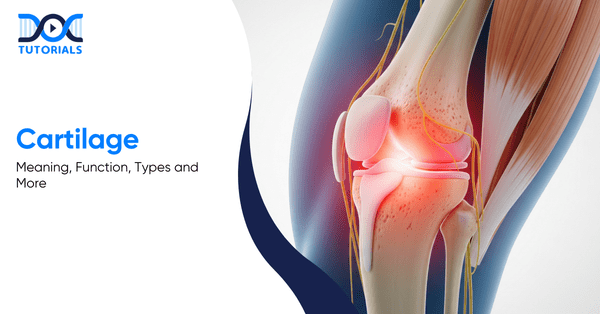
Cartilage is a smooth, resilient connective tissue that supports joints, absorbs shock, and allows friction-free movement. Found in structures like joints, ears, and the spine, it plays a key role in flexibility and skeletal stability.

Cartilage is a kind of flexible connective tissue existing in different parts of the body, including joints, ears, nose, and spacing between different bones of the spine. It gives joints structure, support, and protection, allowing them to move easily. 

The cartilages consist of specialised cells known as chondrocytes, which synthesise collagen, proteoglycans, and other proteins that give the tissue its strength and resilience.

What are the Functions of Cartilages?

Cartilage cushions joints by absorbing shock during movement, reducing friction between bones for smooth motion, and maintaining joint shape and stability. It also provides structural support and attachment points for muscles, tendons, and ligaments, helping the body move efficiently without joint damage.

Cartilage is very critical in cushioning bones and joints. The function of cartilage is pointed out below:

  • Shock Absorption

Cartilage is a natural shock absorber during movement. It cushions the bones when you walk, run, or jump, as sports shoes do for your feet.

  • Reducing Friction

It enables the bones to move more easily against each other by reducing friction at the joints. This enables smooth movement with no grinding and reduces wear and tear.

  • Structural Support

Cartilage helps joints maintain their shape while in motion. It also provides support and connection to other tissues, offering muscle, tendon, and ligament attachments in the entire body.

What are the Different Types of Cartilages? 

The human body has three types of cartilage: hyaline, elastic, and fibrocartilage. Hyaline cartilage provides smooth joint movement, elastic cartilage offers flexibility and shape retention, and fibrocartilage delivers strong cushioning in weight-bearing areas such as the spine and knees.

Three primary types of cartilage are present in the human body, and they are listed as follows:

  • Elastic Cartilage

Elastic cartilage is found in the outer ear, the throat epiglottis, parts of the nose, and the windpipe. Its key role is to provide flexibility along with strength, allowing these structures to maintain their shape while bending easily.

  • Fibrocartilage

Fibrocartilage is found in the menisci, which are special cushioning pads in the knee and between the spinal column in the spine. It aids in the alleviation of friction and pressure in weight-bearing joints. Fibrocartilage is the harder of the two types because it has thick sheets of stiff collagen fibres.

  • Hyaline Cartilage

Hyaline cartilage can be found in the larynx, nose, trachea, and ribs. It can also be found in the thin, smooth covering at the ends of bones in joints, called articular cartilage, which cushions movement. Hyaline cartilage is the weakest of the three types despite the fact that it consists of fine collagen fibres that give it strength.

What are the Common Conditions that Affect Cartilages? 

Cartilage can be affected by injuries, degenerative diseases, and spinal conditions. Some of the common issues are tearing due to sports activities, age-related wear resulting in osteoarthritis, herniated discs in the spine, and osteochondritis dissecans.

Below are some common conditions that affect cartilages:

  • Injuries

Accidents, impacts during sports activities, or sudden trauma can tear or damage cartilage. Common examples include meniscus tears in the knee or shoulder injuries that affect joint cartilage. Damage to a joint may also lead to conditions such as osteochondritis dissecans, which affects the ends of bones in joints. 

  • Osteoarthritis

This is the most widespread form of arthritis. Cartilage in the joints gradually breaks down as people grow older. As a result, joints may become painful, stiff, and inflamed with less cushioning and lubrication.

  • Herniated Discs

The condition is also known as the slipped, bulging or ruptured disc that occurs when the cartilage disc that separates the spinal vertebrae ruptures or becomes damaged. This can have a strain on the other nerves near and bring about discomfort or pain.

What are the Symptoms of Cartilage Damage?

Cartilage damage may cause joint pain, stiffness, swelling, and tenderness over time. Affected individuals may also experience limited motion, inability to perform daily tasks, and joint locking or instability, particularly in weight-bearing joints like the knees and hips.

Damage to cartilages might not be accompanied by immediate or distinct symptoms. Nevertheless, certain common symptoms can develop over time, such as:

  • Pain and Stiffness

Affected weight-bearing joints, such as the knees or hips, may experience discomfort and stiffness during movement.

  • Swelling or Tenderness

Inflammation around the affected area may occur, making the joint feel swollen or sensitive to touch.

  • Restricted Movement

Cartilage damage can limit how freely a joint moves, leading to difficulty with everyday activities.

  • Joint Locking or Instability

In some situations, affected joints can become unstable, or they may even lock up, which can significantly impact movement. 

How to Diagnose Cartilage Damage?

Physical examination, imaging tests such as X-rays and MRI scans, and arthroscopy are the methods used by doctors in the diagnosis of cartilage damage.

In cases of cartilage damage, physicians apply an integrated approach in order to determine the problem. The tests they perform include the following:

  • Physical Examination

The physician will examine the joints to assess swelling, tenderness, and pain due to movement.

  • Imaging Tests

Scans like X-rays and MRI images would assist in visualising the joint structure, such as cartilage, as well as surrounding tissues, to identify any abnormalities.

  • Arthroscopy

It includes inserting a small camera into the joint, enabling the doctor to view the cartilage in question and assess the amount of damage.

What are the Treatment Options for Cartilages?

Treatment for cartilage-based conditions is based on the extent of the damage and can involve physiotherapy, medications, and lifestyle modifications in mild cases. Severe damage may require surgical procedures to restore joint function.

Cartilage issues can be treated based on the cause and severity of damage. The treatment typically starts with a conservative approach and could be followed by surgery in case it is necessary.

Non-Surgical Treatments

  • Physiotherapy involves activities that strengthen the muscles surrounding the affected joint in order to minimise pressure on the cartilage.
  • Anti-inflammatory drugs are commonly prescribed to relieve pain and swelling.
  • Lifestyle changes, such as maintaining a healthy body weight and doing frequent low-impact exercise, can be used to reduce the load on the joints and maintain their long-term health.

Surgical Treatments

The most common treatment methods include: 

  • Abrasion Arthroplasty

A surgical device operates at a very high speed to scrape away the damaged cartilage with some tiny openings in the underlying bone and induce the body to grow new cartilage.

  • Autologous Chondrocyte Implantation

This is a two-stage procedure. Healthy cartilage cells are first collected and grown in a laboratory. The cartilage affected by the disease is removed later via surgery and replaced with the lab-grown cells.

  • Microfracture 

In this operation, the surgeon takes out the damaged cartilage and drills holes in the bone underneath. This improves blood flow, which consequently aids normal healing and cartilage growth.

  • Drilling

Similar to microfracture, drilling creates small channels in the underlying bone to improve blood supply and support the growth of new cartilage.

  • Osteochondral Autograft Transplantation

Healthy cartilage is removed from the patient’s non-weight-bearing joint and implanted into the injured region. This alternative is typically appropriate in small regions because of the lack of donor tissues.

  • Osteochondral Allograft Transplantation

In this procedure, cartilage tissue is sourced from a donor. Allografts are commonly used when there are larger defects and the tissue of the patient is inadequate.

FAQs about Cartilages

  1. Can cartilage repair itself?

The specialised cells, known as chondrocytes, do not regenerate easily, and that is why the cartilage damage rarely heals without medical intervention.

  1. What can athletes do to mitigate the occurrence of cartilage injuries?

Some of the ways athletes can reduce cartilage injury risk are by wearing proper protective gear, using proper training methods, and including strength and conditioning activities in their exercise programmes.

  1. What helps keep cartilage healthy?

The cartilage can be kept healthy by consuming a well-balanced, nutritious diet, engaging in low-impact workouts, and maintaining a healthy body weight, which will help avoid imposing unnecessary loads on the joints.

Conclusion

Being an NEET PG candidate, it is important to have clear and detailed knowledge of the cartilage, as it is a major determinant of joint health, movement, and several clinical conditions. It will help build your clinical reasoning and equip you to tackle orthopaedic and rheumatological cases with a lot of confidence. 

DocTutorials supports your preparation with high-yield video lectures, concise notes, and expert-led discussions tailored for NEET PG.

Start exploring our NEET PG Course today and take a decisive step towards securing your dream rank!

NEET PG Online Live Power Pack
DocTutorials App Download Now

Latest Blogs

Most Repeated Clinical Scenarios for NEET PG

Most repeated clinical scenarios for neet pg

NEET PG preparation can be overwhelming due to its vast syllabus. However, one crucial fact makes the exam a bit easier: the majority of NEET PG questions are repeated. The exam uses set patterns, and the same clinical situations are repeated with slight variations each year.

When you learn how to identify these patterns, you will respond to questions faster and more accurately. This is why understanding the most repeated clinical scenarios is one of the best strategies.

Keep reading to explore the most repeated clinical scenarios asked in NEET PG 2026.

What is the Relevance of NEET PG Clinical Scenarios?

Most of the NEET PG questions are clinical case–based, presenting brief patient scenarios instead of direct theory. Topics like pediatric milestones, thyroid emergencies, portal hypertension, and skin rashes recur yearly. Increasing image-based and data interpretation questions highlight the importance of pattern recognition over rote learning.

Around 65–75% of NEET PG questions are clinical case–based. Rather than direct theory inquiries, the exam does show you a brief patient narrative in which you are required to detect the diagnosis, investigations, or subsequent action in management.

Some topics appear repeatedly every year, such as:

  • Pediatric developmental milestones
  • Thyroid emergencies
  • Portal hypertension and its complications
  • Common skin rashes and image-based questions

In recent years, NEET PG has also increased its focus on image-based questions, research, and data interpretation. This makes pattern recognition even more important than memorising isolated facts.

What is a Repeatable Clinical Scenario?

NEET PG questions follow strict guidelines, focusing on practical, high-yield clinical problems with a single best answer. This leads to repeated clinical patterns built around a clear diagnosis or treatment goal, key signs and symptoms, and a typical patient profile.

NEET PG question setters work with strict guidelines. Each question must test an important and practical clinical problem, be clear and answerable within one question stem, and have a single best answer. Because of these rules, certain clinical patterns are repeated.

A repeatable clinical scenario usually has three key parts:

  1. A Clear Diagnosis or Treatment Goal: The question is designed to lead you toward one specific disease or management step.
  2. Key Signs or Symptoms: These are classic features that strongly point toward a diagnosis and should not be missed.
  3. A Typical Patient Profile: This includes age, lifestyle, habits, or occupation (for example, an elderly smoker, a chronic alcoholic, or a young athlete).

These scenarios act like exam templates. For example, a 45-year-old smoker with chest pain or a young patient with very high blood pressure appears in some form almost every year.

What are the High-Yield Clinical Scenarios by System?

NEET PG repeatedly tests high-yield, system-wise clinical scenarios that act as exam templates across subjects. Questions rely on recognising hallmark symptoms, patient profiles, and investigations, making pattern recognition essential for efficient and focused preparation.

Here are the high-yield clinical scenarios, organised system-wise, that are most frequently tested across subjects and essential for focused NEET PG preparation:

1. Cardiovascular System

  • Young Patient with Severe Hypertension

A 25-year-old man with extremely high blood pressure (180/110) and weaker pulses in his legs than in his arms is a strong indication of coarctation of the aorta. Questions often test the figure of 3 signs, rib notching, and association with a bicuspid aortic valve.

  • Unequal Pulses with Sudden Chest Pain

A middle-aged man with severe, tearing chest pain radiating to the back and different blood pressure (BP) readings in both arms points to aortic dissection. Know the Stanford classification and the finding of a widened mediastinum on X-ray.

  • Irregular Pulse in the Elderly

An elderly patient with palpitations and an irregular pulse is most likely having atrial fibrillation. Common questions focus on rate vs rhythm control and the CHA₂DS₂-VASc score.

  • Syncope During Exercise

A young athlete fainting during physical activity should be diagnosed with hypertrophic cardiomyopathy until it is proven otherwise. ECG findings include deep Q waves and left ventricular hypertrophy.

2. Digestive System

  • Alcoholic with Vomiting Blood

This typical presentation indicates oesophagal variceal bleeding as a result of portal hypertension. Questions commonly asked about terlipressin, band ligation vs sclerotherapy, and the Child-Pugh score.

  • Painless Jaundice in the Elderly

An older patient with weight loss, yellowing of the skin, and a palpable gallbladder points to periampullary carcinoma. This is explained by Courvoisier’s law and appears almost every year.

  • Acute Abdomen with Free Air

Air under the diaphragm on an upright X-ray indicates a perforated peptic ulcer. You should also know other causes of pneumoperitoneum and the saddle-bag sign.

  • Bloody Diarrhoea in Young Adults
    These questions usually compare ulcerative colitis and Crohn’s disease, focusing on:
  1. Continuous lesions vs skip lesions
  2. Crypt abscesses vs granulomas

3. Respiratory System

  • Smoker with Hemoptysis

Most commonly indicates squamous cell carcinoma of the lung. Paraneoplastic syndromes are frequently tested:

  1. SIADH 
  2. Hypercalcemia
  • A Worker with a Chronic Cough

Occupational lung diseases are high-yield.

  • Immigrant with Evening Fever

Tuberculosis is suggested by upper-lobe lung disease, weight loss, and evening fever. Know the difference between primary and post-primary TB.

4. Pediatric System

  • Blue Baby at Birth

Cyanosis that does not improve with oxygen indicates transposition of the great arteries. This is often compared with the Tetralogy of Fallot, which causes cyanotic spells during crying.

  • A Child with Repeated Pneumonia
  • Failure to Thrive in Infants

Clues in the question that relate to timing are significant.

  • Floppy Baby
  • Werdnig-Hoffman Disease

Commonly Tested Pediatric Milestones

MilestoneNormal AgeCommon Question
Social smile6–8 weeks“Cannot sit at 10 months.”
Head holding3 monthsDevelopmental delay
Sitting without support8 months“Cannot sit at 10 months.”
Walking12–15 months“Cannot sit at 10 months.”
Two-word sentences2 yearsSpeech delay

5. Gynaecology and Obstetrics

  • Seizures in Pregnancy

Always treat eclampsia with magnesium sulfate.

  1. Loading dose: 4 g IV
  2. Maintenance dose: 1 g/hour

Toxicity is reversed using calcium gluconate.

  • Postpartum Hemorrhage

Most cases are due to uterine atony. Management follows a stepwise approach:

Uterine massage → oxytocin → ergometrine → carboprost → surgery
Remember the 4 T’s: Tone, Trauma, Tissue, Thrombin.

  • Galactorrhea with Amenorrhea

Think prolactinoma. Check serum prolactin and do a pituitary MRI. Treatment is cabergoline, which is now preferred.

  • Postmenopausal Bleeding

Always assume endometrial cancer until proven otherwise. Key points:

  1. Endometrial thickness > 4 mm → biopsy
  2. Risk factors include obesity, unopposed estrogen, and tamoxifen.

6. Dermatology: Common Image-Based Questions

  • Impetigo

Caused by Streptococcus pyogenes or Staphylococcus aureus. It shows honey-colored crusts.

  1. Local infection → topical mupirocin
  2. Widespread infection → oral antibiotics
  • Pityriasis Rosea

Begins with a herald patch, followed by a Christmas-tree pattern rash. It is self-limiting, so reassurance is the correct answer.

  • Erythema Multiforme

Shows target lesions. Causes include HSV infection and drugs like phenytoin or sulfonamides. Mucosal involvement suggests Stevens-Johnson syndrome.

  • Ash-leaf Macules

Seen in tuberous sclerosis. Look for seizures, intellectual disability, and cardiac rhabdomyomas.

7. Neurology

  • Sudden Weakness on One Side

Always do an urgent CT scan to rule out haemorrhage before treating stroke. Know the basics of NIHSS and the 4.5-hour thrombolysis window.

  • Ascending Paralysis

Suggests Guillain-Barré syndrome, often after a Campylobacter infection. CSF shows high protein with a normal cell count. Treatment is IVIG or plasmapheresis.

  • Morning Headache with Vomiting

Indicates raised intracranial pressure. Check for papilledema. Brain tumour questions focus on symptoms based on location.

  • Unilateral Facial Weakness
  1. Forehead spared → UMN lesion (stroke)
  2. Forehead involved → LMN lesion (Bell’s palsy)

FAQs about Most Repeated Clinical Scenarios for NEET PG 2026

1. How many clinical situations should I learn for NEET PG?

Learning 50–60 core clinical patterns is usually enough. Focus on understanding, not memorising. Repeated practice builds pattern recognition, which matters more than covering too many cases.

2. Which trigger phrases are common in NEET PG questions?

Phrases like rice-water stools, currant jelly sputum, strawberry tongue, and chocolate cyst strongly point to specific diseases. These clues stay constant, so linking them to diagnoses is very helpful.

3. How do I tell apart similar clinical situations?

Focus on key differences, not similarities. For example, raised JVP suggests heart failure, while caput medusae suggests portal hypertension. Use comparison tables to remember confusing pairs.

4. Which subjects have the most repeated clinical questions?

Medicine has the most repeated cases, followed by surgery, pediatrics, and OBG. In medicine, cardiology, GI, and endocrinology are especially important and commonly tested.

5. How old are the repeated clinical scenarios in NEET PG?

Most core clinical patterns have remained unchanged for many years. New topics are added, but solving questions from the last 10 years can cover the most commonly repeated scenarios.

6. What is the pattern of NEET PG 2026?
NEET PG 2026 is a computer-based exam with 180 multiple-choice questions divided into five sections of 40 questions each. Every section has a fixed time limit of 42 minutes, and candidates must complete sections sequentially.

7. What percentage of questions are repeated in NEET PG?
Around 30% of questions are direct or modified repeats from previous years.

8. What is the duration of NEET PG 2026?
The expected duration of the NEET PG 2026 exam is 3 hours and 30 minutes, totaling 210 minutes.

9. What is the marking scheme of NEET PG 2026?
Each correct answer carries +4 marks, while every incorrect answer attracts a negative marking of −1.

10. What type of questions are asked in NEET PG?
NEET PG mainly includes clinical case-based questions, image-based questions, and single-best-response multiple-choice questions.

Conclusion

Most NEET PG clinical questions are standard patient stories that repeat year after year with small changes. By mastering around 50 common clinical scenarios, you can confidently answer 60–70% of the clinical questions in NEET PG 2026.

For structured support, DocTutorials can be your academic companion. With concise video lectures, a clinically oriented Qbank, exam-focused notes, flashcards, and mind maps, we help you tackle high-yield clinical scenarios with confidence.

Join DocTutorials and explore our NEET PG course to excel in your medical career!

NEET PG Online Live Power Pack
DocTutorials App Download Now

Latest Blogs

/20

NEET PG Mock Test

Mini Mock. Major Results. – NEET PG Quick Challenge

1 / 20

A boy presents with fever and chills. A rapid diagnostic test detects the HRP-2 antigen. Which Plasmodium species is most likely responsible for the infection?

2 / 20

During an abdominal surgery, the surgeon accidentally nicks a structure that passes posterior to the epiploic foramen, which results in profuse bleeding. Which of the following structures is most likely to get injured?

3 / 20

A 2-year-old child is brought to the emergency room with a generalised seizure following a high-grade fever. What is the first-line drug of choice for seizure control in this setting?

4 / 20

Upon the delivery of a term new born, a thorough examination of the umbilical cord reveals crucial information. What are the essential components of a healthy umbilical cord?

5 / 20

A 24-year-old male sustained a shaft of femur fracture after a road traffic accident, and internal fixation was done for the same. Two days later, he developed shortness of breath with confusion and a petechial rash. What is the most likely diagnosis?

6 / 20

A female patient with HIV infection (CD4 count 150 cells/mm³) was newly diagnosed with rifampicin-resistant tuberculosis. What anti-tubercular treatment regimen should be initiated, and which HIV-related complication should you look out for?

7 / 20

A patient presents with long limbs and ectopia lentis. His arm span is found to be greater than his height. Marfan syndrome is suspected. Which of the following gene defects is most likely responsible?

8 / 20

Which of the following statements regarding orbital cellulitis is true?

9 / 20

What is the mechanism of renal calculi formation in a patient on Hydrochlorothiazide?

10 / 20

A 19-year-old boy presented to the OPD with shoulder pain. He had been training in the gym and had hyperextended his shoulder. On examination, the avulsion of the long head of the triceps was noticed. What is the site at which avulsion of the long head of triceps occurs?

11 / 20

Which of the options aids in the transportation of fatty acids through the inner mitochondria membrane?

12 / 20

Which of the following enzyme activities can be estimated in red blood cells to diagnose vitamin B2 deficiency?

13 / 20

A 75-year-old male who is a known case of hypertension and type 2 diabetes mellitus presents with epistaxis, which does not respond to conservative measures and nasal packing. Which of the following arteries should be prioritised for ligation?

14 / 20

Which laboratory test is commonly used to confirm the diagnosis of rotavirus-induced diarrhoea?

15 / 20

A patient with tumour lysis syndrome has elevated uric acid levels. What is the mechanism of action of pegloticase?

16 / 20

A patient is diagnosed with paroxysmal supraventricular tachycardia and requires prophylaxis for the same. Which of the following drugs is appropriate for this patient?

17 / 20

What is the term for a single mutation in a nucleotide base pair that leads to formation of termination codon?

18 / 20

In the Pritchard regimen for the management of eclampsia, what is the loading dose of magnesium sulfate?

19 / 20

During eversion of the foot, which of the following ligaments is most likely to be injured?

20 / 20

In a patient presenting with fever and a positive filarial antigen test, what should be the next appropriate step in management?

Your score is

0%

What is the Best Time to Start NEET PG Preparation?

Best Time to Start NEET PG Preparation

The NEET PG is one of the most competitive medical examinations for MBBS graduates and those seeking to transition to postgraduate specialist training. With 19 subjects in the syllabus, every medical student is left with one question: When should I start preparing for NEET PG?

This guide breaks down the NEET PG preparation timeline and proposes specific study plans to students at various phases of the medical career. Continue reading to know more!

Boost Your NEET PG Prep with Grand Test – Download the Brochure Now

When Should You Begin with Your NEET PG Timeline?

Being too early in NEET PG preparation will cause burnout, whereas being too late will result in panic. Strategically, the third year of MBBS is the ideal starting point.

The NEET PG examination is a test of clinical skill and endurance. Many students mistake it for a sprint and exhaust themselves before the actual race starts. Others do not realise the depth of the syllabus (approximately 700 topics covering 19 subjects) and feel they are rushing in the last few months.

It is subjective, though statistically and strategically, toppers and educators have cited the third year of MBBS as the most effective starting point. The students have already gotten beyond the overload of anatomy and physiology and are not yet daunted by the oppressive clinical responsibilities of the internship.

Starting, however, is different at various stages. For a first-year student, it is about constructing concepts. As a post-intern, it involves tough MCQs. It is the initial step in a successful rank to understand this distinction.

Why is the MBBS Third Year Considered Ideal for NEET PG Preparation?

The MBBS third year is the golden period to start NEET PG preparation, as it is when subjects are manageable and clinical exposure is increasing. The reduced pressure during exams allows students to adopt a relaxed yet strategic approach that balances university requirements with competitive exam demands.

By beginning to prepare for NEET PG at this stage, students can combine preparation for university examinations with the requirements of competitive examinations. If you are a third-year student, now is the best time to start.

Here’s why the third year stands to be the golden phase:

  • Manageable Subjects: It has 3 major subjects, namely, ENT, Ophthalmology, and PSM (Community Medicine). These are scoring subjects. Learning them guarantees a significant portion of the marks.
  • Clinical Exposure: Students begin to attend postings. This is the ideal moment to correlate what they observe at the wards with what they read in the preparation notes.
  • No Exam Pressure: In contrast to the last year, when the tension of the professional tests can be immense, there is an easier, absorption-oriented course of study in the third year.

1. Phase 1 (Months 1-4): Finish 3rd Year Subjects

  • Devote the time to ENT, Ophthalmology, and PSM.
  • Resource: Take short video lectures and make your own notes.
  • Goal: By the time you reach your university exams, you should be in a position to answer MCQs that are of NEET PG level in these three subjects without straining.

2. Phase 2 (Months 5-8): Revise 1st & 2nd Year Basics

  • One must have forgotten much about Anatomy and Biochemistry. It is high time to go over them with Rapid Revision modules or QRP (Quick Revision Programs).
  • Do not go through the whole textbook again. Review concepts using notes and videos.

3. Phase 3 (Months 9-12): Touch Base with Final Year Subjects

  • Start with Medicine or Surgery. Although it may not be complete, a glimpse into the systems (e.g., CVS, CNS) will make the final year much smoother.

Subject-Wise Strategy for Third Year

SubjectWeightageStrategy
PSMHighFrequently overlooked but high-ranking. Focus on Epidemiology and Biostatistics.
ENTModerateVisual-heavy. Focus on diagrams and instruments.
OphthalmologyModerateClinical images are key. Study diseases like Cataract, Glaucoma, and Diabetic Retinopathy in depth.

How to Prepare for NEET PG during the Final Year?

Final-year NEET PG preparation demands integrating university learning with exam strategy, focusing on major subjects. Both concepts and clinical skills will be strengthened through smart study, MCQ practice, and the use of standard textbooks.

The last year is also referred to as a marathon on sprint speed. The big four: Medicine, Surgery, OBGYN, and Paediatrics. They are the foundation of the NEET PG Examination (they constitute about 50-60 per cent of the examination, comprising 10 subjects and their corresponding basics).

A common mistake that final-year students make is treating university preparation and NEET PG preparation as separate entities, which is incorrect. Here’s what you can do instead:

  • Read for the Ward, not just the Book: When you are on Surgery, read about the topic concerning the case you observed. Provided that you have a Hernia case, learn the anatomy of the inguinal canal, the categories of repairs, and the complications. This covers Anatomy, Surgery, and clinical skills simultaneously.
  • The Retrograde Method: Rather than picking up a chapter and reading it from beginning to end, what you can do is to answer 10 MCQs on the topic. You will probably make mistakes, but it will also train your brain to search for certain answers as you go through the text.
  • Standard Books are Key: For Medicine (Harrison’s/Davidson’s) and Surgery (Bailey & Love), use regular textbooks on essential subjects. On less important topics, use coaching notes.

Time Management in the Final Year

  • Weekdays: Pay attention to the topic of your current clinical posting.
  • Weekends: Saturday is dedicated to revising a short subject (e.g. Dermatology, Radiology) and Sunday to a Subject-Wise Test.
  • Grand Tests (GTs): Start taking GTs once a month. There is no cause to be worried about that score; you must be concerned about your stamina in sitting for 3.5 hours.

How to do NEET PG Preparation During MBBS Internship?

The preparation for the NEET PG internship is not easy, but it can be done with smart tips. Change focus parameters on thorough reading, Qbank training, and flaky topic modification. Make the most out of limited study hours using early morning, short breaks, and duty-based learning.

One must not panic while starting the preparation during an internship. A high percentage of toppers begin their serious preparation during their internship. However, the strategy should not be comprehensive reading but high-yield targeting.

The students will be subjected to 12-hour shifts, night shifts, and the fatigue that accompanies them. They cannot study for 10 hours a day, but might get 3-4 hours on a comparatively light day.

  • The Qbank is your Best Friend: Even when there is no time to watch a 2-hour video lecture, they can always find time to answer 10 MCQs on an app when it’s time to report to a patient or take a lunch break.
  • Focus on Memory-based Subjects: Other subjects, such as Biochemistry and Microbiology, are memory-based. Have flashcards or online notes on a mobile phone at all times to review them constantly.
  • Duty-Based Learning:
    • Casualty Posting: Learn Management of Trauma, MI, and Poisoning (Forensic Medicine).
    • Labour Room: Master Partograms, stages of labour, and obstetric emergencies (OBGYN).
    • Paediatrics Ward: Learn developmental milestones and immunisation schedules.

Time Management During Internship

  • Early Morning (5 AM – 7 AM): 2 hours of intense study before duty. This can potentially be the golden time if utilised efficiently.
  • During Duty: Solve 20-30 MCQs on your phone.
  • Evening (Post-Duty): You will be tired. Do not try to read heavy theory. Watch a clinical case video or review the MCQs you got wrong during the day.

What are the Recommended Study Resources for NEET PG Preparation?

MBBS textbooks are known to offer a good conceptual base, and short review guides and notes enable fast revision. Question banks and mock tests enhance the accuracy of the exams as they familiarise the students with the pattern and timing.

Standard MBBS textbooks are used to develop the basic knowledge, whereas brief review books are utilised in revision. Mock tests and question banks are used to assist the students with the exam patterns and to develop more accuracy.

  • Online resources and interactive techniques to assist in the exam-oriented practice and concept reinforcement.
  • Practice question banks and mock tests to simulate the exam pattern.
  • Short notes and quick revision guides will be essential during the last few months.

A combination of these in a balanced manner will guarantee strong fundamentals and examination-oriented knowledge. However, it is better to limit resources and revise them several times than to have multifarious sources in a superficial way.

FAQs about What is the Best Time to Start NEET PG Preparation

  1. How much time is required for NEET PG preparation?

On average, 12-18 months of regular study is estimated to be adequate. This involves concept construction, numerous revisions, and frequent mock test practice.

  1. What is the strategic window for maximum success?

The best period would be between the second year of MBBS and the final year, with intense exam-oriented study in the final year and internship. This equilibrium guarantees both the depth of knowledge and the exam preparation.

  1. Should I focus more on clinical or pre-clinical subjects?

Clinical subjects carry more weightage in NEET PG, but both are important. Pre-clinical subjects are to be revised for the basics, whereas clinical subjects require more in-depth attention and practice.

  1. What role do mock tests play in preparation?

The mock tests play a critical role in time management, locating weak points, and exam temperament. Consistent practice will create the simulation of the actual exam setting and increase confidence.

  1. Does starting early guarantee success?

Early learners have a higher chance of revision and better understanding, but it takes a lot of consistent work, clever planning, and practice in exams compared to those who start late, so time alone is not the guarantee.

  1. Does NEET PG preparation follow a different approach in each year of MBBS?

Yes. The preparation plan varies from year to year in MBBS. Early years are best used for building strong concepts, middle years for systematic subject-wise coverage, and final year or internship for revision and clinical integration.

  1. Is it advisable to start NEET PG preparation when clinical subjects are first introduced?

Yes. The introduction of clinical subjects is an ideal phase to begin light, structured NEET PG preparation, as linking theory with clinical practice improves understanding and long-term retention.

  1. How does an early start in NEET PG preparation make an internship more productive?

Early starters tend to join internships with most subjects already undergone once. This will enable use of the internship time to revise, do MCQs, and practice on tests, as opposed to new content under time pressure.

  1. When should I completely stop studying new topics and focus only on revision?

Learning new topics should be cut off approximately 45-60 days prior to the exam. At this stage, the introduction of new content is unhelpful since unrevised material will hardly be retained or remembered during the examination.

  1. How does preparation for university exams support NEET PG preparation?

University examinations foster descriptive knowledge and depth of concepts, whereas NEET PG tests require application and problem-solving. Early NEET PG preparation will allow students to overcome this gap progressively rather than experiencing a high transition at a later date.

Conclusion

The NEET PG exam preparation calls for consistency rather than intensity. over intensity. A student who studies 4 hours a day for 2 years will almost always beat a student who studies 12 hours a day for 2 months.

At DocTutorials, we provide edition notes, clinical QBanks, mindmaps, and flashcards to help you stay well-prepared through efficient studying. Join DocTutorials today and explore our NEET PG course to excel in your medical career!

NEET PG Online Live Power Pack
DocTutorials App Download Now

Latest Blogs

Vaccine Cold Chain: Flowchart, Guiding Parameters & More

Vaccine Cold Chain

The method of storing a vaccine in proper condition is called the cold chain or immunisation supply chain. It refers to a series of procedures designed to maintain the vaccine’s efficacy from manufacturing through administration and to deliver the same in proper condition to the consumer. The vaccine must be kept at the WHO-recommended temperature to maintain its effectiveness throughout the supply chain. 

In this guide, we will discuss the vaccine cold chain in detail to show how vaccine efficiency is maintained across different storage units throughout the supply chain.

Keep reading!

What is a Vaccine Cold Chain?

The vaccine cold chain is a supply chain that ensures the medicine is maintained in proper condition during transportation and delivered to the right place in appropriate condition and on time. This supply chain comprises different stages, such as daily record-keeping, a monthly report to understand demand and calculate quantities, and shipping the vaccine through several levels to end users. 

The vaccine cold chain is depicted as follows:

Daily Record
Monthly Report
Analysis 
Annual Statistics and Estimates
Request For Supply
Manufacturer 
Airport
Central Store
District/Regional Store
Healthcare Centre 
Vaccinator 

What are the Guiding Parameters in the Vaccine Cold Chain?

Light, temperature, and heat are the major factors that control the vaccine’s efficiency as it moves through the vaccine cold chain. The people who handle the medicine in the immunisation chain must know all these parameters and the ideal range to ensure the immunisation is suitable before it is applied to the patient. 

Vaccines are a sensitive medical product. They are sensitive to freezing, heat, and light. Vaccine efficacy, that is, the ability to protect the vaccinated patient, is compromised when they are exposed to improper conditions. Once its efficacy is lost, it cannot be regained. 

To maintain efficacy, it must remain in a suitable condition during movement through the supply chain. Thus, it is mandatory for those who handle the vaccine nationwide to know the conditions for maintaining its quality: 

  • It is crucial to keep the vaccine, which does not contain a preservative, at a temperature between +2°C and +8°C during the immunisation session. 
  • Vaccines produced with virals and/or lyophilised diluents must be stored between -15°C and -25°C.
  • Some vaccines are sensitive to exposure to light. These vaccines must be protected from sunlight and intense artificial light during movement throughout the cold chain. Vaccines, which are light-sensitive, are usually packaged in dark glass containers to maintain their quality.

Norms to Maintain the Vaccine’s Quality During the Vaccine Cold Chain

To ensure the vaccine’s efficacy during movement in the vaccine cold chain, some norms specify where to store it, the conditions under which it must be kept, and how it must be transported. 

Health workers who are involved in the vaccine cold chain have to maintain the following guidelines to ensure the product’s quality:

  • Store the vaccine in the respective refrigeration place.
  • Use a temperature-measuring device to check the temperature and ensure it remains between +2°C and +8°C to preserve quality.
  • The vaccine must be packed in a vaccine carrier along with cooling pads in place to ensure the correct temperature is maintained during transport in the immunisation chain.

What are the Recommended Storage Equipments in the Vaccine Cold Chain?

During the immunisation chain, the vaccine must be stored in various storage units to maintain its efficacy. Recommended storage units for the immunisation chain are refrigerators, vaccine carriers, and cold boxes. To monitor temperature within the specified limits, Vaccine Vial Monitors (VVMs) are recommended.

At different levels of the vaccine cold chain, the product must be stored in various storage units or carriers. These are classified into three categories:

  • Primary Level (National): In this phase, the vaccines are kept in freezers, freezer rooms, refrigerators, cold boxes, and refrigerated trucks (in some cases) for mobilisation.
  • Intermediate Level (Province or District): At this level, vaccines are stored in freezer rooms, refrigerators, cold boxes, carriers, and, in some cases, in refrigerated trucks, depending on capacity.
  • Peripheral Level (Health Centres or Clinics): In the peripheral level, the medicine is stored mainly in refrigerators and also in vaccine carriers and cold boxes.
  1. Refrigerator

Refrigerators in the vaccine cold chain are driven by electricity, solar energy, or gas. Among the three, any one type is chosen based on the intermittent availability of the energy source needed to keep the vaccine within the specified temperature range continuously. All these types of refrigerators are specially designed to maintain the temperature range required for vaccine monitoring.

Domestic refrigerators are not designed to store vaccines continuously because they lack a temperature control device. That is why the World Health Organisation (WHO) does not recommend domestic refrigerators for storing vaccines. Moreover, during a prolonged power cut, domestic refrigerators cannot maintain the WHO-specified temperature range for vaccine immunisation.

  1. Cold Boxes

When the refrigerator is out of order or there is a prolonged power cut, cold boxes are used to store vaccines for up to 2 days. Also, the vaccines are stored in the cold boxes during short-term travel. It is an insulated box lined with water or ice packs to keep the vaccine within the required temperature range. Once packed, the cold boxes must not be opened until the vaccine is needed.

“Cold Life” of a cold box refers to the maximum time it can maintain the temperature of the vaccine vials below +10 °C when packed with ice packs. Usually, the cold life of a cold box is two to seven days when tested at a constant atmospheric temperature of +43 °C.

The “Cool life” of a cold box is the maximum time period during which it can maintain the medicine’s temperature below +20 °C, with cool water packs inside it that have been stored in a refrigerator. A cold box’s cool life is 12 hours to two days when tested at a constant atmospheric temperature of +43 °C.

There are different models of cold boxes available in the market with different vaccine-carrying capacities. Various models require different numbers and sizes of water packs to store the vaccines. It is essential to use the exact number and size of water packs as specified by the manufacturer; otherwise, the cold life will be compromised.

  1. Vaccine Carriers

These are smaller vaccine storage units compared to a cold box. It is easier to carry than the cold box. A prequalified vaccine carrier has a cold life of 18 to 50 hours at +43 °C and a cool life of 3 to 18 hours with cool water packs. The vaccine carriers are used for the following purposes:

  • During mobilisation to other places and for short-term storage in a health centre within the vaccine cold chain.
  • It is used to temporarily store the vaccine at the peripheral level when the refrigerator is out of order or defrosted.
  • For transporting the monthly vaccine demand from the district storage to small health facilities.

The type of vaccine carrier is chosen depending on the following factors:

  • Quantity of the vaccine and type of diluents to be mobilised.
  • The cold life, or cool life, that is required for the longest planned shipment.
  • The type of transport media used.
  1. Vaccine Vial Monitors (VVMs)

It is the sole temperature-monitoring indicator provided with the vaccine vial by the manufacturer. The VVM indicates whether the vaccine’s quality is maintained during movement along the cold supply chain. It is a chemical indicator label placed on the vaccine container. When the product moves through the immunisation chain, it indicates the condition of the vaccine after exposure to heat and light by a change in colour codes during mobilisation in the supply chain.

There are four types of VVMs available to ensure the quality of the heat-sensitive vaccines at every stage in the supply chain. They are: 

  • VVM2 
  • VVM7 
  • VVM14 
  • VVM30 

The number indicates the number of days it takes for the colour of the VVM to reach a discard point when the vaccine is exposed to a constant temperature of +37 °C. The VVM is used with the vaccine container to discard heat-damaged vials and minimise vaccine wastage.

What are the Basic Methods of Maintenance for Cold Chain Equipment?

For different cold chain equipment, you need to perform proper maintenance so that the storage unit remains in good condition and retains the vaccine’s quality. Defrosting a refrigerator, checking and ensuring the availability of the energy source for different energy-driven refrigerators, and properly drying the cold boxes and vaccine carriers are such maintenance methods for the respective equipment.

The basic maintenance methods for cold chain vaccines are as follows:

  1. Defrosting the Vaccine Refrigerator

A vaccine refrigerator remains in optimised condition when it is cleaned, properly installed, and defrosted regularly. When thick ice forms in the freezing compartment and on the evaporator plate, the vaccine refrigerator does not operate correctly, and cooling is not carried out within the required temperature range. 

Additionally, a thick layer of ice increases power consumption from the energy source. The vaccine refrigerator must be defrosted regularly or when there is a 0.5mm thick layer of ice in the storage places, whichever comes first.

  1. Maintenance of the Energy Source for the Vaccine Refrigerators

The energy source for the vaccine refrigerators is electricity, solar power, and fuel (gas/kerosene). You have to check the specific energy consumption data for the energy source as per the manufacturer’s user manual for daily, periodic, and yearly use. 

If the energy source is maintained within the specified consumption limits, the storage unit operates optimally throughout its product life cycle. This optimised running condition of the different energy-driven vaccine refrigerators ensures the vaccine remains healthy without compromising quality.

  1. Maintenance of Cold Boxes and Vaccine Carriers

Cold boxes and vaccine carriers must be dried and the lids opened after use to prevent them from becoming mouldy and moist. A mouldy, moisture-containing cold box or vaccine carrier can contaminate the vaccine vials inside it. All this vaccine storage equipment must be stored with the lid popped open.

Knocks and sunlight cause cracks in the walls of the cold box and vaccine carrier. A crack can compromise insulation and increase the risk of heat exposure during transportation. Also, cracked containers degrade vaccine quality, which is undesirable in the vaccine cold chain. You can use adhesive tape to repair the small crack on the walls until the undamaged cold box or vaccine carrier arrives.

What is a Shake Test?

A shake test is carried out to avoid frozen vaccines that are no longer usable by the vaccinator. By comparing two vials of the same category, one accidentally frozen and the other deliberately frozen, the shake test is done to identify the damaged vaccine, discard it, and reduce wastage.  

The shake test is performed to determine whether a freeze-sensitive vaccine is damaged when exposed to temperatures below 0 °C. After it is stored below 0 °C, a frozen vaccine vial no longer appears cloudy but tends to form flakes that sediment at the bottom of the vial.

To perform the shake test, you need two identical vaccine vials from the same manufacturer with the same batch number. One of them is deliberately frozen overnight, and the other is suspected to be frozen. Shake both vials in one hand, then compare the settlement process by placing them side-by-side. If the suspected frozen vial settles at the same speed as the deliberately frozen vial, then you know it has frozen. And if it settles relatively slowly, then it has not frozen.

Why is the Shake Test Needed?

If a freeze-sensitive vaccine is accidentally thawed, it is likely damaged. In these circumstances, the shake test is required to identify damaged vaccine vials and to discard them before moving them in the vaccine cold chain or using them for patient use.

What is the Importance of the Vaccine Cold Chain?

Storing the vaccine at the WHO-recommended temperature maintains its potency throughout the immunisation chain. This ensures that the vaccine’s effectiveness remains unchanged until it is administered to the patient. 

The significance of the immunisation chain is as follows:

  • Maintain the Vaccine’s Potency: Vaccines are an essential biological product. Through the vaccine cold chain, it is protected from extreme heat, light, and temperature to retain its potency; otherwise, it will become less effective, causing permanent damage to the product.
  • Prevent Disease Outbreaks: An immunised vaccine, when administered to the patient, helps improve immunity, thus preventing various critical illnesses for which it is intended.
  • Less Wastage: A vaccine vial stored at the WHO-recommended temperature throughout the immunisation chain is used for application to the patient. Strictly maintaining such parameters minimises wastage.
  • Healthy Global Population: The vaccine cold chain ensures the supply of the medicine under extreme environmental conditions to the remote areas, as well as to urban regions, ensuring a healthy global population.

FAQs about Vaccine Cold Chain

  1. What is the vaccine cold chain?

The vaccine cold chain is the set of rules to follow during the distribution of the medicine from the national level to the peripheral or local level. The WHO defines a temperature range that must be maintained for the immunisation chain to maintain the vaccine’s potency.

  1. What is the vaccine storage temperature?

Depending on the type of vaccine, two temperature ranges are specified. The freeze-sensitive vaccines must be stored between +2°C and +8°C, and vaccines produced with viral and/or lyophilised diluents must be stored between -15°C and -25°C.

  1. What is the vaccine supply chain?

A vaccine supply chain is defined as the shipment of the vaccine from the national level to the peripheral or local level at the right time, in the right amount, and in the proper condition and temperature.

Conclusion

The vaccine cold chain is required for retaining the medicine’s efficacy at every stage of the supply chain. The vaccine vials are kept in the WHO-recommended temperature range in the suitable storage units. This enables medical personnel to access high-quality vaccines for patient administration, thereby improving public health and the global safety environment. It has a challenging, complex logistics procedure that requires precise temperature control. 

A well-stored vaccine helps achieve the desired immunity for the patient, thereby preventing critical disease outbreaks and keeping the situation under control during epidemics or pandemics.

To help you gain more insight about NEET PG topics, DocTutorials provides crisp visual content, quality question banks, exam-specific mock tests, and roadmaps that help aspirants become more confident and well-prepared for the examination.

Check out our NEET PG courses today!

NEET PG Online Live Power Pack
DocTutorials App Download Now

Latest Blogs

Hamartoma: Causes, Symptoms, Diagnosis, and Treatment

Hamartoma

Hamartomas are non-cancerous growths composed of the same surrounding tissues that grow in an unorganised manner, causing a tumour-like structure. The majority of them do not cause any symptoms and are not harmful. If found to be causing health issues, they are surgically removed by an appropriate surgeon. However, the cause is mainly idiopathic, and genetic syndromes are sometimes included in the aetiology. 

Although a hamartoma is non-cancerous, avoiding treatment may result in dysfunction of other organs. Hence, keep reading to learn about hamartomas, their types, and the best available treatment for managing them. 

What is a Hamartoma? 

A hamartoma is a non-cancerous, benign growth of disorganised mature tissue that can develop anywhere in the body. There are some key characteristics to identify a hamartoma, such as disorganisation in tissue growth, benign nature, location throughout the body, etc.   

Mostly harmless, hamartomas are masses of disorganised tissue that can grow anywhere in the body. However, they can hamper bodily functions if they grow big enough to suppress other organs. If hamartomas grow abnormally large, they need to be removed surgically to relieve the patient. 

Some of the key distinguishing features of a hamartoma are:

  • Benign 
  • Disorganised growth 
  • Symptoms are found incidentally 
  • Developmental error 
  • Can develop anywhere in the body

How are Hamartomas Classified?

Hamartomas do not spread to other parts of the body because they are benign and found in places such as the heart, lungs, brain, and breast, causing cardiac rhabdomyoma, hypothalamic hamartoma, etc.

Hamartomas don’t metastasise like malignant tumours and remain in one place. However, their size increases, putting pressure on other organs in the body. The following section lists the places where hamartomas can grow:

  • Heart (Cardiac Rhabdomyoma): Hamartomas in the heart can result in cardiac arrest and heart failure, which is very common in children. 
  • Brain (Hypothalamic Hamartoma): When hamartomas grow in the brain, the main changes are seen in the behaviour and mood. If they grow in the hypothalamus, epileptic seizures and early puberty are also seen. 
  • Breast: A breast hamartoma is a benign tumour that can develop at any age. However, women above 35 are more prone to breast hamartoma, which can cause breast deformities. 
  • Lungs: Pulmonary hamartomas, or lung hamartomas, are the most common type of tumour that can cause difficulty breathing or trigger pneumonia. In severe cases, tissue damage is also seen. 
  • Spleen: Splenic hamartomas are the rarest form of hamartomas, which are seen more in females than males. Hamartomas on the spleen manifest as abdominal discomfort and pain. 
  • Neck and Chest: Hamartomas in the neck region can cause abnormal swelling and hoarseness, and respiratory issues if they grow in the chest region as well. 
  • Skin: Skin hamartomas can grow anywhere on the skin, with no explicit limitation of the risk area.

What are the Causes of Hamartomas? 

The cause of a hamartoma is idiopathic; however, it is believed to result from various factors such as a genetic mutation, congenital defect, developmental abnormality, genetic defect, etc.

The exact cause of a hamartoma is still unknown, and they are rare. Scientists say that hamartomas result from a genetic mutation in one of the biological parents. However, the following list explains some of the probable causes behind developing a hamartoma:

  • Congenital Defect: In these cases, hamartomas are present at birth but are detected later in life. 
  • Developmental Abnormality: These hamartomas develop as a result of disorganised tissue growth during the foetal stage. 
  • Genetic Defect: Some hamartomas develop as a result of a genetic mutation, or they may be genetically inherited. 
  • Abnormal Tissue Growth: The affected area exhibits tissue growth in an abnormal pattern, resulting in a hamartoma. 

What are the Risk Factors of Hamartomas?

The risk factors behind developing a hamartoma include Neurofibromatosis Type 1, Pallister-Hall Syndrome, Peutz-Jeghers Syndrome, PTEN Hamartoma Tumour Syndrome, and Tuberous Sclerosis.

Although the exact cause behind hamartomas is unknown, certain risk factors make a person more likely to develop them:

  • Neurofibromatosis Type 1: Hamartomas mainly grow in the brain, spinal cord, and skin. 
  • Pallister-Hall Syndrome: Hamartomas grow on the hypothalamus. 
  • Peutz-Jeghers Syndrome: In this condition, hamartomas grow in the bladder, intestines, stomach, and lungs. 
  • PTEN Hamartoma Tumour Syndrome: In PHTS, hamartomas are seen on the breasts, GI tract, uterus, and thyroid. 
  • Tuberous Sclerosis: In this condition, hamartomas grow in various organs, such as the eyes, heart, kidneys, brain, and skin. 

What are the Symptoms of Hamartoma?

In general, hamartomas are asymptomatic; however, symptoms depend on their location, like breast hamartomas cause swelling in the breast and a palpable lump, liver hamartomas cause abdominal discomfort and pain, lung hamartomas cause respiratory distress, etc. Some of these symptoms may cause severe discomfort and pain, depending on their location.

The location of the hamartoma can trigger harmful effects, the main issue being the pressure exerted by the large mass. In severe cases, the hamartoma can turn out to be fatal when it starts pushing on the other organs. 

The following section lists the symptoms depending on the location of the hamartoma:

  • Breast: Swelling in the breast, palpable lump, or deformity
  • Liver: Abdominal discomfort and pain
  • Kidneys: Haematuria (blood in the urine) and flank pain
  • Skin: Discolouration of the skin, visible and palpable growths, and lumps
  • Lungs: Haemoptysis (blood with cough), respiratory distress, shortness of breath, cough, chest pain, and fever
  • Brain: Early puberty, behavioural changes like rage and emotional issues, headaches, visual problems, seizures, and learning difficulties. 
  • Heart: Signs of heart failure include cyanosis and oedema, chest pain, arrhythmia, and palpitation. 

How to Diagnose a Hamartoma?

If a hamartoma is suspected, the physician may order imaging tests, including USG, mammograms, X-rays, CT scans, and MRIs, depending on its location. 

Imaging tests are generally ordered to confirm the diagnosis of a hamartoma. The imaging tests include:

  • Ultrasound: The first investigation advised in cases of suspected hamartoma is ultrasound, which helps identify benign lesions, where the hamartoma appears as a well-defined mass. 
  • Mammogram: Mammography is performed in cases of breast hamartomas, which reveal a well-circumscribed mass that gives a ‘breast within a breast’ appearance. 
  • X-ray: Although not much used in other hamartomas, X-rays are helpful in the case of pulmonary hamartomas, which show a coin-shaped lesion with popcorn-like calcification. 
  • Computed Tomography: Also known as a CT Scan, Computed Tomography is very useful in lung and deeply located hamartomas, providing a detailed view of the internal structures.
  • Magnetic Resonance Imaging (MRI): MRI provides soft-tissue contrast essential for diagnosing a brain or soft-tissue hamartoma and helps develop a better treatment plan.

A biopsy might also be needed to rule out the possibility of a cancerous tumour. 

What are the Available Treatment Options for Hamartoma?

Usually, surgery is not advised; however, severe cases may need ablation therapy and GammaKnife Radiosurgery.

When the hamartoma lies too close to the essential tissues and organs, surgery becomes too risky, causing life-threatening conditions for the patient. In such severe cases, the following two methods are adopted:

  • Ablation Therapy: It uses extreme heat to destroy tissue. 
  • GammaKnife Radiosurgery: A form of radiation therapy that eliminates tissue with high precision.

What are the Complications of Hamartomas?

Generally, hamartomas are not a serious condition but can lead to complications if they grow abnormally and affect nearby organs. 

Developing hamartomas is not a serious condition; however, they can damage the nearby organs if they grow abnormally.

For example,  hamartomas in the hypothalamus can start seizures and lead to developmental delays. 

FAQs about Hamartoma

  1. What is the difference between a tumour and a hamartoma?

A hamartoma is a non-cancerous, benign growth of disorganised tissue, whereas a tumour is an umbrella term encompassing any abnormal growth, benign or malignant. Tumours result from uncontrolled cell division. 

  1. What are hamartoma and choristoma?

Hamartomas are disorganised growths of normal tissues, while choristomas are groups of mature tissues that grow in a completely different location, e.g., pancreatic tissue in the brain.

  1. Should the hamartoma be removed?

Surgical intervention to remove a hamartoma is not always required; however, the symptoms improve with surgery. 

  1. How fast do hamartomas grow?

Generally, hamartomas grow a few millimetres per year, with a volume doubling time (VDT) exceeding 400 days.

  1. Can a hamartoma become cancerous?

Although rare, malignancy can occur in patients with risk factors like Neurofibromatosis Type 1, Pallister-Hall Syndrome, Peutz-Jeghers Syndrome, PTEN Hamartoma Tumour Syndrome, and Tuberous Sclerosis. 

Conclusion

Hamartomas can be scary because they appear out of nowhere. However, they are benign and do not cause serious harm to people. Hamartomas can be managed without surgery, but surgery is more effective at improving symptoms. If you feel any abnormal lump in your body, do not hesitate to consult the appropriate physician and seek treatment.

At DocTutorials, you will find several other topics related to NEET PG, along with concise video lectures, high-yield notes, expert guidance, and more. Check out our NEET PG courses today!

NEET PG Online Live Power Pack
DocTutorials App Download Now

Latest Blogs

Venous Drainage of the Upper and Lower Limbs: Anatomy, Functions, and Clinical Insights

Venous Drainage

Understanding the venous drainage of the upper and lower limbs and the function of major superficial and deep veins, their connections, and valves is key to procedures such as venipuncture, catheterisation, and the management of venous disorders. Abnormalities or blockages in limb veins can cause varicose veins, deep vein thrombosis, and chronic venous insufficiency, posing significant health risks.

This guide covers the anatomy, functions of limb venous drainage, and clinical relevance techniques to assist students and healthcare professionals in developing a solid knowledge base.

Keep reading to learn more!

What is Venous Drainage?

Venous drainage is the return of oxygen-poor blood from body tissues, particularly the arms and legs, to the heart. It depends on one-way valves and muscle movement to push blood upward against gravity. Together, superficial and deep veins support healthy circulation and prevent blood pooling or tissue congestion.

The venous vessels channel deoxygenated blood from the tissues and drain it back into the heart. In limbs, venous drainage refers to the return flow from the arms and legs. The veins are responsible for pumping oxygen-depleted blood to the heart, with the help of a one-directional valve and the pumping effects of the muscles. 

The venous system in each of the limbs has been bisected into the superficial veins (the veins beneath the superficial fascia, or directly beneath the skin) and the deep veins (that is, veins beneath the deep fascia). Collectively, they preserve the circulatory balance through the draining of the blood in the muscles, skin, and bones into the central circulation.

The differences between veins and arteries are that the arteries pump the blood away under high pressure, whereas the veins pump it back under low pressure. Veins have valves (more particularly leg veins) that do not allow the blood to be pushed back, and skeletal muscles squeeze deep veins as pumps force the blood upwards. Proper venous drainage is essential to avoid the stagnation of blood and congestion of the tissue.

Anatomy and Functions of Venous Drainage in Upper and Lower Limbs

There are complex venous systems in both the lower and upper extremities. Major superficial veins are located within the subcutaneous tissue, whereas paired deep veins accompany major arteries found beneath the deep fascia.

The venous drainage systems in the upper and lower limbs are connected to each other through perforating veins. One-way flow towards the heart is guaranteed by valves, and pumping is facilitated by muscle movements. This anatomy aids in the explanation of how blood is brought back to the extremities as well as the relevance of some veins in clinical practices.

Here is a detailed overview:

Upper Limb Venous Anatomy

The veins in the arm have a superficial network (basilic and cephalic) and arterial veins. The cephalic vein and the axillary/subclavian vein are lateral to one another, but the basilic vein is used as the median of the two, and the brachial veins are linked with these two veins to create the axillary vein. They are united by the median cubital vein at the elbow, which is a usual venous access site.

Superficial veins of the upper extremity consist of the medial basilic vein and the lateral cephalic vein, which are joined together at the elbow by the median cubital vein. The basilic vein originates in the dorsal venous arch of the hand and runs up the medial forearm, underneath the fascia close to the arm, to join the brachial veins to form the axillary vein.

The cephalic vein originates at the hand, passes along the anterolateral side of the forearm and arm, over the deltoid and pectoralis major muscles, and empties into the axillary or subclavian vein. The cubital fossa in the basilic vein is a junction between the basilic and distal veins and is a popular site of venous access.

  • Major Superficial Veins (upper limb): The primary superficial veins of the arm are the cephalic (lateral) and basilic (medial) veins and are directly beneath the skin. The cephalic vein empties into the axillary vein above the shoulder, and the basilic vein is deep and empties into the axillary vein above the shoulder. They are connected to each other at the elbow by a position known as the median cubital vein.
  • Deep Veins (upper limb): The deep veins are venae comitantes, which accompany arteries under the fascia. As an illustration, the brachial veins go hand in hand with the brachial artery. These extensive veins are sustained by the arm muscles and end up joining to produce the axillary vein. Notably, the pulses in arteries aid in the movement of blood in such veins.
  • Connections (perforators): In the arm, small perforating veins connect the deep and superficial ones, through which the blood enters the deep veins. The upper limb venous system offers a good way of blood drainage throughout the hand and the forearm. The deep veins are the ones of high capacity, and the superficial veins, which are numerous in valves, play important clinical roles in venipuncture and grafting.

Mechanisms Aiding Venous Return 

The veins of the upper limbs depend on a number of factors to be drained efficiently:

  • Muscle Pump: The muscles on the forearm and arm contract, causing veins to squeeze, forcing blood back upwards.
  • Venous Valves: One-way valves avoid back-flow during relaxation of muscles.
  • Gravity Assistance: As the arm is moved or raised up, gravity will assist in pumping the blood to the heart.

Lower Limb Venous Anatomy

The veins in the leg comprise the superficial (great and small saphenous) and the deep (tibial, popliteal, and femoral) veins. The dorsal venous arch supplies the great saphenous and small saphenous veins, respectively, and empties into the popliteal and femoral veins. Deep veins follow the arteries, and the valves, muscles, and calf pump assist in the effective circulation of blood.

The view of the upper thigh (femoral triangle) demonstrates the great saphenous vein in the middle and the femoral vein under the inguinal ligament. The longest vein in the body is the great saphenous, which begins at the dorsal venous arch of the foot and proceeds along the medial leg and medial thigh and empties into the femoral vein below the inguinal ligament. The small saphenous vein is a dorsal lateral vein of the foot, which develops behind the lateral malleolus distal to the calf into the popliteal vein at the knee.

  • Superficial Veins (lower limb): The great saphenous (medial) and small saphenous (posterior) are the main components of the superficial leg veins that are located right beneath the skin of the lower limb above the deep fascia. They drain the foot and superficial tissues, and the great saphenous vein empties into the femoral vein, and the small saphenous vein empties into the popliteal vein.
  • Deep Veins (lower limb): Deep veins are located isotopically to the deep fascia and are typically accompanied by arteries. Posterior tibial, fibular, and anterior tibial veins are located correspondingly to the arteries and converge at the knee, forming the popliteal vein that joins the femoral vein in the thigh. The profunda femoris vein empties into the distal section of the femoral vein, known as the external iliac vein. The superior gluteal vein and the inferior gluteal vein empty into the internal iliac vein in the pelvis and the gluteal region, respectively.
  • Venous Valves and Calf Pump: The veins in the legs contain a higher number and stronger valves than those in the arms to prevent backflow caused by gravity. Calf muscles are a sort of muscle pump that contracts, compressing deep veins to push the blood upwards, but relaxes to allow the backflow to be prevented by valves. The venous return of the legs depends on this mechanism.
  • Pressure Gradients: Breathing movements also help the leg venous return: the lowered thoracic pressure and the increased abdominal pressure, which occur in the course of inspiration, promote the flow of blood in the legs towards the chest.

Superficial and Deep Veins of the Upper and Lower Limbs: Key Differences

The deep veins accompany the arteries and are the primary high-capacitance pathway since they occur within the subcutaneous tissue, whereas superficial veins run in isolation within subcutaneous tissue and tend to support thermoregulation. Superficial veins get emptied into a deep system, which is under the fascia. 

The following is a comparison between the superficial and deep venous systems in the limbs:

AspectSuperficial VeinsDeep Veins
LocationJust under the skin (subcutaneous)Muscles followed beneath the deep fascia
Relationship to ArteriesNo independent course of associationMajor arteries have venae comitantes, which accompany them
FunctionDrain the skin and subcutaneous structures; thermoregulation.Drain distribution of muscle and bone; principal recapillary, facilitated by the movements of the arteries
Examples (Upper Limb)Cephalic, BasilicBrachial Vectors, Axillary Vein
Examples (Lower Limb)Great and Small SaphenousPosterior/Anterior Tibial, Popliteal, Femoral veins

Functions of the Limb Venous System

The limb venous system carries deoxygenated blood back to the heart, stores blood, and helps regulate body temperature. It depends on muscle movement, one-way valves, breathing-related pressure changes, and perforator veins to maintain smooth, upward blood flow from the limbs to the central circulation.

The major role of the limb venous system is to pump the deoxygenated blood into the heart to be oxygenated. Veins are also used as reservoirs of blood within the limbs and assist in maintaining body temperature (superficial veins are capable of dilating to get rid of the heat). In veins, there are venous valves that ensure unidirectional movement, which is an essential attribute in the low-pressure venous system. 

Mechanisms involved are functional, and they include:

  • Muscle Contraction: The contraction of limb muscles (particularly, calf muscles in the legs) squeezes neighbouring deep veins, forcing the blood towards the heart. This type of muscle pump is one of the significant sources of venous return during activity.
  • Venous Valves: The walls of each segment of the veins include valves, which close as the blood attempts to fall backwards, to maintain a unidirectional flow towards the heart. Numerous valves are available in the legs to counter the force of gravity.
  • Pressure Gradients: The variations in thoracic and abdominal pressures at inhalation produce suction that pulls venous blood to the chest.
  • Perforator Veins: The limbs have the perforating veins that interconnect the superficial and deep veins and carry blood in the superficial reservoirs to the larger deep veins to maintain the proper flow of blood.

The combination of these characteristics ensures that blood returns to the central circulation, even at the opposite end of a hand or foot.

Common Disorders of the Limb Venous System

Disorders of limb veins are characterised by clots or dystrophy of valves, such as DVT, varicose veins, and chronic venous insufficiency. Reasons are stagnant flow, incompetence of the valve, or trauma, causing swelling, pain, and potential complications.

Limb vein disorders are frequent and may vary between superficial and severe complications, which may be life-threatening. They are usually caused by stagnation of blood flow, valve failure, or vessel damage, leading to pain, swelling, and permanent complications. It is imperative to understand these conditions to detect them early, prevent them, and manage them effectively.

Here is a detailed overview:

  • Deep Vein Thrombosis (DVT): A DVT is the clotting of blood in a deep vein of a limb, mostly the legs. It impedes blood flow in veins, resulting in pain, swelling, and tenderness. Among risk factors are immobility, surgery, or hypercoagulability. Pulmonary embolism is the primary risk, where a piece of clot is carried to the lungs, and this may be fatal. Management aims at anticoagulation, compression, and prevention, and high-risk patients are subjected to prophylaxis.
  • Varicose Veins: Varicose veins are dilations of twisted superficial veins that typically occur in the lower extremities due to chronic failure of valves, which cause an accumulation of blood and elevation of pressure. The symptoms are painful, heavy, itchy, and bulging veins. These include risk factors such as heredity, pregnancy, obesity, and prolonged standing. When left unattended, they may lead to chronic venous insufficiency, skin alterations, and ulcers. Therapy involves compression stockings, endovenous ablation, or vein stripping.
  • Chronic Venous Insufficiency (CVI): CVI is long-term impaired venous return, often after DVT or chronic varicose veins. Ruptured valves result in blood stasis, which results in ankle ulcers, swelling of the legs, and discolouration of the skin. Approximately 20-50 % of patients with DVT develop post-thrombotic syndrome (a type of CVI) several years later. Treatment involves leg raising, compression, and ulcer management.
  • Other Conditions: Superficial thrombophlebitis (clot in a superficial vein) may be known to cause local pain and redness, but it is typically not as harmful as DVT. Venous disease may be connected with lymphoedema (however, the condition does not involve veins but lymph vessels). Another thing to note is that there are certain systemic pathologies (e.g., heart failure) that may cause generalised venous oedema in the extremities.

The typical symptoms of venous disorders are pain in the legs, swelling, cramping, heaviness, and skin changes (itching, hyperpigmentation, and ulcers), and can be detected through a reflux vein patency test (e.g., Doppler ultrasound). The goals of treatment of venous disorders usually include the restoration of valves or removal/occlusion of diseased veins and the prevention of clot formation.

FAQs about Venous Drainage

  1. Why do limb veins have valves?

Valves maintain the flow of blood towards the heart, and they prevent pooling and reverse movement. They play a significant role in the legs, where the blood opposes gravity. Muscle contractions push blood upward, and valves close during relaxation to stop backflow. Sitting or standing might result in oedema without valves.

  1. Why is the great saphenous vein clinically important?

The great saphenous vein (GSV) is the longest superficial vein, and it has a course that runs down the medial leg and the thigh and empties into the femoral vein. It is commonly utilised as a bypass graft to the coronary artery, and it is a common location for varicose veins. Its anatomy knowledge is useful in leg and groin surgeries.

  1. What is the median cubital vein used for?

At the inner elbow, the cephalic and basilic veins are joined together in order to form the median cubital vein. It is superficial, large, and easily accessible, and thus, suitable for venipuncture and insertion of IV.

  1. Can veins become diseased?

Yes. Some of the common issues are thrombosis (DVT in deep veins, superficial thrombophlebitis in superficial veins) and valve failure to the point of reflux and varicose veins. This could cause chronic venous insufficiency and leg ulcers with time.

  1. How does venous return differ in arms vs. legs?

Mechanisms are similar, but leg veins face higher hydrostatic pressure. Legs have more valves and rely on the calf muscle pump, while arm veins experience less gravity and are less prone to varicosities.

Conclusion

The venous drainage of the upper and lower limbs is a well-organised system of superficial (cephalic, basilic, great and small saphenous) and deep veins (brachial, femoral, etc.) that efficiently return blood to the heart. Valves and muscle pumps maintain one-way flow, while disruptions can cause conditions like DVT and varicose veins. A solid understanding of limb venous anatomy is essential for medical students and clinicians for procedures like vein grafting and managing venous disorders.

DocTutorials offers structured resources—concise notes, case-based Qbanks, flashcards, and expert mentorship—designed to help students master limb venous anatomy, clinical correlations, and procedural skills. Leverage these tools to strengthen your clinical knowledge, enhance practical skills, and confidently navigate exams and patient care.

Check out our NEET PG study materials for more such content!

NEET PG Online Live Power Pack
DocTutorials App Download Now

Latest Blogs

NEET PG Online Live Power Pack
DocTutorials App Download Now