guide Archives -

Cell Injury: Mechanism, Types, Causes, and Cell Adaptation
Cell Injury | DocTutorials

A cell is the fundamental unit of an organism, and a collection of cells forms tissues that make various important parts of the organism, which together give a living being its characteristics. An injured cell is a case study for various malfunctioning states of a living being. Cell injury results from endogenous and exogenous stimuli, leading to significant changes in the cell and, often, cell death. Therefore, the basis of different pathological findings starts with the study of an abnormal or injured cell.

This guide will help you understand the various aspects of cell injury, its causes, mechanisms, types, and more!

What is the Definition of Cell Injury?

A cell can be damaged by internal and external influences that cause reversible or irreversible injuries and often lead to cell death. Reversible injuries allow the cell to regain its original shape and properties after temporary damage, whereas irreversible injuries do not, leading to permanent damage and sometimes cell death.

Cell injury is defined as the loss of homeostasis in any cellular constituent caused by various exogenous and endogenous factors, including microorganisms, genetic changes, invasion by toxic substances, and metabolic changes. Cells have an inbuilt mechanism for recovering from injury and maintaining homeostasis. Still, persistent injuries, if irrecoverable, lead to adaptation, degeneration, and cell death. 

What are the Types of Cell Injury?

Depending on the extent of cell recovery after injury, cell injury is categorised into two types: reversible and irreversible. In the first type, the cell recovers due to its own homeostatic mechanisms, whereas in the latter type, the cell’s original state and properties are not restored.

Cell injury is divided into two types as follows:

  1. Reversible Cell Injury 

Reversible cell injury is a type of injury in which the cell can regain homeostasis and a functionally normal state after temporary internal or external influences are resolved. Acute swelling is the basic morphological change in reversible cell injury. 

In this case, regardless of the nature of the initial injury, hypoxia is the ultimate cause of cell swelling because it leads to depletion of adenosine triphosphate (ATP). In many situations, this is the early change of irreversible cell injury.

  1. Irreversible Cell Injury

In the case of irreversible cell injury, if the internal or external factor causing the damage persists for a long time, the cell fails to maintain homeostasis and a functionally steady state, and its response to the injury may be adaptive or degenerative, sometimes causing cell death. 

What is the Difference between Reversible and Irreversible Cell Injury?

The reversible cell injury is mild and temporary. Upon removal of the adverse situation, the cell recovers and attains the functionally steady state. But irreversible cell injury does not recover; it leads to adaptation or degeneration and often to permanent cell rupture, resulting in cell death.

What are Myelin Figures?

It is the cytoplasmic figures that are visible in the cell by electron microscopy after injury. They are composed of whorls of membrane formed by damaged cytoplasmic constituents, such as mitochondria or RER. 

In Tay-Sachs disease of the neuron, the myelin figures are prominent, as are other inborn errors of metabolism that rupture the cytoplasmic membrane. Myelin figures are taken up by autophagosomes, similar to other residues of harmed organelles. 

What is the Mechanism of Cell Injury?

The fundamental pathogenesis of cell injury is disturbed homeostasis. Initiation of cell injury begins at the molecular level, and the specific causes are numerous. The mechanism is categorised into four types: mitochondrial dysfunction, disruption of cellular membranes, impaired protein synthesis, and disturbed calcium homeostasis.   

The different mechanisms that result in cell injury are as follows: 

  1. Mitochondrial Dysfunction

The well-being of a cell is dependent on the healthy function of mitochondria, which is also called the powerhouse of a healthy cell and is primarily responsible for adenosine triphosphate (ATP) production. 

Mitochondrial dysfunction leads to ATP depletion, causing cell damage and swelling. These damaged cells due to an abnormality in mitochondrial function cannot be reversed.

  1. Disruption of Cellular Membranes

When the cell membrane is exposed to adverse conditions, such as free radical formation or oxygen deprivation, its permeability increases. This change results in an increase in the sodium or water levels in the cell, leading to swelling, and is recognised as reversible cell injury.

  1. Impaired Protein Synthesis

Protein synthesis is an essential process of a cell to maintain its function. Adverse influences that harm the cell disrupt protein synthesis, including RNA and ribosome production, leading to irreversible cell injury and, in some cases, cell death. 

  1. Disturbed Calcium Homeostasis

Disturbance in calcium homeostasis reduces ATP generation and leads to cell swelling due to an imbalance in ion equilibrium. This imbalance causes irreversible cell injury by disrupting energy-dependent ion pumps that slow sodium ion influx.

What are the Causes of Cell Injury?

Exogenous and endogenous factors can harm a cell. Factors responsible for cell injury from outside the organism are referred to as exogenous, and disturbances in certain constituents and internal processes of the cell that cause cell injury are referred to as endogenous factors. 

The causes of cell injury can be classified as 

  1. Exogenous factors responsible for cell injury include physical, chemical, and biological agents such as heat and cold, drugs and toxins, and bacteria and viruses.
  2. Endogenous factors that are responsible for cell injury include genetic defects, metabolites, hormones, cytokines, and other ‘bioactive’ substances.

What is Cell Adaptation?

When a cell undergoes irreversible injury, it sometimes undergoes adaptive changes. Changes due to injury may involve structural, morphological, or functional aspects. There are several types of adaptation, including atrophy, hypertrophy, hyperplasia, metaplasia, and dysplasia.

Cellular adaptation is the physiological changes that occur in response to a new environment, including physical/chemical factors and exposure to other stimuli. This is reflected in changes in cell size and number, as well as in morphological and functional changes. 

It is divided into two categories: reversible damage resulting from temporary changes induced by exposure to influencing factors, and irreversible damage resulting from continuous exposure to adverse stimuli. 

The adaptations are of different types as listed below:

  1. Atrophy.
  2. Hypertrophy.
  3. Hyperplasia.
  4. Metaplasia.
  5. Dysplasia.

FAQs about Cell Injury

  1. What is the key reason for cell injury?

Hypoxia is the primary cause of cell injury. It is a deficiency of oxygen, which is caused by disturbed aerobic oxidative respiration.

  1. How does cell injury affect organs?

In living organisms, cell injury triggers various vascular, inflammatory, and immune-mediated responses, as well as disrupting growth.

  1. What are the types of cell injury?

There are two types of cell injury: reversible and irreversible. 

  1. Which ions are important in cell injury?

Calcium ions are responsible for toxic cell injury and are implicated as mediators of cell injury mechanisms under a variety of pathological conditions.

  1. What is released when a cell suffers injury?

Different parts of an organism behave differently when they are injured. For example, damaged tissues release inflammatory chemicals that trigger dilation of blood vessels.

  1. How does cell injury affect cellular function?

While a cell sustains injury, it elicits a variety of vascular, inflammatory, and immune-mediated responses, as well as disturbances in growth. 

  1. Why does cell swelling occur in cell injury?

Cell swelling is a degenerative change characterised by an influx of sodium and water into the cell, mainly due to the malfunction of sodium and potassium ion pumps. If not reversed, it results in cell lysis and death. 

  1. How does the body repair cell injury?

Oxygen-rich red blood cells arrive at injured tissues to build new cells and maintain homeostasis. 

  1. What are the irreversible forms of cell injury?

All injuries that lead to cell death are irreversible forms of cell injury. There are six types of irreversible injury, such as Apoptosis, Necrosis, Autophagy, Oxidative Stress, Ischaemia, and Hypoxia. 

  1. Which cell never regenerates after injury?

The most common cell that does not regenerate after injury is Neurons (nerve cells). Most neurons in the central nervous system never regenerate after injury.

Conclusion

Monitoring cell injury, such as DNA damage, cell membrane damage, or tissue damage, can provide insights into the disease and aid its diagnosis and progression. Molecular imaging methods combined with biochemical analysis are essential tools to identify individual cell injury with great precision. Thus, gaining knowledge and an in-depth understanding of cell injury can deepen your understanding of the illness and help you improve your clinical skills throughout your medical career.  

For more detailed information, updated reading materials, informative animated videos, and expert guidance, join DocTutorials’ FMGE courses today!

DocTutorials App Download Now

Latest Blogs

Vocal Cord and Supraglottic Diseases: Common Disorders, Diagnosis & Treatment

Vocal Cord and Supraglottic Diseases

The vocal cords are an essential structural component of our body that helps in the production of sound and is an important part of the airway tract. They are covered by a superior cartilaginous structure called the supraglottis or the epiglottis. Any form of abnormality in the vocal cord and supraglottic structure and function can lead to serious complications, which can turn life-threatening. 

These abnormalities constitute the vocal cord and supraglottic diseases. Learning about these conditions is essential for both early detection and FMGE preparation. 

Keep scrolling for a detailed guide covering the types, disorders, diagnosis, treatment, and other crucial aspects related to vocal cord and supraglottic diseases.

What are the Types of Vocal Cord Disorders?

Vocal cord disorders refer to any anatomical or physiological anomaly that affects the vocal cords. These can be due to multiple causes, which divide them into various types.

Vocal cord disorders are mainly organic, neurological, and functional. These types have been further described below:

  1. Organic: These are due to anomalies in the structure of the vocal cords, larynx, or lungs. These can include disorders like vocal cord polyps.
  2. Neurological: Disorders due to abnormalities in the nerves controlling the vocal cords.
  3. Functional: Mainly occurs due to decreased or absent function of the vocal cords. It is mostly due to problems with the vocal cord muscles.

What are the Common Vocal Cord Disorders?

Common vocal cord disorders include conditions caused by inflammation or stress on the vocal cords. In this regard, vocal cord paralysis is one of the most dreaded conditions, as it can be life-threatening if ignored.

Some of the common vocal cord problems have been tabulated below:

Vocal Cord DisorderFeature
LaryngitisInflammation of the vocal cords that leads to a raspy and hoarse voice.
Vocal NodulesNon-malignant calluses that are usually formed due to continuous pressure on the vocal cords. They generally occur in pairs, one in each cord. This condition can be commonly seen in professional singers.
Vocal PolypsThese are soft, non-malignant growths, similar to a blister. The blisters may contain blood, which turns clear with time.
Vocal Cord ParalysisIt refers to paralysis of one or both cords, which can lead to weakening of voice, problems in swallowing, aspiration of food or fluid, and more.

What is Vocal Cord Paralysis?

Vocal cord paralysis usually occurs when one or both of the cords do not open or close properly. This can cause the voice to be weak. Other symptoms of vocal cord paralysis include: 

  • Aspiration of food or liquid into the trachea or the lungs 
  • Choking or coughing while eating 
  • Dyspnoea (trouble in breathing)

The causes for vocal cord paralysis include:

  • Head, neck, or chest injury
  • Problem during surgery
  • Stroke
  • Tumour
  • Lung or thyroid cancer
  • Certain neurological disorders, such as multiple sclerosis or Parkinson’s disease
  • Viral infection

Treatment for vocal cord paralysis includes surgery and speech therapy. Many times, the person may recover spontaneously and require no treatment at all.

What are the Common Supraglottic Diseases?

Supraglottic diseases commonly constitute inflammation and abnormalities in the supraglottis, which can go from inflammation to even scarring.

The common supraglottic diseases include:

  1. Supraglottitis

It is defined as an acute bacterial infection of the epiglottis and other supraglottic structures. It can cause acute, severe airway obstruction, making breathing difficult. The most common causative organism of supraglottitis is Haemophilus influenzae type b. 

  1. Supraglottic Stenosis

Supraglottic stenosis is a disorder characterised by fibrotic changes and/or scarring of the supraglottic structures. Common symptoms for supraglottic stenosis include dysphagia, dyspnoea, stridor, and airway obstruction. The major causes for supraglottic stenosis primarily include laryngotracheal stenoses and can be secondary to autoimmune diseases, trauma, infections, or irradiation.

  1. Supraglottic Cancer

Supraglottic cancer is mostly a squamous cell type of cancer. This is a rare condition and can be life-threatening, as it causes disruption in the normal mechanisms of swallowing, respiration, and speech. 

  1. Autoimmune Conditions of the Supraglottis

IgG4-related disease is an autoimmune condition in which there are fibroinflammatory changes in the supraglottis and laryngeal structures. It is mediated by IgG4, and these antibodies attack the self-tissues, leading to inflammation and scarring.

What are the Symptoms of Vocal Cord and Supraglottic Disorders?

The symptoms of vocal cord disorders most commonly affect the breathing and swallowing process of an individual. They are sufficient for the diagnosis of a vocal cord disorder. Supraglottic disease is usually characterised by symptoms like dysphagia, odynophagia, dyspnoea and a mass in the neck. 

The common symptoms of a vocal cord disorder include changes in the voice, which may be:

  • Strained
  • Gurgly or wet
  • Raspy or hoarse
  • Breathy or airy
  • Uneven or shaky, with breaks or gaps in sound
  • Weak and hard to project
  • Too high or too low
  • Too loud or too soft

In case of a supraglottic disease, the symptoms are similar to those of a vocal cord disorder. In addition to that, there are symptoms like

  • Dysphagia
  • Odynophagia
  • Dyspnoea
  • Mass in the neck

What are the Causes of Vocal Cord and Supraglottic Disorders?

The causes of vocal cord disorders include overuse, infection, allergy, or autoimmune conditions. In cases of supraglottic disease, the aetiology remains similar.

The most common cause of voice disorders is overuse of the voice, such as excessive yelling, singing, or talking. Additionally, voice disorders sometimes result from acute, short-term conditions like a cold, allergic reactions, or sinusitis. 

Such disorders cause inflammation of the laryngopharyngeal area and change airflow. Once resolved, a return to a normal voice usually occurs within days to weeks. More severe voice disorders occur when an impairment affects the structure, muscles, or nerves of the larynx or the vocal cords.

What Diagnostic Tests are Available for Vocal Cord and Supraglottic Disorders?

The diagnostic modalities for vocal cord and supraglottic disorders include detailed history taking and clinical examination before proceeding with diagnostic tests, like laryngoscopy, videostroboscopy, laryngeal electromyography (LEMG) or imaging studies.    

Vocal cord and supraglottic disorders are mostly evaluated using clinical examination. This is usually done by a specialist (laryngologist) by taking a detailed history and performing a clinical examination. The specialist can then suggest some diagnostic tests. These tests include:

  • Laryngoscopy: It uses a thin, flexible scope with an attached video camera to view the back wall of the pharynx and larynx. Clinicians can take a biopsy of nodules, polyps, or cysts for disease testing.
  • Videostroboscopy: Utilises a scope attached to a special camera to visualise the slow-motion movement of the vocal cord during phonation.
  • Laryngeal electromyography (LEMG): Applies an electrode to measure the neural electrical activity of the laryngeal muscles during voice use.
  • Imaging: Includes obtaining an MRI or CT scan that allows detailed views of the vocal cords and other laryngeal structures to be visualised for masses or other pathology.

What are the Treatment Options Available for the Vocal Cord and Supraglottic Disorders?

The treatment options available for the vocal cord and supraglottic disorders include speech therapy, drugs, and surgical procedures. The doctor first evaluates the disorder, its severity, and its course, and then prescribes the most suitable treatment method.

Some acute voice disorders may respond to vocal rest. Clinicians may recommend avoiding shouting, singing, or straining the voice for a few days and reducing voice use when possible.

Other therapeutic methods include: 

  • Voice Therapy: Speech-language pathologists provide techniques and exercises to achieve effective and efficient voice use. 
  • Pharmacologic Treatment: Medication like proton pump inhibitors or other drugs may be needed if LPR/GERD is affecting the condition. Corticosteroids or antibiotics may be prescribed in selected cases. 
  • Medical Procedures or Surgery: May be required to remove the lesion from the vocal cords. Botulinum toxin injections can be used to relax hypertonic laryngeal muscles. In the event of vocal cord paralysis, injectable fillers or medialization laryngoplasty may be used to decrease the distance between the laryngeal cords and improve glottal closure.

FAQs about Vocal Cord and Supraglottic Disorders

  1. What are the diseases affecting the vocal cords?

The vocal cord disorders include vocal nodules, vocal polyps, laryngitis, and vocal cord paralysis.

  1. What are the different types of vocal cord disorders?

The various types of vocal cord disorders include structural, neurogenic, and functional ones.

  1. Which disease attacks the vocal cords?

Laryngitis, inflammation of the larynx, is a swelling that affects the vocal cords.

  1. What are the early signs of vocal cord damage?

The early signs of vocal cord disorders include changes in voice, like raspiness, hoarseness, or a strained or weak voice.

  1. What are Supraglottic Airways or Supraglottic Airway Devices?

Supraglottic airways/airway devices are one of the most commonly used instruments to establish an airway in case of an obstruction in the original one. They are used to maintain the airway patency and enable ventilation as they are placed in the supraglottic region. These devices are an accepted standard of airway management and fall between the range of facemasks and tracheal tubes.

Conclusion

Vocal cord and supraglottic diseases are common across all age groups. In some cases, they can lead to severe airway obstruction, which can lead to life-threatening conditions. Moreover, these conditions form a major topic for the FMGE examination. 

If you are looking for guidance regarding other similar topics that can aid your preparation, look no further than DocTutorials. Our concise video lectures, clinical Qbanks, exam-focused notes, and mind maps aid aspirants and guide them towards a better understanding of the medical concepts.  Explore our FMGE courses today!

DocTutorials App Download Now

Latest Blogs

Carbohydrate Metabolism: A Comprehensive Guide

Carbohydrate Metabolism

Carbohydrate metabolism is a high-yield topic for the FMGE examination. It is a sure-shot topic from which multiple questions can arise. Therefore, it is crucial to understand the concept of carbohydrate metabolism to achieve an excellent score in the FMGE examination.

In this guide, we will cover all the important aspects of carbohydrate metabolism, including the processes, the enzymes involved, and the disorders caused by abnormalities in the pathways.

Keep reading to know more!

What are Carbohydrates?

Carbohydrates are the fundamental source of energy in our bodies. These are made up of various constituent elements and have different types, which have been mentioned below.

Carbohydrates refer to organic molecules that consist of elemental components like carbon, hydrogen, and oxygen atoms. They can be simple sugars and complex sugars. Simple sugars consist of a single molecule of carbohydrate known as a monosaccharide. These include sugars like glucose and fructose.

Complex sugars, on the other hand, are made up of multiple monosaccharide units and are known as polysaccharides. These include sugars like starch and glycogen. Another type between the monosaccharide and the polysaccharide is the oligosaccharide. These compounds are composed of 2 to 10 monosaccharide units and include sucrose, maltose, etc.

The breakdown of carbohydrates is the first step in carbohydrate metabolism, and it begins in the mouth. The process begins with the breakdown of carbohydrates and ends with the absorption of simple sugars by the intestinal epithelium. After the breakdown and absorption, the process of utilising carbohydrates for energy begins, which is cellular respiration.

What is Glycolysis in Carbohydrate Metabolism?

Glycolysis is the first breakdown reaction in the body. It helps convert glucose into energy and forms an important end product, pyruvate, which is later used in other processes to produce energy.

Glycolysis is a cytosolic, ten-step pathway by which one glucose molecule is converted to two pyruvate molecules, each containing three carbons. The process produces 2 ATP (Adenosine Triphosphate) and 2 NADH (Nicotinamide Adenine Dinucleotide) per glucose molecule under aerobic conditions.

Conceptually, glycolysis can be divided into an investment phase, in which ATP is used to phosphorylate and activate glucose, and a payoff phase, in which ATP is made, and NAD⁺ is reduced. In the investment phase, hexokinase phosphorylates glucose to glucose-6-phosphate, thereby trapping the sugar intracellularly and committing it to metabolism.

In addition, PFK-1 catalyses an irreversible, rate-limiting phosphorylation of fructose-6-phosphate to fructose-1,6-bisphosphate, utilising ATP.

The payoff phase of glycolysis includes the NADH-producing glyceraldehyde-3-phosphate dehydrogenase step and two substrate-level phosphorylation steps, catalysed by phosphoglycerate kinase and pyruvate kinase, respectively, which generate ATP.

The product, pyruvate, has multiple potential fates depending on oxygen availability and tissue type: under anaerobic conditions, lactate dehydrogenase reduces pyruvate to lactate, replenishing the NAD⁺ required for glycolysis to continue. 

Under aerobic conditions, pyruvate is converted to acetyl-CoA by PDH (Pyruvate dehydrogenase) to enter the TCA (tricarboxylic acid) cycle; or, in selected tissues, pyruvate is carboxylated to oxaloacetate for gluconeogenesis.

How is Glycolysis Regulated?

Glycolysis is highly regulated to balance energy production with biosynthetic demands.

PFK-1 is the main control point and integrates cellular energy status and hormonal signals: it is activated by AMP, fructose-2,6-bisphosphate—a potent allosteric activator made by the bifunctional enzyme PFK-2/FBPase-2—and inhibited by ATP and citrate to prevent futile cycling in the energy-sufficient state.

There are different isoforms of hexokinase with different affinities for glucose and different regulatory properties; glucokinase has a higher Km and is not inhibited by G6P so that the liver can act as a glucose sensor and take up glucose after meals.

Pyruvate kinase catalyses the last irreversible step and is subject to feed-forward activation by fructose-1,6-bisphosphate, while it is inhibited by ATP and by phosphorylation under glucagon signalling in the liver.

These multiple layers of allosteric and covalent regulation combine to ensure that the flux through glycolysis adaptively responds to variations in the demand for ATP, substrate availability, and hormonal environment.

What is the Pyruvate Cycle in Carbohydrate Metabolism?

The pyruvate cycle is an important step in carbohydrate metabolism that helps the body produce energy. After glycolysis, pyruvate is converted into acetyl-CoA by the pyruvate dehydrogenase (PDH) complex.

Pyruvate dehydrogenase (PDH) catalyses the conversion of pyruvate to acetyl-CoA. This is the link reaction between glycolysis and the TCA cycle. The activity of PDH is controlled by reversible phosphorylation:

  • PDH kinase phosphorylates and inactivates the complex upon high levels of ATP, NADH, and Acetyl CoA.
  • PDH phosphatase reactivates the complex when ADP (Adenosine Diphosphate) and pyruvate levels remain high.
  • The product Acetyl CoA from PDH is taken up by the mitochondrial TCA cycle, where FAD and NAD⁺ are converted into FADH₂ and NADH. Here, CO₂ is also formed as a by-product.
  • These reducing equivalents are then fed into the ETC (Electron Transport Chain), which results in the production of energy (ATP) via oxidative phosphorylation.

What is Gluconeogenesis in Carbohydrate Metabolism?

Another important step in carb metabolism is gluconeogenesis. This simply refers to the process of glucose synthesis. This is one of the first synthetic steps in carbohydrate metabolism.

Gluconeogenesis is a multi-step process that results in the biosynthesis of glucose from substrates such as lactate, glycerol, and amino acids such as alanine. It primarily occurs in the liver, and a small portion occurs in the renal cortex when the person is on a prolonged fast.

In gluconeogenesis, 3 new steps occur, which include:

  1. Formation of PEP (Phosphoenolpyruvate) from pyruvate due to the action of PEP carboxylase and pyruvate carboxylase.
  2. Formation of fructose-6-phosphate from fructose-1,6-bisphosphate by the action of fructose-1,6-bisphosphatase.
  3. Formation of glucose occurs when glucose-6-phosphate is hydrolysed by glucose-6-phosphatase.

The above-mentioned steps specifically include the formation of glucose from pyruvate. In the case of lactate, the lactate undergoes the Cori cycle, whose major enzyme is lactate dehydrogenase (LDH).

Similarly, for alanine, there is the glucose-alanine cycle, and for glycerol, the glycerol-3-phosphate dehydrogenase is the main cycle for the glycerol-phosphate shuttle.

What are the Other Reactions Involved in Carbohydrate Metabolism?

There are some primary steps involved in carbohydrate metabolism. However, there are further steps involved in the overall metabolism, which are mentioned below.

Many more reactions are involved in carbohydrate metabolism, which form a short portion of the total process. These processes include:

  • Glycogenolysis: It is the breakdown of glycogen and the formation of glucose from it. Glycogen is the primary storage form of carbohydrates in our bodies.
  • Glycogenesis: The formation of glycogen from glucose refers to glycogenesis. The main enzymes involved include hexokinase, phosphoglucomutase, glucoronyl-1-phosphate-uridyl transferase, etc.
  • Pentose Phosphate Pathway: It serves as an alternative to glycolysis and the TCA cycle. It takes place in all cells, and the enzymes for it are located in the cytosol itself. 

Other reactions include the glucuronic acid cycle and the fructose metabolism pathway.

What is the Clinical Significance of Carbohydrate Metabolism?

Carbohydrate metabolism is an important source of energy for our body. If there are any defects in the pathways, it can lead to serious disorders in the body.

Any abnormalities in the pathways of carb metabolism can lead to serious illnesses. Hence, understanding the cycle is of utmost importance, especially for endocrinologists. The various disorders related to carbohydrate metabolism have been tabulated below:

DiseaseFeature
Diabetes MellitusExcessive gluconeogenesis in the liver and peripheral insulin resistance.
Von-Gierke’s DiseaseGlucose-6-phosphatase deficiency
McArdle’s DiseaseMyophosphorylase deficiency
Pyruvate Dehydrogenase Complex DefectLeads to lactic acidosis and neurological defects.

Apart from these, there are specific carbohydrate disorders like glycogen storage disorders, galactosemia, essential fructosuria, essential pentosuria, etc.

FAQs about Carbohydrate Metabolism

  1. What is meant by carbohydrate metabolism?

Carbohydrate metabolism refers to the breakdown of complex sugars into simple monosaccharides for ease of intestinal absorption.

  1. What are the main types of carbohydrates?

These include monosaccharides, oligosaccharides, and polysaccharides.

  1. Which is the primary organ for carbohydrate metabolism?

The liver is the major organ where the majority of the reactions of carbohydrate metabolism take place.

  1. What are the main metabolic reactions involved in carbohydrate metabolism?

The major metabolic reactions involved in carbohydrate metabolism include glycolysis, glycogenesis, gluconeogenesis, and glycogenolysis.

  1. What are the primary functions of carbohydrates?

The carbohydrates are mainly important for the immediate requirements of energy, storage of energy, sparing of protein for muscle buildup, and helping in fat metabolism.

Conclusion

Carbohydrate metabolism is an essential process in the human body. It is important for the cycles to function properly for the proper supply of energy and replenishment of the energy stores. Moreover, it is also a crucial topic to understand, as it serves as a high-yield topic for the FMGE examination.

For added guidance in such complex FMGE concepts, DocTutorials can be your study partner. We offer V5 integrated videos, quick revision program, crisp Qbank with 8000+ MCQs, and mind maps to aid aspirants and guide them towards a better understanding of medical concepts.
Join DocTutorials today and explore our FMGE course to excel in your medical career!

DocTutorials App Download Now

Latest Blogs

What is the Tympanic Membrane?

Tympanic Membrane

The tympanic membrane (medical term for the eardrum) is a thin circular layer of tissue that separates the outer ear from the middle ear. It acts as a protective layer against bacteria, dirt, and debris. This tissue contains various blood vessels and nerves that contribute to hearing.

If the tympanic membrane is damaged, you can lose your hearing. This article covers the function and anatomy of the tympanic membrane, what happens when it is perforated, and other details. Keep reading to learn more.

What is the Anatomy of the Tympanic Membrane?

The tympanic membrane has three layers and is supplied by specific blood vessels and nerves that support its sensory and protective functions. The eardrum’s location, parts, and structure are crucial in the human body are crucial, serving as tools for hearing and as barriers against foreign bodies.

The eardrum and tympanic membrane are located at the end of the ear canal, and they separate your outer ear from the middle ear and play a significant role in hearing.

The ossicles, also known as the three tiny bones, are placed on the other side of the membrane. This membrane is a circular piece of tissue measuring about 1 centimetre in diameter. A normal eardrum is pearly white or grey in colour, but you can see through it.

Layers of the Tympanic Membrane

1. An Outer Layer: Made of epithelial tissues. This is the same type of tissue that lies on the outer surface of the human body.

2. A Middle Layer: It is composed of flexible, fibrous tissue containing radial and circular collagen fibres. This layer consists of nerves and blood vessels.

3. An Inner Layer: This layer is made of mucosa. This tissue is the same as the tissues that cover the surface of specific organs in your body, such as your digestive tract.

Blood Vessels Attached to the Tympanic Membrane

The blood supply to the eardrum is taken from the same source that provides blood to both the external auditory meatus (outer layer) and the middle ear.

These blood vessels form an anastomosis near the connective tissue layers of the fibrous middle layer. They are connected to the tympanic membrane, including:

  1. Epidermal Vessels: Arising from the part of the auricular branch of the maxillary artery.
  2. Mucosal Vessels: These vessels arise from the anterior tympanic branch of the maxillary artery, stylomastoid branch of the posterior auricular artery, and the meningeal artery.

Nerves Connected to the Tympanic Membrane

The nerves connected to the normal tympanic membrane include:

  1. Auriculotemporal Nerve: This is a branch of the mandibular division of the trigeminal nerve. This is connected to the anterior half of the lateral surface of the eardrum.
  2. Auricular Branch of the Vagus Nerve: This is connected to the posterior half of the lateral surface of the eardrum.

What are the Key Parts of the Tympanic Membrane?

The eardrum mainly consists of two parts, namely the pars tensa & pars flaccida. These parts are necessary for hearing sensation and are examined for eardrum damage. It features landmarks such as the umbo and the cone of light.

The tympanic membrane or the eardrum consists of 2 major parts, namely:

1. Pars Tensa

The pars tensa is the principal constituent of the tympanic membrane, and it is the stiff tensile portion. At the peripheral part of the pars tensa, the eardrum becomes thickened, forming a fibrocartilaginous ring-like structure that is called the annulus tympanicus.

This ring fits into the groove of the canal, called the tympanic sulcus. The central part of the tympanic membrane, near the tip of the malleus, is tented inward. This portion is called the umbo.

During a clinical examination, a cone of light is visible, radiating from the umbo. A cone of light, when visible, denotes a normal tympanic membrane.

2. Pars Flaccida

The pars flaccida is situated at the lateral area of the malleus. This region is less tense in comparison with the pars tensa, and it sometimes appears slightly pinkish.

What is the Function of a Tympanic Membrane?

The tympanic membrane performs various functions, such as preventing the entry of bacteria, dirt, and debris, and, most importantly, is involved in hearing. The hearing process has multiple stages, from receiving sound waves in the outer ear to their effect on the brain, and the eardrum plays a significant role.

The eardrum plays a pivotal role in our hearing process. The process goes as follows:

  • The sound wave first reaches the tympanic membrane with vibration.
  • This vibration travels to the ossicles, three tiny bones inside the ear.
  • Then it travels to the fluid-filled cochlea in the inner ear, where the vibration is captured and converted into electrical signals.
  • Last of all, the auditory nerve transmits this electric signal to the brain, where it creates the impression of the sound.

In addition to hearing, the eardrum protects the ear from bacteria, dirt, and debris, helping prevent infection and keep the ear clean.

What is a Perforated Tympanic Membrane?

A ruptured tympanic membrane, referred to as tympanic membrane perforation, is a hole or tear in the eardrum that causes hearing loss. It can recover on its own in a few weeks. But it may need treatment or surgery to repair it if it does not recover on its own.

A perforated tympanic membrane or eardrum, due to a hole or tear in the membrane, prevents sound waves from moving to the middle ear as they should. This results in symptoms such as ear pain, mucus or bloody fluid from the ear, hearing loss, ringing in the ears, a spinning feeling (vertigo), and nausea or vomiting due to vertigo.

Causes of a Perforated Tympanic Membrane

The causes of rupture of the eardrum include:

  1. Otitis Media (Middle Ear Infection): This infection in the middle ear is caused by fluid accumulation. This fluid pressure often damages the tympanic membrane.
  2. Barotrauma: When the air pressure in the middle ear and outside the ear is out of balance, the mismatch causes barotrauma. Barotrauma leads to pressure on the tympanic membrane, causing a tear in the eardrum. It usually happens during air travel or scuba diving.
  3. Acoustic Trauma: High-decibel sounds, such as an explosion or a gunshot, can cause (even though in rare scenarios) a tear in the tympanic membrane.
  4. Putting Foreign Objects Inside the Ear: Small objects, such as a hairpin or a cotton swab, used to clean the wax from the ear, can poke through or tear the eardrum.
  5. Serious Head Injury: A severe head trauma, such as a fracture of the skull, can often damage the tympanic membrane or the eardrum.

What are the Complications of Perforation of the Tympanic Membrane?

A tympanic membrane perforation can lead to hearing loss, middle-ear infections, and, in rare cases, cholesteatoma. These complications may arise without proper treatment and can require surgical intervention.

There are several medical complications of a perforated tympanic membrane. The complications that arise are as follows:

  • Hearing Loss: Sometimes, a small tear that recovers on its own can cause temporary hearing loss. But major injuries to the tympanic membrane can lead to long-term hearing loss. This cannot be cured without proper treatment and often requires a surgery called tympanoplasty.
  • Infection in the Middle Ear (Otitis Media): A ruptured eardrum allows bacteria and fungi to enter the middle ear, leading to infection. In this case, bloody fluid or pus can ooze out from the ear repeatedly.
  • Middle Ear Cyst (Cholesteatoma): Although rare, an unhealed tympanic membrane can form a middle-ear cyst composed of skin cells and debris. This condition may also require surgery.

FAQs about the Tympanic Membrane

  1. What part of the human ear contains the tympanic membrane?

The tympanic membrane is located in your middle ear, at the end of the ear canal and just in front of the ossicles.

  1. What happens to the tympanic membrane when a sound wave reaches it?

The tympanic membrane vibrates when a sound wave arrives, and this vibration is passed on to the ossicles and then to the inner ear. In the inner ear, the auditory nerves convert the vibration to electrical signals, which are transmitted to the brain to create the perception of sound.

  1. What happens if you damage your eardrum?

Minor damage can recover on its own within several weeks, but a severe injury can result in permanent hearing loss.

  1. Can the tympanic membrane recover on its own?

In many situations, the answer is yes. Oftentimes, the tympanic membrane or the eardrum can heal on its own within a few weeks in case of small tears.

  1. Can you see the tympanic membrane with the naked eye?

No, you can not see the tympanic membrane or the eardrum with the naked eye. You need an otoscope, which magnifies the ear canal and eardrum so they can be seen.

  1. Can you hear without the tympanic membrane?

Without the tympanic membrane, you can not hear properly. With a damaged eardrum, temporary hearing loss persists until it heals on its own.

Conclusion

It is important for medical aspirants to understand the anatomy of the tympanic membrane so they can examine it properly during treatment. Familiarising yourself with the parts, blood supply, arteries, and ear nerves improves your clinical observation as a medical practitioner.

Therefore, the concept of a perforated tympanic membrane and its causes helps you understand your patients and their concerns. To better understand complex NEET PG concepts, DocTutorials can be your study partner.

We offer high-quality integrated videos, quick revision programs, crisp Qbank with 8000+ MCQs, and mind maps for retention. Join DocTutorials today and explore our NEET PG Course to excel in your medical career!

NEET PG Online Live Power Pack
DocTutorials App Download Now

Latest Blogs

Cartilage: Meaning, Function, Types, and More

Cartilages

Cartilages are essential connective tissues that support movement, protect joints, and maintain the structure of several body parts. These tissues may get damaged or become degenerative due to several factors, which can have a significant impact on mobility and quality of life. As a result, cartilages have become a topic of significant medical research and clinical practice. 

Among medical aspirants, particularly those preparing for NEET PG, it is essential to clearly understand cartilages. Knowledge of its meaning, functions, types, associated disorders, methods of diagnosis, and treatment can aid proper clinical evaluation and sound decision-making.

Read on to gain an in-depth understanding of cartilage and its importance in the human body!

What are Cartilages?

Cartilage is a smooth, resilient connective tissue that supports joints, absorbs shock, and allows friction-free movement. Found in structures like joints, ears, and the spine, it plays a key role in flexibility and skeletal stability.

Cartilage is a kind of flexible connective tissue existing in different parts of the body, including joints, ears, nose, and spacing between different bones of the spine. It gives joints structure, support, and protection, allowing them to move easily. 

The cartilages consist of specialised cells known as chondrocytes, which synthesise collagen, proteoglycans, and other proteins that give the tissue its strength and resilience.

What are the Functions of Cartilages?

Cartilage cushions joints by absorbing shock during movement, reducing friction between bones for smooth motion, and maintaining joint shape and stability. It also provides structural support and attachment points for muscles, tendons, and ligaments, helping the body move efficiently without joint damage.

Cartilage is very critical in cushioning bones and joints. The function of cartilage is pointed out below:

  • Shock Absorption

Cartilage is a natural shock absorber during movement. It cushions the bones when you walk, run, or jump, as sports shoes do for your feet.

  • Reducing Friction

It enables the bones to move more easily against each other by reducing friction at the joints. This enables smooth movement with no grinding and reduces wear and tear.

  • Structural Support

Cartilage helps joints maintain their shape while in motion. It also provides support and connection to other tissues, offering muscle, tendon, and ligament attachments in the entire body.

What are the Different Types of Cartilages? 

The human body has three types of cartilage: hyaline, elastic, and fibrocartilage. Hyaline cartilage provides smooth joint movement, elastic cartilage offers flexibility and shape retention, and fibrocartilage delivers strong cushioning in weight-bearing areas such as the spine and knees.

Three primary types of cartilage are present in the human body, and they are listed as follows:

  • Elastic Cartilage

Elastic cartilage is found in the outer ear, the throat epiglottis, parts of the nose, and the windpipe. Its key role is to provide flexibility along with strength, allowing these structures to maintain their shape while bending easily.

  • Fibrocartilage

Fibrocartilage is found in the menisci, which are special cushioning pads in the knee and between the spinal column in the spine. It aids in the alleviation of friction and pressure in weight-bearing joints. Fibrocartilage is the harder of the two types because it has thick sheets of stiff collagen fibres.

  • Hyaline Cartilage

Hyaline cartilage can be found in the larynx, nose, trachea, and ribs. It can also be found in the thin, smooth covering at the ends of bones in joints, called articular cartilage, which cushions movement. Hyaline cartilage is the weakest of the three types despite the fact that it consists of fine collagen fibres that give it strength.

What are the Common Conditions that Affect Cartilages? 

Cartilage can be affected by injuries, degenerative diseases, and spinal conditions. Some of the common issues are tearing due to sports activities, age-related wear resulting in osteoarthritis, herniated discs in the spine, and osteochondritis dissecans.

Below are some common conditions that affect cartilages:

  • Injuries

Accidents, impacts during sports activities, or sudden trauma can tear or damage cartilage. Common examples include meniscus tears in the knee or shoulder injuries that affect joint cartilage. Damage to a joint may also lead to conditions such as osteochondritis dissecans, which affects the ends of bones in joints. 

  • Osteoarthritis

This is the most widespread form of arthritis. Cartilage in the joints gradually breaks down as people grow older. As a result, joints may become painful, stiff, and inflamed with less cushioning and lubrication.

  • Herniated Discs

The condition is also known as the slipped, bulging or ruptured disc that occurs when the cartilage disc that separates the spinal vertebrae ruptures or becomes damaged. This can have a strain on the other nerves near and bring about discomfort or pain.

What are the Symptoms of Cartilage Damage?

Cartilage damage may cause joint pain, stiffness, swelling, and tenderness over time. Affected individuals may also experience limited motion, inability to perform daily tasks, and joint locking or instability, particularly in weight-bearing joints like the knees and hips.

Damage to cartilages might not be accompanied by immediate or distinct symptoms. Nevertheless, certain common symptoms can develop over time, such as:

  • Pain and Stiffness

Affected weight-bearing joints, such as the knees or hips, may experience discomfort and stiffness during movement.

  • Swelling or Tenderness

Inflammation around the affected area may occur, making the joint feel swollen or sensitive to touch.

  • Restricted Movement

Cartilage damage can limit how freely a joint moves, leading to difficulty with everyday activities.

  • Joint Locking or Instability

In some situations, affected joints can become unstable, or they may even lock up, which can significantly impact movement. 

How to Diagnose Cartilage Damage?

Physical examination, imaging tests such as X-rays and MRI scans, and arthroscopy are the methods used by doctors in the diagnosis of cartilage damage.

In cases of cartilage damage, physicians apply an integrated approach in order to determine the problem. The tests they perform include the following:

  • Physical Examination

The physician will examine the joints to assess swelling, tenderness, and pain due to movement.

  • Imaging Tests

Scans like X-rays and MRI images would assist in visualising the joint structure, such as cartilage, as well as surrounding tissues, to identify any abnormalities.

  • Arthroscopy

It includes inserting a small camera into the joint, enabling the doctor to view the cartilage in question and assess the amount of damage.

What are the Treatment Options for Cartilages?

Treatment for cartilage-based conditions is based on the extent of the damage and can involve physiotherapy, medications, and lifestyle modifications in mild cases. Severe damage may require surgical procedures to restore joint function.

Cartilage issues can be treated based on the cause and severity of damage. The treatment typically starts with a conservative approach and could be followed by surgery in case it is necessary.

Non-Surgical Treatments

  • Physiotherapy involves activities that strengthen the muscles surrounding the affected joint in order to minimise pressure on the cartilage.
  • Anti-inflammatory drugs are commonly prescribed to relieve pain and swelling.
  • Lifestyle changes, such as maintaining a healthy body weight and doing frequent low-impact exercise, can be used to reduce the load on the joints and maintain their long-term health.

Surgical Treatments

The most common treatment methods include: 

  • Abrasion Arthroplasty

A surgical device operates at a very high speed to scrape away the damaged cartilage with some tiny openings in the underlying bone and induce the body to grow new cartilage.

  • Autologous Chondrocyte Implantation

This is a two-stage procedure. Healthy cartilage cells are first collected and grown in a laboratory. The cartilage affected by the disease is removed later via surgery and replaced with the lab-grown cells.

  • Microfracture 

In this operation, the surgeon takes out the damaged cartilage and drills holes in the bone underneath. This improves blood flow, which consequently aids normal healing and cartilage growth.

  • Drilling

Similar to microfracture, drilling creates small channels in the underlying bone to improve blood supply and support the growth of new cartilage.

  • Osteochondral Autograft Transplantation

Healthy cartilage is removed from the patient’s non-weight-bearing joint and implanted into the injured region. This alternative is typically appropriate in small regions because of the lack of donor tissues.

  • Osteochondral Allograft Transplantation

In this procedure, cartilage tissue is sourced from a donor. Allografts are commonly used when there are larger defects and the tissue of the patient is inadequate.

FAQs about Cartilages

  1. Can cartilage repair itself?

The specialised cells, known as chondrocytes, do not regenerate easily, and that is why the cartilage damage rarely heals without medical intervention.

  1. What can athletes do to mitigate the occurrence of cartilage injuries?

Some of the ways athletes can reduce cartilage injury risk are by wearing proper protective gear, using proper training methods, and including strength and conditioning activities in their exercise programmes.

  1. What helps keep cartilage healthy?

The cartilage can be kept healthy by consuming a well-balanced, nutritious diet, engaging in low-impact workouts, and maintaining a healthy body weight, which will help avoid imposing unnecessary loads on the joints.

Conclusion

Being an NEET PG candidate, it is important to have clear and detailed knowledge of the cartilage, as it is a major determinant of joint health, movement, and several clinical conditions. It will help build your clinical reasoning and equip you to tackle orthopaedic and rheumatological cases with a lot of confidence. 

DocTutorials supports your preparation with high-yield video lectures, concise notes, and expert-led discussions tailored for NEET PG.

Start exploring our NEET PG Course today and take a decisive step towards securing your dream rank!

NEET PG Online Live Power Pack
DocTutorials App Download Now

Latest Blogs

Vaccine Cold Chain: Flowchart, Guiding Parameters & More

Vaccine Cold Chain

The method of storing a vaccine in proper condition is called the cold chain or immunisation supply chain. It refers to a series of procedures designed to maintain the vaccine’s efficacy from manufacturing through administration and to deliver the same in proper condition to the consumer. The vaccine must be kept at the WHO-recommended temperature to maintain its effectiveness throughout the supply chain. 

In this guide, we will discuss the vaccine cold chain in detail to show how vaccine efficiency is maintained across different storage units throughout the supply chain.

Keep reading!

What is a Vaccine Cold Chain?

The vaccine cold chain is a supply chain that ensures the medicine is maintained in proper condition during transportation and delivered to the right place in appropriate condition and on time. This supply chain comprises different stages, such as daily record-keeping, a monthly report to understand demand and calculate quantities, and shipping the vaccine through several levels to end users. 

The vaccine cold chain is depicted as follows:

Daily Record
Monthly Report
Analysis 
Annual Statistics and Estimates
Request For Supply
Manufacturer 
Airport
Central Store
District/Regional Store
Healthcare Centre 
Vaccinator 

What are the Guiding Parameters in the Vaccine Cold Chain?

Light, temperature, and heat are the major factors that control the vaccine’s efficiency as it moves through the vaccine cold chain. The people who handle the medicine in the immunisation chain must know all these parameters and the ideal range to ensure the immunisation is suitable before it is applied to the patient. 

Vaccines are a sensitive medical product. They are sensitive to freezing, heat, and light. Vaccine efficacy, that is, the ability to protect the vaccinated patient, is compromised when they are exposed to improper conditions. Once its efficacy is lost, it cannot be regained. 

To maintain efficacy, it must remain in a suitable condition during movement through the supply chain. Thus, it is mandatory for those who handle the vaccine nationwide to know the conditions for maintaining its quality: 

  • It is crucial to keep the vaccine, which does not contain a preservative, at a temperature between +2°C and +8°C during the immunisation session. 
  • Vaccines produced with virals and/or lyophilised diluents must be stored between -15°C and -25°C.
  • Some vaccines are sensitive to exposure to light. These vaccines must be protected from sunlight and intense artificial light during movement throughout the cold chain. Vaccines, which are light-sensitive, are usually packaged in dark glass containers to maintain their quality.

Norms to Maintain the Vaccine’s Quality During the Vaccine Cold Chain

To ensure the vaccine’s efficacy during movement in the vaccine cold chain, some norms specify where to store it, the conditions under which it must be kept, and how it must be transported. 

Health workers who are involved in the vaccine cold chain have to maintain the following guidelines to ensure the product’s quality:

  • Store the vaccine in the respective refrigeration place.
  • Use a temperature-measuring device to check the temperature and ensure it remains between +2°C and +8°C to preserve quality.
  • The vaccine must be packed in a vaccine carrier along with cooling pads in place to ensure the correct temperature is maintained during transport in the immunisation chain.

What are the Recommended Storage Equipments in the Vaccine Cold Chain?

During the immunisation chain, the vaccine must be stored in various storage units to maintain its efficacy. Recommended storage units for the immunisation chain are refrigerators, vaccine carriers, and cold boxes. To monitor temperature within the specified limits, Vaccine Vial Monitors (VVMs) are recommended.

At different levels of the vaccine cold chain, the product must be stored in various storage units or carriers. These are classified into three categories:

  • Primary Level (National): In this phase, the vaccines are kept in freezers, freezer rooms, refrigerators, cold boxes, and refrigerated trucks (in some cases) for mobilisation.
  • Intermediate Level (Province or District): At this level, vaccines are stored in freezer rooms, refrigerators, cold boxes, carriers, and, in some cases, in refrigerated trucks, depending on capacity.
  • Peripheral Level (Health Centres or Clinics): In the peripheral level, the medicine is stored mainly in refrigerators and also in vaccine carriers and cold boxes.
  1. Refrigerator

Refrigerators in the vaccine cold chain are driven by electricity, solar energy, or gas. Among the three, any one type is chosen based on the intermittent availability of the energy source needed to keep the vaccine within the specified temperature range continuously. All these types of refrigerators are specially designed to maintain the temperature range required for vaccine monitoring.

Domestic refrigerators are not designed to store vaccines continuously because they lack a temperature control device. That is why the World Health Organisation (WHO) does not recommend domestic refrigerators for storing vaccines. Moreover, during a prolonged power cut, domestic refrigerators cannot maintain the WHO-specified temperature range for vaccine immunisation.

  1. Cold Boxes

When the refrigerator is out of order or there is a prolonged power cut, cold boxes are used to store vaccines for up to 2 days. Also, the vaccines are stored in the cold boxes during short-term travel. It is an insulated box lined with water or ice packs to keep the vaccine within the required temperature range. Once packed, the cold boxes must not be opened until the vaccine is needed.

“Cold Life” of a cold box refers to the maximum time it can maintain the temperature of the vaccine vials below +10 °C when packed with ice packs. Usually, the cold life of a cold box is two to seven days when tested at a constant atmospheric temperature of +43 °C.

The “Cool life” of a cold box is the maximum time period during which it can maintain the medicine’s temperature below +20 °C, with cool water packs inside it that have been stored in a refrigerator. A cold box’s cool life is 12 hours to two days when tested at a constant atmospheric temperature of +43 °C.

There are different models of cold boxes available in the market with different vaccine-carrying capacities. Various models require different numbers and sizes of water packs to store the vaccines. It is essential to use the exact number and size of water packs as specified by the manufacturer; otherwise, the cold life will be compromised.

  1. Vaccine Carriers

These are smaller vaccine storage units compared to a cold box. It is easier to carry than the cold box. A prequalified vaccine carrier has a cold life of 18 to 50 hours at +43 °C and a cool life of 3 to 18 hours with cool water packs. The vaccine carriers are used for the following purposes:

  • During mobilisation to other places and for short-term storage in a health centre within the vaccine cold chain.
  • It is used to temporarily store the vaccine at the peripheral level when the refrigerator is out of order or defrosted.
  • For transporting the monthly vaccine demand from the district storage to small health facilities.

The type of vaccine carrier is chosen depending on the following factors:

  • Quantity of the vaccine and type of diluents to be mobilised.
  • The cold life, or cool life, that is required for the longest planned shipment.
  • The type of transport media used.
  1. Vaccine Vial Monitors (VVMs)

It is the sole temperature-monitoring indicator provided with the vaccine vial by the manufacturer. The VVM indicates whether the vaccine’s quality is maintained during movement along the cold supply chain. It is a chemical indicator label placed on the vaccine container. When the product moves through the immunisation chain, it indicates the condition of the vaccine after exposure to heat and light by a change in colour codes during mobilisation in the supply chain.

There are four types of VVMs available to ensure the quality of the heat-sensitive vaccines at every stage in the supply chain. They are: 

  • VVM2 
  • VVM7 
  • VVM14 
  • VVM30 

The number indicates the number of days it takes for the colour of the VVM to reach a discard point when the vaccine is exposed to a constant temperature of +37 °C. The VVM is used with the vaccine container to discard heat-damaged vials and minimise vaccine wastage.

What are the Basic Methods of Maintenance for Cold Chain Equipment?

For different cold chain equipment, you need to perform proper maintenance so that the storage unit remains in good condition and retains the vaccine’s quality. Defrosting a refrigerator, checking and ensuring the availability of the energy source for different energy-driven refrigerators, and properly drying the cold boxes and vaccine carriers are such maintenance methods for the respective equipment.

The basic maintenance methods for cold chain vaccines are as follows:

  1. Defrosting the Vaccine Refrigerator

A vaccine refrigerator remains in optimised condition when it is cleaned, properly installed, and defrosted regularly. When thick ice forms in the freezing compartment and on the evaporator plate, the vaccine refrigerator does not operate correctly, and cooling is not carried out within the required temperature range. 

Additionally, a thick layer of ice increases power consumption from the energy source. The vaccine refrigerator must be defrosted regularly or when there is a 0.5mm thick layer of ice in the storage places, whichever comes first.

  1. Maintenance of the Energy Source for the Vaccine Refrigerators

The energy source for the vaccine refrigerators is electricity, solar power, and fuel (gas/kerosene). You have to check the specific energy consumption data for the energy source as per the manufacturer’s user manual for daily, periodic, and yearly use. 

If the energy source is maintained within the specified consumption limits, the storage unit operates optimally throughout its product life cycle. This optimised running condition of the different energy-driven vaccine refrigerators ensures the vaccine remains healthy without compromising quality.

  1. Maintenance of Cold Boxes and Vaccine Carriers

Cold boxes and vaccine carriers must be dried and the lids opened after use to prevent them from becoming mouldy and moist. A mouldy, moisture-containing cold box or vaccine carrier can contaminate the vaccine vials inside it. All this vaccine storage equipment must be stored with the lid popped open.

Knocks and sunlight cause cracks in the walls of the cold box and vaccine carrier. A crack can compromise insulation and increase the risk of heat exposure during transportation. Also, cracked containers degrade vaccine quality, which is undesirable in the vaccine cold chain. You can use adhesive tape to repair the small crack on the walls until the undamaged cold box or vaccine carrier arrives.

What is a Shake Test?

A shake test is carried out to avoid frozen vaccines that are no longer usable by the vaccinator. By comparing two vials of the same category, one accidentally frozen and the other deliberately frozen, the shake test is done to identify the damaged vaccine, discard it, and reduce wastage.  

The shake test is performed to determine whether a freeze-sensitive vaccine is damaged when exposed to temperatures below 0 °C. After it is stored below 0 °C, a frozen vaccine vial no longer appears cloudy but tends to form flakes that sediment at the bottom of the vial.

To perform the shake test, you need two identical vaccine vials from the same manufacturer with the same batch number. One of them is deliberately frozen overnight, and the other is suspected to be frozen. Shake both vials in one hand, then compare the settlement process by placing them side-by-side. If the suspected frozen vial settles at the same speed as the deliberately frozen vial, then you know it has frozen. And if it settles relatively slowly, then it has not frozen.

Why is the Shake Test Needed?

If a freeze-sensitive vaccine is accidentally thawed, it is likely damaged. In these circumstances, the shake test is required to identify damaged vaccine vials and to discard them before moving them in the vaccine cold chain or using them for patient use.

What is the Importance of the Vaccine Cold Chain?

Storing the vaccine at the WHO-recommended temperature maintains its potency throughout the immunisation chain. This ensures that the vaccine’s effectiveness remains unchanged until it is administered to the patient. 

The significance of the immunisation chain is as follows:

  • Maintain the Vaccine’s Potency: Vaccines are an essential biological product. Through the vaccine cold chain, it is protected from extreme heat, light, and temperature to retain its potency; otherwise, it will become less effective, causing permanent damage to the product.
  • Prevent Disease Outbreaks: An immunised vaccine, when administered to the patient, helps improve immunity, thus preventing various critical illnesses for which it is intended.
  • Less Wastage: A vaccine vial stored at the WHO-recommended temperature throughout the immunisation chain is used for application to the patient. Strictly maintaining such parameters minimises wastage.
  • Healthy Global Population: The vaccine cold chain ensures the supply of the medicine under extreme environmental conditions to the remote areas, as well as to urban regions, ensuring a healthy global population.

FAQs about Vaccine Cold Chain

  1. What is the vaccine cold chain?

The vaccine cold chain is the set of rules to follow during the distribution of the medicine from the national level to the peripheral or local level. The WHO defines a temperature range that must be maintained for the immunisation chain to maintain the vaccine’s potency.

  1. What is the vaccine storage temperature?

Depending on the type of vaccine, two temperature ranges are specified. The freeze-sensitive vaccines must be stored between +2°C and +8°C, and vaccines produced with viral and/or lyophilised diluents must be stored between -15°C and -25°C.

  1. What is the vaccine supply chain?

A vaccine supply chain is defined as the shipment of the vaccine from the national level to the peripheral or local level at the right time, in the right amount, and in the proper condition and temperature.

Conclusion

The vaccine cold chain is required for retaining the medicine’s efficacy at every stage of the supply chain. The vaccine vials are kept in the WHO-recommended temperature range in the suitable storage units. This enables medical personnel to access high-quality vaccines for patient administration, thereby improving public health and the global safety environment. It has a challenging, complex logistics procedure that requires precise temperature control. 

A well-stored vaccine helps achieve the desired immunity for the patient, thereby preventing critical disease outbreaks and keeping the situation under control during epidemics or pandemics.

To help you gain more insight about NEET PG topics, DocTutorials provides crisp visual content, quality question banks, exam-specific mock tests, and roadmaps that help aspirants become more confident and well-prepared for the examination.

Check out our NEET PG courses today!

NEET PG Online Live Power Pack
DocTutorials App Download Now

Latest Blogs

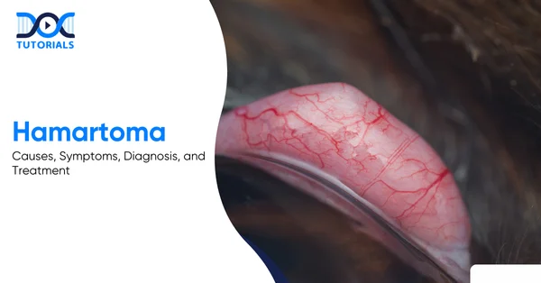
Hamartoma: Causes, Symptoms, Diagnosis, and Treatment

Hamartoma

Hamartomas are non-cancerous growths composed of the same surrounding tissues that grow in an unorganised manner, causing a tumour-like structure. The majority of them do not cause any symptoms and are not harmful. If found to be causing health issues, they are surgically removed by an appropriate surgeon. However, the cause is mainly idiopathic, and genetic syndromes are sometimes included in the aetiology. 

Although a hamartoma is non-cancerous, avoiding treatment may result in dysfunction of other organs. Hence, keep reading to learn about hamartomas, their types, and the best available treatment for managing them. 

What is a Hamartoma? 

A hamartoma is a non-cancerous, benign growth of disorganised mature tissue that can develop anywhere in the body. There are some key characteristics to identify a hamartoma, such as disorganisation in tissue growth, benign nature, location throughout the body, etc.   

Mostly harmless, hamartomas are masses of disorganised tissue that can grow anywhere in the body. However, they can hamper bodily functions if they grow big enough to suppress other organs. If hamartomas grow abnormally large, they need to be removed surgically to relieve the patient. 

Some of the key distinguishing features of a hamartoma are:

  • Benign 
  • Disorganised growth 
  • Symptoms are found incidentally 
  • Developmental error 
  • Can develop anywhere in the body

How are Hamartomas Classified?

Hamartomas do not spread to other parts of the body because they are benign and found in places such as the heart, lungs, brain, and breast, causing cardiac rhabdomyoma, hypothalamic hamartoma, etc.

Hamartomas don’t metastasise like malignant tumours and remain in one place. However, their size increases, putting pressure on other organs in the body. The following section lists the places where hamartomas can grow:

  • Heart (Cardiac Rhabdomyoma): Hamartomas in the heart can result in cardiac arrest and heart failure, which is very common in children. 
  • Brain (Hypothalamic Hamartoma): When hamartomas grow in the brain, the main changes are seen in the behaviour and mood. If they grow in the hypothalamus, epileptic seizures and early puberty are also seen. 
  • Breast: A breast hamartoma is a benign tumour that can develop at any age. However, women above 35 are more prone to breast hamartoma, which can cause breast deformities. 
  • Lungs: Pulmonary hamartomas, or lung hamartomas, are the most common type of tumour that can cause difficulty breathing or trigger pneumonia. In severe cases, tissue damage is also seen. 
  • Spleen: Splenic hamartomas are the rarest form of hamartomas, which are seen more in females than males. Hamartomas on the spleen manifest as abdominal discomfort and pain. 
  • Neck and Chest: Hamartomas in the neck region can cause abnormal swelling and hoarseness, and respiratory issues if they grow in the chest region as well. 
  • Skin: Skin hamartomas can grow anywhere on the skin, with no explicit limitation of the risk area.

What are the Causes of Hamartomas? 

The cause of a hamartoma is idiopathic; however, it is believed to result from various factors such as a genetic mutation, congenital defect, developmental abnormality, genetic defect, etc.

The exact cause of a hamartoma is still unknown, and they are rare. Scientists say that hamartomas result from a genetic mutation in one of the biological parents. However, the following list explains some of the probable causes behind developing a hamartoma:

  • Congenital Defect: In these cases, hamartomas are present at birth but are detected later in life. 
  • Developmental Abnormality: These hamartomas develop as a result of disorganised tissue growth during the foetal stage. 
  • Genetic Defect: Some hamartomas develop as a result of a genetic mutation, or they may be genetically inherited. 
  • Abnormal Tissue Growth: The affected area exhibits tissue growth in an abnormal pattern, resulting in a hamartoma. 

What are the Risk Factors of Hamartomas?

The risk factors behind developing a hamartoma include Neurofibromatosis Type 1, Pallister-Hall Syndrome, Peutz-Jeghers Syndrome, PTEN Hamartoma Tumour Syndrome, and Tuberous Sclerosis.

Although the exact cause behind hamartomas is unknown, certain risk factors make a person more likely to develop them:

  • Neurofibromatosis Type 1: Hamartomas mainly grow in the brain, spinal cord, and skin. 
  • Pallister-Hall Syndrome: Hamartomas grow on the hypothalamus. 
  • Peutz-Jeghers Syndrome: In this condition, hamartomas grow in the bladder, intestines, stomach, and lungs. 
  • PTEN Hamartoma Tumour Syndrome: In PHTS, hamartomas are seen on the breasts, GI tract, uterus, and thyroid. 
  • Tuberous Sclerosis: In this condition, hamartomas grow in various organs, such as the eyes, heart, kidneys, brain, and skin. 

What are the Symptoms of Hamartoma?

In general, hamartomas are asymptomatic; however, symptoms depend on their location, like breast hamartomas cause swelling in the breast and a palpable lump, liver hamartomas cause abdominal discomfort and pain, lung hamartomas cause respiratory distress, etc. Some of these symptoms may cause severe discomfort and pain, depending on their location.

The location of the hamartoma can trigger harmful effects, the main issue being the pressure exerted by the large mass. In severe cases, the hamartoma can turn out to be fatal when it starts pushing on the other organs. 

The following section lists the symptoms depending on the location of the hamartoma:

  • Breast: Swelling in the breast, palpable lump, or deformity
  • Liver: Abdominal discomfort and pain
  • Kidneys: Haematuria (blood in the urine) and flank pain
  • Skin: Discolouration of the skin, visible and palpable growths, and lumps
  • Lungs: Haemoptysis (blood with cough), respiratory distress, shortness of breath, cough, chest pain, and fever
  • Brain: Early puberty, behavioural changes like rage and emotional issues, headaches, visual problems, seizures, and learning difficulties. 
  • Heart: Signs of heart failure include cyanosis and oedema, chest pain, arrhythmia, and palpitation. 

How to Diagnose a Hamartoma?

If a hamartoma is suspected, the physician may order imaging tests, including USG, mammograms, X-rays, CT scans, and MRIs, depending on its location. 

Imaging tests are generally ordered to confirm the diagnosis of a hamartoma. The imaging tests include:

  • Ultrasound: The first investigation advised in cases of suspected hamartoma is ultrasound, which helps identify benign lesions, where the hamartoma appears as a well-defined mass. 
  • Mammogram: Mammography is performed in cases of breast hamartomas, which reveal a well-circumscribed mass that gives a ‘breast within a breast’ appearance. 
  • X-ray: Although not much used in other hamartomas, X-rays are helpful in the case of pulmonary hamartomas, which show a coin-shaped lesion with popcorn-like calcification. 
  • Computed Tomography: Also known as a CT Scan, Computed Tomography is very useful in lung and deeply located hamartomas, providing a detailed view of the internal structures.
  • Magnetic Resonance Imaging (MRI): MRI provides soft-tissue contrast essential for diagnosing a brain or soft-tissue hamartoma and helps develop a better treatment plan.

A biopsy might also be needed to rule out the possibility of a cancerous tumour. 

What are the Available Treatment Options for Hamartoma?

Usually, surgery is not advised; however, severe cases may need ablation therapy and GammaKnife Radiosurgery.

When the hamartoma lies too close to the essential tissues and organs, surgery becomes too risky, causing life-threatening conditions for the patient. In such severe cases, the following two methods are adopted:

  • Ablation Therapy: It uses extreme heat to destroy tissue. 
  • GammaKnife Radiosurgery: A form of radiation therapy that eliminates tissue with high precision.

What are the Complications of Hamartomas?

Generally, hamartomas are not a serious condition but can lead to complications if they grow abnormally and affect nearby organs. 

Developing hamartomas is not a serious condition; however, they can damage the nearby organs if they grow abnormally.

For example,  hamartomas in the hypothalamus can start seizures and lead to developmental delays. 

FAQs about Hamartoma

  1. What is the difference between a tumour and a hamartoma?

A hamartoma is a non-cancerous, benign growth of disorganised tissue, whereas a tumour is an umbrella term encompassing any abnormal growth, benign or malignant. Tumours result from uncontrolled cell division. 

  1. What are hamartoma and choristoma?

Hamartomas are disorganised growths of normal tissues, while choristomas are groups of mature tissues that grow in a completely different location, e.g., pancreatic tissue in the brain.

  1. Should the hamartoma be removed?

Surgical intervention to remove a hamartoma is not always required; however, the symptoms improve with surgery. 

  1. How fast do hamartomas grow?

Generally, hamartomas grow a few millimetres per year, with a volume doubling time (VDT) exceeding 400 days.

  1. Can a hamartoma become cancerous?

Although rare, malignancy can occur in patients with risk factors like Neurofibromatosis Type 1, Pallister-Hall Syndrome, Peutz-Jeghers Syndrome, PTEN Hamartoma Tumour Syndrome, and Tuberous Sclerosis. 

Conclusion

Hamartomas can be scary because they appear out of nowhere. However, they are benign and do not cause serious harm to people. Hamartomas can be managed without surgery, but surgery is more effective at improving symptoms. If you feel any abnormal lump in your body, do not hesitate to consult the appropriate physician and seek treatment.

At DocTutorials, you will find several other topics related to NEET PG, along with concise video lectures, high-yield notes, expert guidance, and more. Check out our NEET PG courses today!

NEET PG Online Live Power Pack
DocTutorials App Download Now

Latest Blogs

Venous Drainage of the Upper and Lower Limbs: Anatomy, Functions, and Clinical Insights

Venous Drainage

Understanding the venous drainage of the upper and lower limbs and the function of major superficial and deep veins, their connections, and valves is key to procedures such as venipuncture, catheterisation, and the management of venous disorders. Abnormalities or blockages in limb veins can cause varicose veins, deep vein thrombosis, and chronic venous insufficiency, posing significant health risks.

This guide covers the anatomy, functions of limb venous drainage, and clinical relevance techniques to assist students and healthcare professionals in developing a solid knowledge base.

Keep reading to learn more!

What is Venous Drainage?

Venous drainage is the return of oxygen-poor blood from body tissues, particularly the arms and legs, to the heart. It depends on one-way valves and muscle movement to push blood upward against gravity. Together, superficial and deep veins support healthy circulation and prevent blood pooling or tissue congestion.

The venous vessels channel deoxygenated blood from the tissues and drain it back into the heart. In limbs, venous drainage refers to the return flow from the arms and legs. The veins are responsible for pumping oxygen-depleted blood to the heart, with the help of a one-directional valve and the pumping effects of the muscles. 

The venous system in each of the limbs has been bisected into the superficial veins (the veins beneath the superficial fascia, or directly beneath the skin) and the deep veins (that is, veins beneath the deep fascia). Collectively, they preserve the circulatory balance through the draining of the blood in the muscles, skin, and bones into the central circulation.

The differences between veins and arteries are that the arteries pump the blood away under high pressure, whereas the veins pump it back under low pressure. Veins have valves (more particularly leg veins) that do not allow the blood to be pushed back, and skeletal muscles squeeze deep veins as pumps force the blood upwards. Proper venous drainage is essential to avoid the stagnation of blood and congestion of the tissue.

Anatomy and Functions of Venous Drainage in Upper and Lower Limbs

There are complex venous systems in both the lower and upper extremities. Major superficial veins are located within the subcutaneous tissue, whereas paired deep veins accompany major arteries found beneath the deep fascia.

The venous drainage systems in the upper and lower limbs are connected to each other through perforating veins. One-way flow towards the heart is guaranteed by valves, and pumping is facilitated by muscle movements. This anatomy aids in the explanation of how blood is brought back to the extremities as well as the relevance of some veins in clinical practices.

Here is a detailed overview:

Upper Limb Venous Anatomy

The veins in the arm have a superficial network (basilic and cephalic) and arterial veins. The cephalic vein and the axillary/subclavian vein are lateral to one another, but the basilic vein is used as the median of the two, and the brachial veins are linked with these two veins to create the axillary vein. They are united by the median cubital vein at the elbow, which is a usual venous access site.

Superficial veins of the upper extremity consist of the medial basilic vein and the lateral cephalic vein, which are joined together at the elbow by the median cubital vein. The basilic vein originates in the dorsal venous arch of the hand and runs up the medial forearm, underneath the fascia close to the arm, to join the brachial veins to form the axillary vein.

The cephalic vein originates at the hand, passes along the anterolateral side of the forearm and arm, over the deltoid and pectoralis major muscles, and empties into the axillary or subclavian vein. The cubital fossa in the basilic vein is a junction between the basilic and distal veins and is a popular site of venous access.

  • Major Superficial Veins (upper limb): The primary superficial veins of the arm are the cephalic (lateral) and basilic (medial) veins and are directly beneath the skin. The cephalic vein empties into the axillary vein above the shoulder, and the basilic vein is deep and empties into the axillary vein above the shoulder. They are connected to each other at the elbow by a position known as the median cubital vein.
  • Deep Veins (upper limb): The deep veins are venae comitantes, which accompany arteries under the fascia. As an illustration, the brachial veins go hand in hand with the brachial artery. These extensive veins are sustained by the arm muscles and end up joining to produce the axillary vein. Notably, the pulses in arteries aid in the movement of blood in such veins.
  • Connections (perforators): In the arm, small perforating veins connect the deep and superficial ones, through which the blood enters the deep veins. The upper limb venous system offers a good way of blood drainage throughout the hand and the forearm. The deep veins are the ones of high capacity, and the superficial veins, which are numerous in valves, play important clinical roles in venipuncture and grafting.

Mechanisms Aiding Venous Return 

The veins of the upper limbs depend on a number of factors to be drained efficiently:

  • Muscle Pump: The muscles on the forearm and arm contract, causing veins to squeeze, forcing blood back upwards.
  • Venous Valves: One-way valves avoid back-flow during relaxation of muscles.
  • Gravity Assistance: As the arm is moved or raised up, gravity will assist in pumping the blood to the heart.

Lower Limb Venous Anatomy

The veins in the leg comprise the superficial (great and small saphenous) and the deep (tibial, popliteal, and femoral) veins. The dorsal venous arch supplies the great saphenous and small saphenous veins, respectively, and empties into the popliteal and femoral veins. Deep veins follow the arteries, and the valves, muscles, and calf pump assist in the effective circulation of blood.

The view of the upper thigh (femoral triangle) demonstrates the great saphenous vein in the middle and the femoral vein under the inguinal ligament. The longest vein in the body is the great saphenous, which begins at the dorsal venous arch of the foot and proceeds along the medial leg and medial thigh and empties into the femoral vein below the inguinal ligament. The small saphenous vein is a dorsal lateral vein of the foot, which develops behind the lateral malleolus distal to the calf into the popliteal vein at the knee.

  • Superficial Veins (lower limb): The great saphenous (medial) and small saphenous (posterior) are the main components of the superficial leg veins that are located right beneath the skin of the lower limb above the deep fascia. They drain the foot and superficial tissues, and the great saphenous vein empties into the femoral vein, and the small saphenous vein empties into the popliteal vein.
  • Deep Veins (lower limb): Deep veins are located isotopically to the deep fascia and are typically accompanied by arteries. Posterior tibial, fibular, and anterior tibial veins are located correspondingly to the arteries and converge at the knee, forming the popliteal vein that joins the femoral vein in the thigh. The profunda femoris vein empties into the distal section of the femoral vein, known as the external iliac vein. The superior gluteal vein and the inferior gluteal vein empty into the internal iliac vein in the pelvis and the gluteal region, respectively.
  • Venous Valves and Calf Pump: The veins in the legs contain a higher number and stronger valves than those in the arms to prevent backflow caused by gravity. Calf muscles are a sort of muscle pump that contracts, compressing deep veins to push the blood upwards, but relaxes to allow the backflow to be prevented by valves. The venous return of the legs depends on this mechanism.
  • Pressure Gradients: Breathing movements also help the leg venous return: the lowered thoracic pressure and the increased abdominal pressure, which occur in the course of inspiration, promote the flow of blood in the legs towards the chest.

Superficial and Deep Veins of the Upper and Lower Limbs: Key Differences

The deep veins accompany the arteries and are the primary high-capacitance pathway since they occur within the subcutaneous tissue, whereas superficial veins run in isolation within subcutaneous tissue and tend to support thermoregulation. Superficial veins get emptied into a deep system, which is under the fascia. 

The following is a comparison between the superficial and deep venous systems in the limbs:

AspectSuperficial VeinsDeep Veins
LocationJust under the skin (subcutaneous)Muscles followed beneath the deep fascia
Relationship to ArteriesNo independent course of associationMajor arteries have venae comitantes, which accompany them
FunctionDrain the skin and subcutaneous structures; thermoregulation.Drain distribution of muscle and bone; principal recapillary, facilitated by the movements of the arteries
Examples (Upper Limb)Cephalic, BasilicBrachial Vectors, Axillary Vein
Examples (Lower Limb)Great and Small SaphenousPosterior/Anterior Tibial, Popliteal, Femoral veins

Functions of the Limb Venous System

The limb venous system carries deoxygenated blood back to the heart, stores blood, and helps regulate body temperature. It depends on muscle movement, one-way valves, breathing-related pressure changes, and perforator veins to maintain smooth, upward blood flow from the limbs to the central circulation.

The major role of the limb venous system is to pump the deoxygenated blood into the heart to be oxygenated. Veins are also used as reservoirs of blood within the limbs and assist in maintaining body temperature (superficial veins are capable of dilating to get rid of the heat). In veins, there are venous valves that ensure unidirectional movement, which is an essential attribute in the low-pressure venous system. 

Mechanisms involved are functional, and they include:

  • Muscle Contraction: The contraction of limb muscles (particularly, calf muscles in the legs) squeezes neighbouring deep veins, forcing the blood towards the heart. This type of muscle pump is one of the significant sources of venous return during activity.
  • Venous Valves: The walls of each segment of the veins include valves, which close as the blood attempts to fall backwards, to maintain a unidirectional flow towards the heart. Numerous valves are available in the legs to counter the force of gravity.
  • Pressure Gradients: The variations in thoracic and abdominal pressures at inhalation produce suction that pulls venous blood to the chest.
  • Perforator Veins: The limbs have the perforating veins that interconnect the superficial and deep veins and carry blood in the superficial reservoirs to the larger deep veins to maintain the proper flow of blood.

The combination of these characteristics ensures that blood returns to the central circulation, even at the opposite end of a hand or foot.

Common Disorders of the Limb Venous System

Disorders of limb veins are characterised by clots or dystrophy of valves, such as DVT, varicose veins, and chronic venous insufficiency. Reasons are stagnant flow, incompetence of the valve, or trauma, causing swelling, pain, and potential complications.

Limb vein disorders are frequent and may vary between superficial and severe complications, which may be life-threatening. They are usually caused by stagnation of blood flow, valve failure, or vessel damage, leading to pain, swelling, and permanent complications. It is imperative to understand these conditions to detect them early, prevent them, and manage them effectively.

Here is a detailed overview:

  • Deep Vein Thrombosis (DVT): A DVT is the clotting of blood in a deep vein of a limb, mostly the legs. It impedes blood flow in veins, resulting in pain, swelling, and tenderness. Among risk factors are immobility, surgery, or hypercoagulability. Pulmonary embolism is the primary risk, where a piece of clot is carried to the lungs, and this may be fatal. Management aims at anticoagulation, compression, and prevention, and high-risk patients are subjected to prophylaxis.
  • Varicose Veins: Varicose veins are dilations of twisted superficial veins that typically occur in the lower extremities due to chronic failure of valves, which cause an accumulation of blood and elevation of pressure. The symptoms are painful, heavy, itchy, and bulging veins. These include risk factors such as heredity, pregnancy, obesity, and prolonged standing. When left unattended, they may lead to chronic venous insufficiency, skin alterations, and ulcers. Therapy involves compression stockings, endovenous ablation, or vein stripping.
  • Chronic Venous Insufficiency (CVI): CVI is long-term impaired venous return, often after DVT or chronic varicose veins. Ruptured valves result in blood stasis, which results in ankle ulcers, swelling of the legs, and discolouration of the skin. Approximately 20-50 % of patients with DVT develop post-thrombotic syndrome (a type of CVI) several years later. Treatment involves leg raising, compression, and ulcer management.
  • Other Conditions: Superficial thrombophlebitis (clot in a superficial vein) may be known to cause local pain and redness, but it is typically not as harmful as DVT. Venous disease may be connected with lymphoedema (however, the condition does not involve veins but lymph vessels). Another thing to note is that there are certain systemic pathologies (e.g., heart failure) that may cause generalised venous oedema in the extremities.

The typical symptoms of venous disorders are pain in the legs, swelling, cramping, heaviness, and skin changes (itching, hyperpigmentation, and ulcers), and can be detected through a reflux vein patency test (e.g., Doppler ultrasound). The goals of treatment of venous disorders usually include the restoration of valves or removal/occlusion of diseased veins and the prevention of clot formation.

FAQs about Venous Drainage

  1. Why do limb veins have valves?

Valves maintain the flow of blood towards the heart, and they prevent pooling and reverse movement. They play a significant role in the legs, where the blood opposes gravity. Muscle contractions push blood upward, and valves close during relaxation to stop backflow. Sitting or standing might result in oedema without valves.

  1. Why is the great saphenous vein clinically important?

The great saphenous vein (GSV) is the longest superficial vein, and it has a course that runs down the medial leg and the thigh and empties into the femoral vein. It is commonly utilised as a bypass graft to the coronary artery, and it is a common location for varicose veins. Its anatomy knowledge is useful in leg and groin surgeries.

  1. What is the median cubital vein used for?

At the inner elbow, the cephalic and basilic veins are joined together in order to form the median cubital vein. It is superficial, large, and easily accessible, and thus, suitable for venipuncture and insertion of IV.

  1. Can veins become diseased?

Yes. Some of the common issues are thrombosis (DVT in deep veins, superficial thrombophlebitis in superficial veins) and valve failure to the point of reflux and varicose veins. This could cause chronic venous insufficiency and leg ulcers with time.

  1. How does venous return differ in arms vs. legs?

Mechanisms are similar, but leg veins face higher hydrostatic pressure. Legs have more valves and rely on the calf muscle pump, while arm veins experience less gravity and are less prone to varicosities.

Conclusion

The venous drainage of the upper and lower limbs is a well-organised system of superficial (cephalic, basilic, great and small saphenous) and deep veins (brachial, femoral, etc.) that efficiently return blood to the heart. Valves and muscle pumps maintain one-way flow, while disruptions can cause conditions like DVT and varicose veins. A solid understanding of limb venous anatomy is essential for medical students and clinicians for procedures like vein grafting and managing venous disorders.

DocTutorials offers structured resources—concise notes, case-based Qbanks, flashcards, and expert mentorship—designed to help students master limb venous anatomy, clinical correlations, and procedural skills. Leverage these tools to strengthen your clinical knowledge, enhance practical skills, and confidently navigate exams and patient care.

Check out our NEET PG study materials for more such content!

NEET PG Online Live Power Pack
DocTutorials App Download Now

Latest Blogs

Necrotising Enterocolitis: Causes, Risk Factors, Symptoms, Diagnosis, and Treatment Options

Necrotising Enterocolitis

Necrotising enterocolitis (NEC) refers to a gastrointestinal emergency that is common in preterm neonates in the NICU. Some infants experience mild NEC, while others can experience life-threatening severe symptoms. NEC can also occur in term babies who are born with asphyxia, heart disease, polycythaemia, sepsis, and hypotension.

Necrotising enterocolitis is a crucial and high-yielding topic for NEET-PG aspirants and can help you achieve a good score in the NEET-PG exam. In this guide, we are going to cover all the information about necrotising enterocolitis, including causes and risk factors, symptoms and diagnosis, treatment options, and other crucial aspects.  

Keep reading to learn more!

What is Necrotising Enterocolitis?

NEC is a common gastrointestinal condition in premature babies and can lead to perforations as well. Babies with low birth weight and those fed enterally have a higher risk of developing this condition. It can be life-threatening if left untreated. 

Necrotising enterocolitis is a neonatal gastrointestinal disorder characterised by ischaemic and inflammatory necrosis of the intestinal wall, leading to systemic sepsis and potential perforation.

Necrotising enterocolitis is a common disease in premature babies. Almost 90% of all babies who suffer from NEC are premature babies. The incidence of NEC is about 1 in 1000 premature babies. Moreover, low birth weight babies and enterally fed babies are at a higher risk of developing necrotising enterocolitis.

What are the Causes and Risk Factors Involved?

As of now, the exact cause of necrotising enterocolitis is not known. Healthcare providers usually link this condition to the underdeveloped immunity and circulatory systems of premature babies, which provide an ideal environment for intestinal pathogens to affect the intestines. Major causes include premature birth, low birth weight, term babies born with asphyxia or heart disease, etc. 

The exact cause of necrotising enterocolitis is not yet known. However, healthcare providers believe it is due to the underdeveloped immunity of the premature babies. The immune system takes a long time to develop to its full potential, usually crossing infancy and childhood. 

This gives the intestinal pathogens an opportunity to infest the intestines. Since the immunity is underdeveloped, the body faces a harder time fighting the pathogens.

Moreover, the circulatory system of a premature baby is not fully effective. This deters the flow of blood to the intestines, leading to a decreased extent of the immunoglobulins and ischaemic damage to the intestines.

What are the Major Risk Factors of Necrotising Enterocolitis?

The risk factors of necrotising enterocolitis do not cause the disease directly, but make it more likely to develop:

  • Prematurity is the most consistent risk factor
  • Hypoxic-ischaemic injury leading to gut ischaemia and inflammation
  • Enteral feeding 
  • Lack of breastfeeding
  • Use of acid suppression drugs
  • Patent ductus arteriosus
  • Term babies born with asphyxia, heart disease, polycythaemia, sepsis, and hypotension
  • Low birthweight babies

What are the Symptoms and Diagnostic Modalities for NEC?

NEC has both systemic and GI manifestations, and the course can be very rapidly progressive. Clinical symptoms generally include feeding intolerance and increased gastric residuals, with bloody stools, lethargy, and abdominal wall erythema being the advanced symptoms. Diagnostic methods generally include lab tests and radiological diagnosis. 

Clinical Features

The clinical features, including signs and symptoms for NEC, are tabulated below:

Early symptoms– Feeding intolerance- Increased gastric residuals- Abdominal distension- Vomiting
Advanced symptoms– Bloody stools- Abdominal wall erythema- Apnoea and bradycardia- Lethargy- Temperature instability- Shock in severe cases
Physical Examination– Tense, distended abdomen- Decreasing bowel sounds- Abdominal tenderness- Palpable abdominal mass (late)

Diagnostic Testing

Laboratory Findings– Thrombocytopenia- Metabolic acidosis- Leukocytosis or leukopenia- Increased levels of C-reactive protein
Radiological Diagnosis (Cornerstone)– Abdominal radiography- Pneumatosis intestinalis is pathognomonic- Portal venous gas – Dilated bowel loops – Free air under the diaphragm (perforation) 

Bell’s Staging (Essential for Examination) 

Bell’s staging is used to assess the severity of necrotising enterocolitis in a newborn. This helps to decide the management for the affected baby.

StageFeature
Stage ISuspected NEC
Stage IIDefinite NEC
Stage IIIAdvanced NEC with perforation

What are the Treatment Options for NEC?

NEC can be a life-threatening condition. Hence, early diagnosis, prompt treatment, and prevention of complications can help the baby recover faster and prevent mortality. The main aims of treatment include maintenance of nutrition, clearing of the pathogenic load, and prevention of recurrence.

Your baby’s intestines need time to rest and heal. The first part of treating NEC is to stop all feedings by tube and mouth. Instead, your baby will get the fluids and nutrition he or she needs through an intravenous line (IV).

Your baby may also receive several other therapies, like:

  1. Nasogastric Tube 

A provider gently places a long, narrow tube through the nose and sometimes through the mouth, down to the stomach, allowing removal of accumulated gas and fluids.

  1. Antibiotics 

These help in combating bacterial infections. May include ampicillin, clindamycin, or metronidazole, and gentamicin. 

  1. Surgery

About one quarter of babies with NEC need surgery to remove sections of dead intestinal tissue and to repair any holes. Sometimes, your baby’s provider may do an ostomy procedure. This surgery involves:

  • Creation of a small opening, called a stoma, in the abdomen.
  • Connecting the large intestine to the stoma.
  • Allows waste to leave the body through the stoma into a bag worn outside the body.

When your baby gets a bit stronger, the intestines can be reattached and returned to their original place inside the abdomen. If your baby is tiny, less than 1 pound, or too sick for surgery right away, your provider may insert a drain or a catheter into the abdomen. 

A drain can help your baby feel better sooner by draining out unhealthy and infected fluids and gas. If more surgery is needed, it will be scheduled at a later time when your baby is larger and healthier.

Prevention Methods for NEC

If you have a risk of having a premature baby, the doctors would prescribe you some corticosteroid injections. These steroids help in boosting the foetus’s development, thus reducing the chances of intestinal or lung issues.  

Also, initiating early breastfeeding in a baby helps to develop their immune system. The breast milk contains a lot of essential immunoglobulins, which help your baby develop their passive immune system.

FAQs about Necrotising Enterocolitis (NEC)

  1. What is the major causative factor of NEC?

While the exact causes of NEC are not yet known, it generally occurs in premature infants, whose underdeveloped intestinal and circulatory systems create an ideal environment for intestinal pathogens. 

  1. What is the treatment modality for NEC?

Treatment of NEC involves supportive care, nasogastric feeding, surgical treatment, IV fluids and antibiotics, and close monitoring of the baby.

  1. What is the mortality rate for NEC?

The overall mortality rate for NEC usually ranges between 10% and 50%.

  1. What are the long-term complications of NEC?

Even after surgery, some infants can face problems like short bowel syndrome, inadequate digestion, and poor growth.

  1. What early signs appear in NEC?

The early signs of NEC are abdominal distension, bloody stools, poor feeding, and bilious vomiting or stools.

Conclusion

Necrotising enterocolitis is a serious disease as well as a hot topic for the NEET-PG examination. It is a high-incidence disease in newborns, especially in premature babies. It is a curable and preventable condition as well.

What is important to know about NEC is its early detection and prompt cure. To understand more in detail about NEC, and other conditions, check out DocTutorials. We offer concise and crisp video lectures, clinical Qbanks, exam-focused notes, and mind maps to aid aspirants and guide them towards a better understanding of medical concepts. Explore our NEET PG courses today!

NEET PG Online Live Power Pack
DocTutorials App Download Now

Latest Blogs

Lead Poisoning: Causes, Symptoms, Diagnosis, and Treatment

Lead Poisoning

Lead poisoning is a silent enemy that has remained with us despite great victories for the environment over the last few decades. One of the sneakiest things about lead is that it quite successfully mimics certain vital minerals, so brain dysfunction, blood disorders, and renal diseases go rather unnoticed for some time. 

For the NEET PG exam candidates, plumbism is a high-yielding topic in toxicology, which is well known for its classic type of presentation and very straightforward treatment regimens. This guide covers lead poisoning symptoms, causes, diagnosis, treatment, and other crucial aspects. 

Keep scrolling!

What is Lead Poisoning?

Lead poisoning is a toxic condition that results from the accumulation of lead in a person’s body over time, mostly due to long-term environmental, occupational, or household exposure. Lead imitates the qualities of minerals that are essential for the body, like calcium, iron, and zinc, and thus it interferes with the enzymatic pathways, the nervous system, blood formation, kidneys, and brain development of the affected individual (usually children).

Lead poisoning, or plumbism, is one such clinical condition that is toxic in nature and is the result of the gradual buildup of lead (Pb) in the human body. It is a case of long-term exposure to environmental, workplace, or household sources of lead, like lead-based paint, drinking water, batteries, and cosmetics.

Lead is a divalent metal that has a very close similarity to the three essential minerals, namely calcium, iron, and zinc. For this reason, it competes with these ions in biological pathways, thus disrupting multiple enzyme systems and impairing cellular functions.

After lead absorption, it is carried to the brain, peripheral nerves, bones, blood, liver, and kidneys, where it interferes with neurotransmission, inhibits heme synthesis, and damages renal tubules. Most of its symptoms originate from the nervous system, and neurotoxicity is one of the features that the organ system expresses most distinctly.

Children are highly vulnerable, as they absorb lead more readily through the gut and their blood-brain barriers are still immature. Even minimal exposure may cause permanent cognitive deficits, behavioural changes, and developmental delays.

What are the Causes of Lead Poisoning?

Lead exposure is usually a result of existing surroundings, routine activities, and certain workplaces. It is very important to understand these common sources to be able to intervene early and reduce the risk of chronic harm.

Here are the common causes of lead poisoning: 

  • Occupational Exposure 

Workplaces engaged in battery production, smelting, plumbing, printing presses, and cable production, and the paint industry prominently involve lead-containing materials. The prolonged inhalation of fumes or dust is the primary mode of exposure for people who work in such industries, making them the group at the highest risk and accounting for the largest number of cases.

  • Household Sources 

There might be lead-based paints in older homes, which, as they crumble, release lead-containing dust. The presence of lead may also be inferred from the use of old plumbing systems, soldered household items, children’s toys, and traditional cosmetics such as surma/kohl. People are daily exposed to these things, and such exposure usually goes unnoticed.

  • Food and Water Contamination 

Lead can go to the food if it’s stored in a lead-soldered can or kept in a traditional lead container or on ceramics that are not properly glazed. The release of heavy metals from contaminated groundwater is likewise a major cause of lead intake in the human body over time, especially if the area is going through industrialisation or mining.

  • Traditional Practices 

Some Ayurvedic or herbal medicines might deliberately contain heavy metals such as lead or be contaminated as a result of pollution. Moreover, certain generational heritage-based folk remedies that have been passed down throughout the years may also be the source of your unnoticeable exposure.

  • Environmental Pollution 

The lead components of the industrial waste, petrol residues, and other such leftover material may finally settle on the ground (on soil and/or crops). Thus, children playing in lead-contaminated soils and people living near industrial areas are the ones who are mostly at risk.

What are the Risk Factors of Lead Poisoning?

Certain groups and living conditions may raise the chances of lead absorption and toxicity to a great extent. Knowing the risk factors associated with it is helpful for prioritising screening and preventive measures for the most affected people.

Now, let’s see the risk factors of lead poisoning: 

  • Young Children Aged Between 1 and 6: Kids at an early age habitually put all kinds of things in their mouths, and their nervous systems are still in the process of development; therefore, they are the most susceptible to even small amounts of lead taken from dust, paint chips, soil, toys, etc.
  • Pregnant Women: Lead is one of the harmful substances that the placenta does not filter out as it reaches the foetal development stage. If a mother has lead in her bones, which is stored from earlier exposures, the lead may be released during pregnancy, thus increasing the risks of miscarriage, preterm birth, developmental deficits, and other problems.
  • Poor Nutrition: When one is iron, calcium, or zinc deficient, the lead absorption through the gastrointestinal tract increases. Malnourished people have a greater chance of being exposed to a higher level of the toxin.
  • Living in Older Houses with Lead Paint: The homes that were built before the regulations that limit the use of lead were put in place are likely to have been painted with lead-based paints. Such paint may deteriorate, especially during renovations, thus creating dust that is contaminated with lead and can easily be inhaled or ingested.
  • Workers Without Adequate Safety Measures: People engaged in roles such as battery factories, smelting units, construction, painting, or cable manufacturing may be exposed to lead dust and fumes. Lack of protective equipment or improper use significantly escalates their risk.

What are the Symptoms of Lead Poisoning?

Lead toxicity produces a very diverse range of clinical manifestations as the metal builds up in different organs of the body over time. The intensity and signs of the condition vary according to the dose, time of exposure, and the patient’s age.

  1. Neurological Symptoms

When lead interferes with neurotransmission and injures both central and peripheral nervous structures, it results in behavioural, cognitive, and motor control changes:

  • Irritability and Behaviour Changes: Behavioural changes associated with neurotoxicity are illustrated by mood changes, hyperactivity, shortened attention span, and social withdrawal, especially in children.
  • Cognitive Decline and Poor Concentration: Lead interferes with synaptic formation and neural signalling, which causes difficulties in learning, memory, and concentration.
  • Peripheral Neuropathy (wrist drop, foot drop): Over time, motor nerves are damaged due to chronic exposure; thus, weakness of the extensor muscles and the development of typical nerve palsies occur.
  • Encephalopathy: Headache, vomiting, seizures, and altered sensorium are some of the symptoms of lead toxicity, along with increased intracranial pressure, and it may even progress to coma if not treated.
  1. Gastrointestinal Symptoms

These symptoms appear when lead stops the function of smooth muscles in the digestive tract and inflames the gastrointestinal mucosa, which is why the patient experiences continuous digestive discomfort. 

  • Lead Colic (severe abdominal pain): A typical case involves extremely severe, cramping pain caused by spasm of intestinal smooth muscle.
  • Constipation, Anorexia, Vomiting: Difficulty in bowel movements, vomiting, and loss of appetite occur as digestive tract motility gradually decreases.
  • Metallic Taste, Weight Loss: Changed taste sensation leads to less food intake and slow weight loss.
  1. Haematological Symptoms

They happen because lead inhibits the key enzymes of heme synthesis and disturbs red blood cell maturation, which eventually leads to defective erythropoiesis.

  • Microcytic Hypochromic Anaemia: The failure of heme production causes the blood cells to be small and pale, which means the oxygen-carrying capacity of the cells is lowered.
  • Basophilic Stippling of RBCs: Yet another diagnostic sign is the presence of typical coarse granules in the blood cells due to impaired RNA degradation.
  • Fatigue, Pallor: The reduction of haemoglobin causes the patient to feel tired all the time, experience a lack of energy during physical activities, and see the skin and mucous membranes becoming pale.
  1. Renal and Musculoskeletal Effects

These symptoms are a result of damage to renal tubules due to chronic lead buildup in the kidneys and deposition in bones and joints, thus causing metabolic and structural changes. They include:

  • Interstitial Nephritis: The kidney damage caused by lead in the tubules is the main reason for renal filtration impairment, proteinuria, and gradual kidney failure.
  • Hyperuricemia (“saturnine gout”): Decreased uric acid excretion causes increased serum urate levels, resulting in joint inflammation similar to gout.

How is Lead Poisoning Diagnosed?

Diagnosis of lead poisoning is based on a mixture of lab tests, imaging results, and probes that help figure out how deeply the patient has been poisoned and how many organs have been affected.

  1. Laboratory Findings

The tests mentioned here directly detect lead levels in the blood and reveal typical haematological changes due to impaired heme synthesis.

  • Blood Lead Level (BLL):
    • 5 g/dL: Points to exposure to a worrying level, and the source needs to be evaluated.
    • 45 g/dL: In such a case, where a patient is showing symptoms, the use of chelation therapy is advised.
    • 70 g/dL: A medical emergency is signalled because of the risk of severe neurological damage.
  • Zinc Protoporphyrin (ZPP): An elevated ZPP level reflects the concept of inhibited heme synthesis and aligns with the idea of chronic lead exposure.
  • Free Erythrocyte Protoporphyrin Raised: A marker of heme production disruption, most of the time it is at a high level before the onset of anaemia.
  • CBC Reveals Microcytic Anaemia with Basophilic Stippling: A peripheral smear shows small, pale RBCs and coarse granules, both of which are the typical features of lead toxicity.
  1. Imaging

Radiological studies serve the purpose of locating lead-induced changes in the skeletal system, helping clinicians, especially with diagnoses of children suffering from long-term lead exposure.

  • Children’s Radiographs: May show thickened metaphyseal lead lines at growth plates, which are areas where lead has been accumulated for a long time in bones.
  1. Additional Tests

The aim here is to determine how well the organs work and to keep an eye on the excretion process of lead during treatment and after it has finished.

  • Renal Function Tests: For the evaluation of kidney damage that can be a consequence of chronic tubular injury. 
  • Urine Lead Levels (post-chelation): It is a measurement taken after chelation therapy to evaluate the amount of lead that is being released and excreted. 

What are the Treatment Options for Lead Poisoning?

Lead poisoning management largely depends on removing the patient from the lead source, stabilising their condition, limiting lead absorption, and finally chelating the lead.

Let’s take a look at the first line and supportive treatment options for lead poisoning:

  1. Immediate Measures

These measures remove the patient from the lead source as fast as possible and stabilise the patient’s physiology to prevent further damage:

  • Remove Patient from Exposure Source: Immediate stopping of the patient’s contact with the occupational, environmental, or household source is the very first and most vital step. 
  • Rehydrate and Maintain Nutrition: Proper hydration and dietary intake (particularly iron and calcium) will not only lower lead absorption but also be supportive of the general recovery process.
  1. Lead Poisoning Antidotes (Chelating Agents)
DrugIndicationKey Notes
EDTA (Calcium Disodium EDTA)Moderate-severe toxicityIV: May cause nephrotoxicity
Dimercaprol (BAL)Severe toxicity, especially with encephalopathyOften combined with EDTA for severe cases
D-PenicillamineMild-moderate chronic poisoningOral: Used for long-term therapy
Succimer (DMSA)Preferred in childrenOral: Well-tolerated
  1. Supportive Care

Supportive treatment deals with the complications of lead poisoning in the nervous system, blood, and kidneys, and also prepares the patient for chelation therapy, which may be required.

  • Control of Seizures in Encephalopathy: Seizures associated with lead intoxication can be controlled with the administration of benzodiazepines or other anticonvulsants.
  • Treatment of Anaemia: Iron therapy or transfusion may be necessary to correct microcytic anaemia that is caused by the inhibition of the heme synthesis pathway.
  • Monitoring of Renal Function: It should be regularly monitored because long-term lead exposure may lead to renal tubular dysfunction, and this is a very common side effect of chelation therapy.

What are the Complications of Untreated Lead Poisoning?

When lead poisoning persists for a prolonged period without treatment, the metal builds up in the body and causes permanent damage, in many cases, organ failures as well. Children remain the most vulnerable to this condition, as their bodies absorb more lead when their nervous systems are still in the development phase.

Below is a detailed discussion on the complications caused by untreated lead poisoning: 

  • Permanent Cognitive Impairment in Children: Chronic lead exposure alters synaptic development and neuronal maturation, resulting in permanent decreases of IQ, learning ability, attention, and behaviour.
  • Developmental Delay: Infants and young children may be less mobile, have delayed speech, show less interest in social interaction, and have impaired cognitive functioning.
  • Chronic Nephropathy: The tubular injury caused by lead can evolve into interstitial nephritis, a urine concentrating defect, proteinuria, and eventually chronic kidney disease.
  • Infertility and Miscarriage: Lead impairs reproductive hormones, damages gametes, and crosses the placenta; the risks of miscarriage, preterm birth, and decreased fertility in both men and women are elevated.
  • Peripheral Neuropathy: Exposure over a long period damages peripheral nerves, thus the patient experiences motor weakness, sensory deficits, and typical wrist or foot drop.
  • Stunted Growth: Lead disrupts bone metabolism and endocrine regulation; thus, children who are chronically exposed have reduced height growth.
  • Cardiovascular Hypertension due to Renal Damage: Chronic nephropathy changes renin-angiotensin regulation and causes vasoconstriction, thus leading to persistent hypertension.

How to Prevent Lead Poisoning?

Preventing lead poisoning involves different layers of strategies that focus on the root causes in homes, workplaces, and nature. Moreover, early testing and public health interventions are essential in lessening the risk, particularly to sensitive groups like kids and expectant mothers. 

The following actions aim to eliminate or at least minimise lead exposure from the daily environment, the workplace, and the community:

  • The inspection of homes, especially old buildings, for deteriorated lead-based paint is a must, as the dust coming off the paint is nowadays the main source of lead exposure. 
  • Children should be discouraged from playing in the ground near roads, old buildings, and industries, which are always areas of lead deposition.
  • Children within the age bracket of 1 to 5 years should have their blood lead levels checked regularly, especially if they reside in an old house or a contaminated area. 
  • The use of personal protective equipment and safety procedures by workers in industries such as battery manufacturing, smelting, and construction is a must. 
  • Lead pipes, solder, and fixtures are the main culprits of lead in drinking water that develops in most cases over a long period of time. So, the replacement of these parts with lead-free ones greatly contributes to the prevention of prolonged consumption, especially in old houses and public buildings.
  • Disposal of industrial waste in an environmentally friendly manner is not only good for the planet but also for the health of people living nearby.

FAQs about Lead Poisoning

1. What’s the most common symptom of lead poisoning in children?

Children with lead poisoning usually show crankiness, fall behind in their development, experience abdominal pain, and have difficulties in learning.

2. What’s the antidote for lead poisoning?

Doctors use chelating agents like EDTA, BAL, DMSA (succimer), or D-penicillamine to treat lead poisoning. The selection of an antidote depends on the severity of the symptoms.

3. How do you diagnose plumbism?

To diagnose plumbism, you need to check blood lead levels, run a CBC and ZPP, and sometimes rely on radiological findings.

4. Can you reverse lead poisoning?

Neurological damage may be irreversible, especially in children. Diagnosing it early and treating it fast significantly improves outcomes.

5. Who’s most at risk for lead poisoning?

Children, pregnant women, and anyone working in industries dealing with batteries, paint, smelting, or manufacturing face the highest risk of lead poisoning.

Conclusion

Lead poisoning is usually thought of as a toxicological curiosity from the past, but in reality, it remains an active clinical and public health problem. It retains equal importance for NEET PG candidates. Aetiology, symptomatology, diagnosis, and treatment based on proof are highly relevant toward fetching good marks in the examination paper and identifying actual cases where treatment can make a difference.

DocTutorials assists students who intend to prepare basic and conceptual knowledge about Forensic Medicine through organised learning and preparation for exams. Lead poisoning is a high-yield topic because it is continuously asked in entrance examinations. 

Instead of cramming notes, understanding the concept with our video lectures and expert guidance will ensure long-term memory retention and application in real scenarios with confidence.

Check out our NEET PG courses today!

NEET PG Online Live Power Pack
DocTutorials App Download Now

Latest Blogs

NEET PG Online Live Power Pack
DocTutorials App Download Now